AI Generated Quiz
Secondary 3 Biology Human Physiology Quiz
Free Sec 3 Biology Human Physiology quiz, Kimi2.6 AI version, with questions, answers, and O Level-style practice for Singapore students.
These static practice materials are generated from the site's syllabus and paper-generation workflow, with source and model context shown so students and parents can evaluate the material before use.
Questions
Secondary 3 Biology Quiz - Human Physiology
Name: _________________________________ Class: _______________
Date: _______________ Score: ______ / 40
Duration: 35 minutes Total Marks: 40 marks
Instructions: Answer all questions. Write your answers in the spaces provided. For questions involving diagrams, label clearly. Marks are shown in brackets [ ].
Section A: Multiple Choice (Questions 1–5) [5 marks]
Choose the correct answer for each question. Write your answer (A, B, C, or D) in the box provided.
1. Which blood vessel carries oxygenated blood from the lungs to the heart?
| A | pulmonary artery | | B | pulmonary vein | | C | vena cava | | D | aorta |
Answer: [ ]
[1 mark]
2. The diagram below shows a section through the human heart.
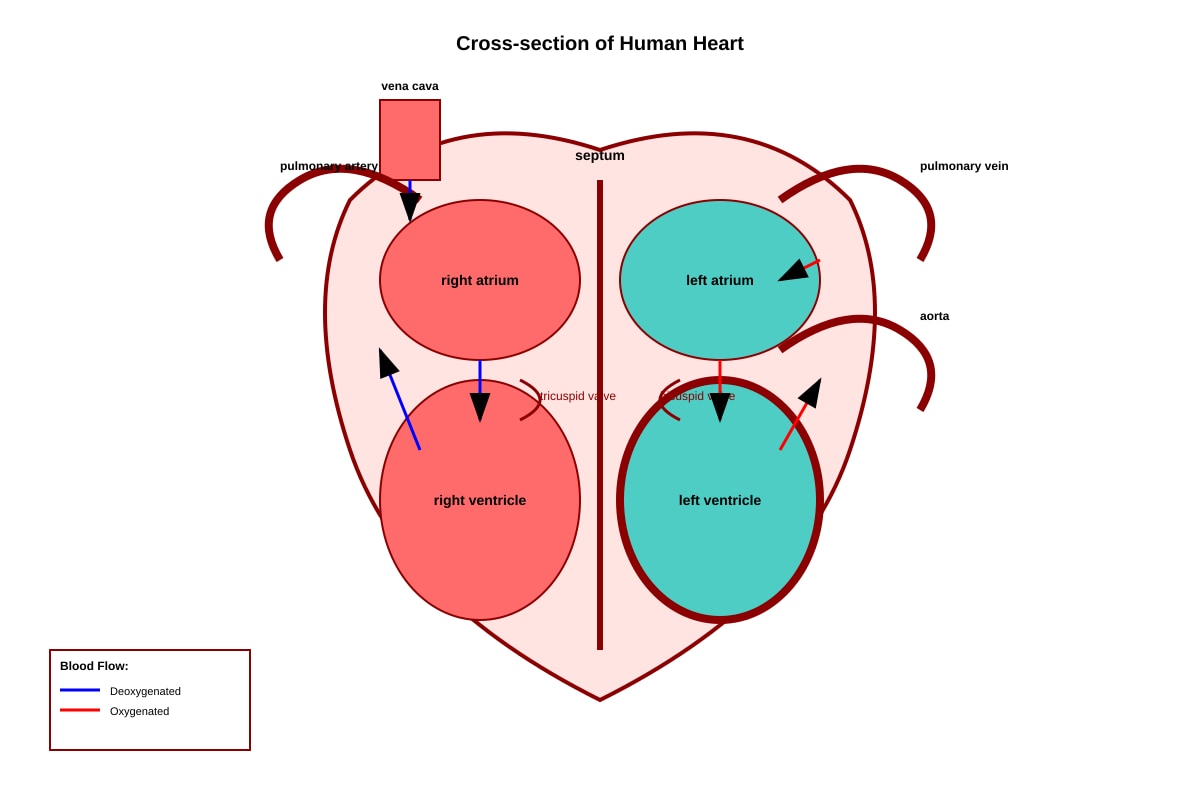
Generated diagram for Q2.
Based on the diagram, which chamber has the thickest muscular wall?
| A | right atrium | | B | right ventricle | | C | left atrium | | D | left ventricle |
Answer: [ ]
[1 mark]
3. Which of the following correctly describes the function of red blood cells?
| A | They produce antibodies to fight pathogens | | B | They engulf bacteria by phagocytosis | | C | They transport oxygen from the lungs to tissues | | D | They contain nuclei to direct cellular activities |
Answer: [ ]
[1 mark]
4. During exercise, a person's breathing rate increases. Which statement best explains why?
| A | Less carbon dioxide is produced by muscles | | B | More oxygen is needed and more carbon dioxide must be removed | | C | The heart stops pumping blood to the muscles | | D | The diaphragm becomes less flexible |
Answer: [ ]
[1 mark]
5. The graph shows changes in blood glucose concentration after a meal.
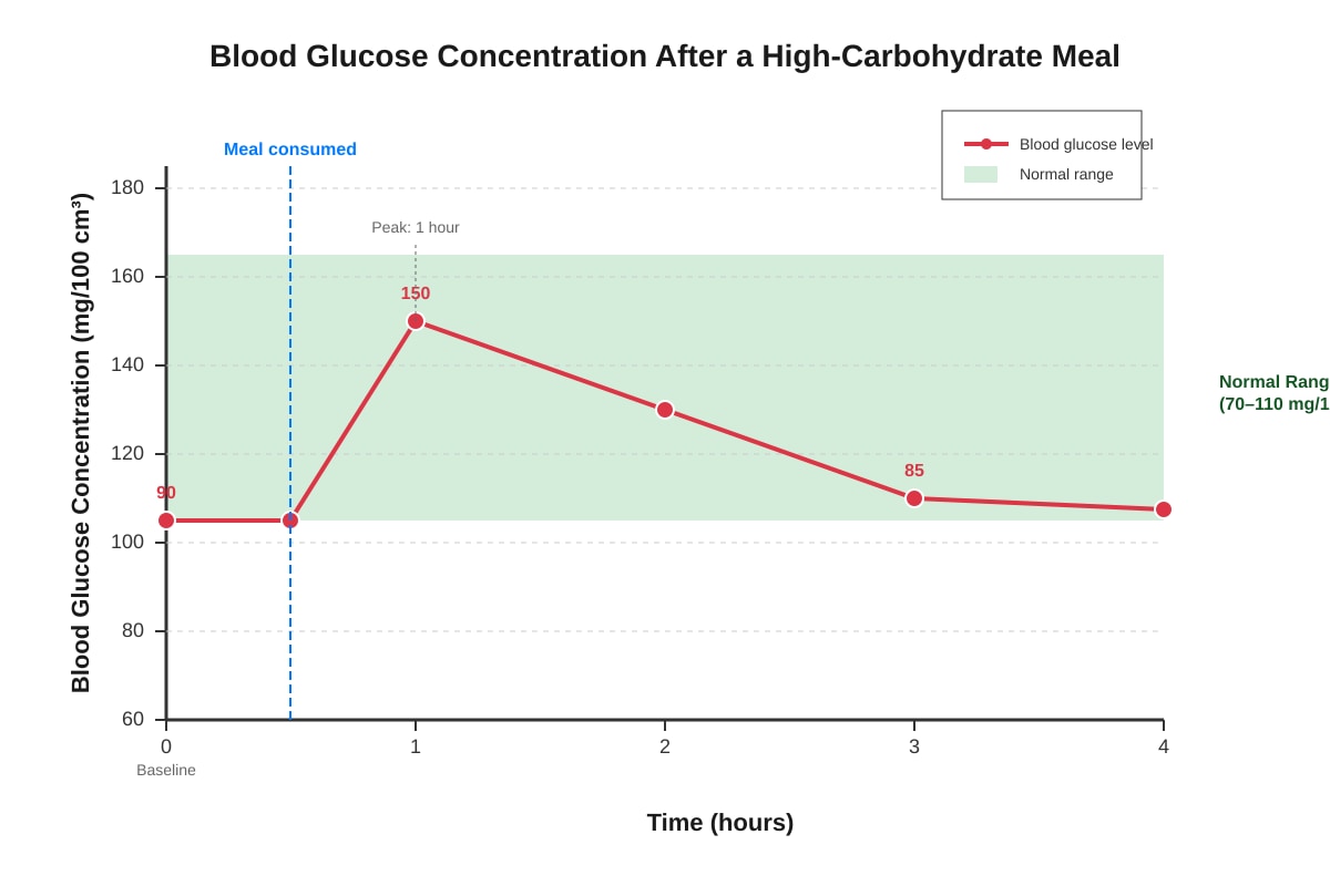
Generated graph for Q5.
At which time point is insulin secretion most likely to be highest?
| A | 0 hours (before the meal) | | B | 1 hour after the meal | | C | 3 hours after the meal | | D | 4 hours after the meal |
Answer: [ ]
[1 mark]
Section B: Short Answer and Structured Response (Questions 6–15) [22 marks]
6. State two features of alveoli that make them efficient for gas exchange.
[2 marks]
7. The table below shows some components of blood and their characteristics.
| Component | Presence of nucleus | Main function |
|---|---|---|
| Red blood cell | Absent | (a) |
| White blood cell | Present | (b) |
| Platelet | Absent | (c) |
Complete the table by stating the main function of each component.
(a) _________________________________________________________________
(b) _________________________________________________________________
(c) _________________________________________________________________
[3 marks]
8. Describe the pathway of air from the external environment to the alveoli, naming three structures in the correct order.
[2 marks]
9. Explain why the walls of capillaries are only one cell thick.
[1 mark]
10. The diagram shows the structure of a nephron from the human kidney.
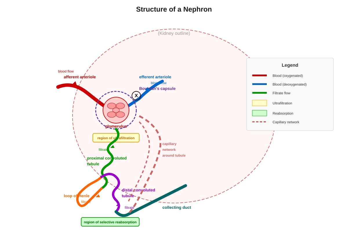
Generated diagram for Q10.
(a) Name the structure labelled X in the diagram where ultrafiltration occurs.
[1 mark]
(b) Explain why protein molecules do not normally pass through the filtration membrane during ultrafiltration.
[2 marks]
11. A student measured their pulse rate before and after running 400 metres. The results are shown below.
| Condition | Pulse rate (beats per minute) |
|---|---|
| At rest | 72 |
| Immediately after running | 156 |
| 2 minutes after running | 102 |
| 5 minutes after running | 78 |
(a) Calculate the percentage increase in pulse rate from rest to immediately after running. Show your working.
[2 marks]
(b) Explain why the pulse rate takes several minutes to return to the resting value after exercise stops.
[2 marks]
12. Distinguish between the processes of inhalation and exhalation in terms of diaphragm movement and volume of the thoracic cavity.
| Inhalation | Exhalation | |
|---|---|---|
| Diaphragm movement | (a) | (b) |
| Thoracic cavity volume | (c) | (d) |
[2 marks]
13. Explain how the structure of a red blood cell is related to its function of transporting oxygen.
[2 marks]
14. The graph shows the effect of different concentrations of glucose on the rate of glucose uptake by kidney tubule cells.
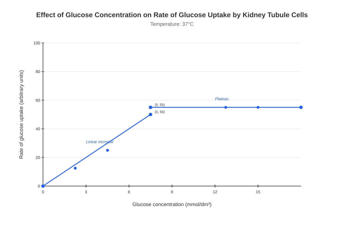
Generated graph for Q14.
(a) Describe the relationship between glucose concentration and rate of uptake between 0 and 5 mmol/dm³.
[1 mark]
(b) Suggest why the rate of uptake plateaus at concentrations above 5 mmol/dm³.
[2 marks]
15. State the role of the liver in regulating blood glucose concentration when levels are too high, and name the hormone involved.
[2 marks]
Section C: Data Analysis and Extended Response (Questions 16–20) [13 marks]
16. The diagram shows a synapse between two neurones.
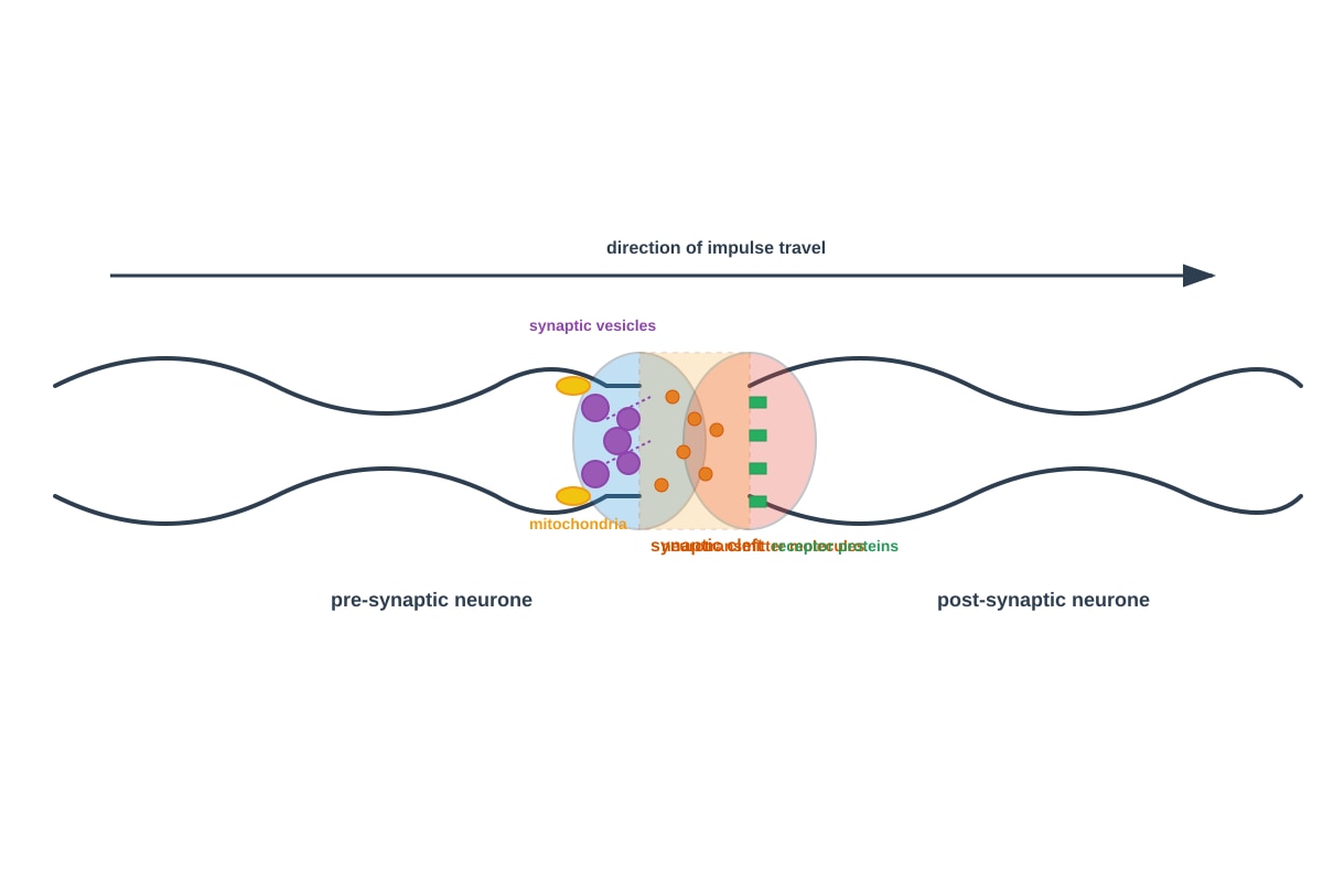
Generated diagram for Q16.
(a) Name the chemical substance released from the vesicles at the synapse.
[1 mark]
(b) Explain why the synapse ensures that nerve impulses travel in only one direction.
[3 marks]
17. The table shows measurements of breathing in an individual at rest and during exercise.
| Measurement | At rest | During exercise |
|---|---|---|
| Breathing rate (breaths per minute) | 12 | 30 |
| Tidal volume (cm³ per breath) | 500 | 2500 |
| Oxygen percentage in expired air | 16% | 14% |
(a) Calculate the total volume of air breathed in per minute during exercise (the respiratory minute volume). Show your working.
[2 marks]
(b) Explain why the percentage of oxygen in expired air is lower during exercise than at rest.
[2 marks]
18. Describe the role of the skin in thermoregulation when body temperature rises above normal. Include reference to blood vessels and sweat glands in your answer.
[3 marks]
19. Explain the importance of tissue matching before organ transplantation, with reference to antigens and antibodies.
[3 marks]
20. The diagram shows the feedback mechanism involving thyroxine.
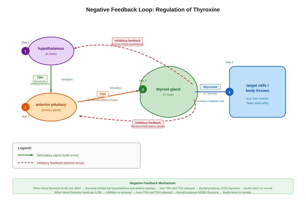
Generated diagram for Q20.
(a) State one effect of thyroxine on the body.
[1 mark]
(b) Explain how the negative feedback mechanism controls thyroxine levels when thyroxine concentration in the blood becomes too high.
[3 marks]
END OF QUIZ
Total Marks: 40
Answers
Secondary 3 Biology Quiz - Human Physiology (Answer Key)
Total Marks: 40 marks Note: This answer key provides teaching explanations for each question, suitable for students new to human physiology.
Section A: Multiple Choice (Questions 1–5)
1. Answer: B (pulmonary vein)
Mark: [1]
Explanation: The pulmonary vein is the only vein in the body that carries oxygenated blood. It transports blood from the lungs, where oxygen has been loaded, back to the left atrium of the heart. Students often confuse this with the pulmonary artery, which carries deoxygenated blood from the heart to the lungs. Remember the pattern: arteries carry blood away from the heart, veins carry blood toward the heart — the oxygenation state depends on which circuit (pulmonary or systemic) we're in.
Common mistake: Selecting "aorta" (D) — the aorta carries oxygenated blood but to the body, not from the lungs to the heart.
2. Answer: D (left ventricle)
Mark: [1]
Explanation: The left ventricle pumps oxygenated blood to the entire body through the aorta. This requires generating much higher pressure than the right ventricle, which only pumps blood to the nearby lungs. The thicker muscular wall of the left ventricle develops this greater force — approximately 5× thicker than the right ventricle wall. This is a key structural adaptation for function.
Visual check: In the diagram, the left ventricle should appear as the chamber with the thickest wall, positioned lower left when viewing the heart in anatomical position (as if in the chest, not as you face the diagram).
3. Answer: C (They transport oxygen from the lungs to tissues)
Mark: [1]
Explanation: Red blood cells contain haemoglobin, which binds reversibly with oxygen in the lungs (where oxygen concentration is high) and releases oxygen in tissues (where oxygen concentration is low). Mammalian red blood cells lose their nucleus during maturation, which maximises space for haemoglobin — this is why they cannot reproduce or perform complex metabolic functions.
- A describes lymphocytes (white blood cells)
- B describes phagocytes (white blood cells)
- D is incorrect because mature red blood cells are anucleate (have no nucleus)
4. Answer: B (More oxygen is needed and more carbon dioxide must be removed)
Mark: [1]
Explanation: During exercise, skeletal muscles respire more rapidly to produce ATP for contraction. Aerobic respiration requires oxygen and produces carbon dioxide as a waste product. The increased breathing rate serves two purposes:
- Increased oxygen intake — to meet elevated metabolic demand
- Increased carbon dioxide removal — to prevent dangerous pH changes in blood (CO₂ forms carbonic acid)
This is controlled by chemoreceptors in the medulla oblongata that detect changes in blood CO₂, O₂, and pH.
Why other options are wrong:
- A is opposite of truth (more CO₂ is produced, not less)
- C is biologically impossible — the heart must pump more, not stop
- D is incorrect — the diaphragm works harder, not less
5. Answer: B (1 hour after the meal)
Mark: [1]
Explanation: Insulin is secreted by the β-cells of the islets of Langerhans in the pancreas when blood glucose rises above normal. The graph shows blood glucose peaking at 1 hour (150 mg/100 cm³, well above the normal shaded range of 70–110). This high concentration stimulates insulin release, which promotes:
- Glucose uptake by cells (especially muscle and fat)
- Glycogenesis (converting glucose to glycogen in liver and muscle)
- Inhibition of gluconeogenesis
By 3–4 hours, glucose returns toward baseline and insulin secretion decreases. Glucagon would become relevant if levels dropped too low.
Section B: Short Answer and Structured Response (Questions 6–15)
6. Two features of alveoli for efficient gas exchange:
Marking points (any 2): [2]
| Feature | Why it improves efficiency |
|---|---|
| Large surface area (approximately 70–100 m² in total) | More area for O₂ and CO₂ to diffuse |
| Thin walls (one cell thick, ~0.5 μm) | Short diffusion distance for gases |
| Moist lining | Allows gases to dissolve before diffusing |
| Rich blood supply (extensive capillary network) | Maintains concentration gradient by removing O₂ and bringing CO₂ |
| Wall is one cell thick | [same as thin walls — do not double-credit] |
Teaching note: Alveoli epithelial cells are extremely thin (squamous epithelium). The fusion of alveolar and capillary basement membranes creates a very short diffusion path: alveolar air → type I pneumocyte → fused basement membrane → capillary endothelium → blood.
Common mistake: Saying "alveoli have cilia" — cilia are found in bronchi and bronchioles, not alveoli.
7. Table completion:
| Component | Main function |
|---|---|
| (a) Red blood cell | Transport oxygen (and some carbon dioxide) [1] |
| (b) White blood cell | Fight infection / produce antibodies / engulf pathogens (phagocytosis) [1] |
| (c) Platelet | Blood clotting / thrombosis / formation of platelet plug [1] |
Teaching notes:
- (a) Oxygen binds to haemoglobin forming oxyhaemoglobin; about 23% of CO₂ is transported bound to haemoglobin as carbaminohaemoglobin
- (b) Three main types: phagocytes (neutrophils, macrophages) engulf pathogens; lymphocytes produce antibodies
- (c) Platelets are cell fragments (not whole cells) that release clotting factors and form temporary plugs; they contain mitochondria but no nucleus
8. Pathway of air to alveoli:
Marking points (any 3 in correct order): [2]
Correct order: Nostril/nose/mouth → pharynx → larynx → trachea → bronchi → bronchioles → alveoli
Accept any three consecutive structures, or all structures with one error.
Teaching note: Air is:
- Warmed by blood vessels in nasal mucosa
- Moistened by mucus secretions
- Filtered by cilia trapping particles in mucus (mucociliary escalator)
The epiglottis prevents food entering the larynx during swallowing.
9. Why capillary walls are one cell thick:
Answer: To provide a short diffusion distance / to allow rapid exchange of materials between blood and tissues [1]
Teaching note: This is a classic "structure relates to function" question. The endothelial cells of capillaries are joined by very thin cellar junctions with small gaps (pores) that allow small molecules, ions, and water to pass. In some tissues (like the blood-brain barrier), these junctions are tighter.
10. Nephron structure and function:
(a) Structure X (where ultrafiltration occurs): glomerulus / renal capsule (Bowman's capsule) [1]
Accept "glomerular capillaries" or "Bowman's capsule" — the functional unit is the glomerulus inside Bowman's capsule.
(b) Why proteins don't normally pass through:
Marking points: [2]
- Protein molecules are too large relative to the size of the pores in the basement membrane / filtration slits [1]
- The basement membrane and podocytes act as a molecular sieve [1]
Explanation: Ultrafiltration is based on size and charge. The glomerular filtration barrier consists of:
- Fenestrated endothelium of glomerular capillaries (large pores, but plasma proteins held back by charge)
- Basement membrane (type IV collagen, negatively charged — repels negatively charged proteins)
- Podocyte foot processes with slit diaphragms
Small molecules (water, glucose, amino acids, urea, salts) pass through. Plasma proteins (albumin ~66 kDa) and blood cells are retained. Glomerular disease can damage this barrier, causing proteinuria (protein in urine).
11. Pulse rate and exercise:
(a) Percentage increase calculation:
Marking: [2]
- Correct formula: [(156 − 72) / 72] × 100 [1] for method
- Correct answer: 116.7% or 117% or 116.67% [1]
Working shown: 72156−72×100=7284×100=116.7%
Teaching note: The pulse rate increases because the heart must pump more blood to deliver oxygen and glucose to working muscles and remove metabolic wastes. Cardiac output = heart rate × stroke volume; both increase during exercise.
(b) Why pulse rate takes time to return to normal:
Marking points: [2]
- Oxygen debt / anaerobic respiration has occurred [1]
- Lactic acid produced in muscles needs to be removed / needs to be converted back to glucose in liver (Cori cycle) or fully oxidised [1]
- OR: Body temperature needs to be brought down, so blood continues to be circulated to skin for heat loss [1]
- OR: Hormones (adrenaline) persist in blood for some time [1]
Explanation: During intense exercise, when oxygen supply is insufficient, muscle cells respire anaerobically, producing lactic acid. After exercise, excess post-exercise oxygen consumption (EPOC) occurs — the "oxygen debt" is repaid as:
- Lactic acid is transported to the liver for gluconeogenesis
- Creatine phosphate stores are replenished
- Elevated body temperature is normalised
12. Inhalation vs. exhalation:
| Inhalation | Exhalation | |
|---|---|---|
| Diaphragm movement | (a) Contracts and flattens / moves downward | (b) Relaxes and domes upward / moves upward |
| Thoracic cavity volume | (c) Increases / becomes larger | (d) Decreases / becomes smaller |
Marking: [2] — 0.5 marks per cell, round down; or full marks for all four correct.
Teaching note: This is about pressure changes:
- Inhalation: Diaphragm contracts (dome → flat) AND external intercostal muscles contract → ribs move up and out → volume increases → pressure decreases (Boyle's law) → air rushes in
- Exhalation: Diaphragm relaxes, external intercostals relax → elastic recoil of lungs → volume decreases → pressure increases → air forced out
Forced exhalation (during exercise) also uses internal intercostal and abdominal muscles.
13. Structure of red blood cell related to oxygen transport:
Marking points: [2]
- Biconcave shape → increases surface area to volume ratio for faster/more efficient oxygen diffusion [1]
- Contains haemoglobin → can bind/reversibly combine with oxygen to form oxyhaemoglobin [1]
- No nucleus / no mitochondria → more space for haemoglobin / no oxygen used by cell itself [1]
Any two points for full marks.
Teaching note: The biconcave disc shape also makes red blood cells flexible, allowing them to squeeze through capillaries narrower than their diameter. The lack of mitochondria means they rely on anaerobic glycolysis for their own ATP, preserving all oxygen for tissues.
14. Glucose uptake by kidney tubule cells:
(a) Relationship 0–5 mmol/dm³:
Answer: The rate of glucose uptake increases proportionally / linearly with glucose concentration / as concentration increases, rate increases [1]
(b) Why plateaus above 5 mmol/dm³:
Marking points: [2]
- Carrier proteins / transport proteins / facilitated diffusion channels are saturated [1]
- All binding sites on transport proteins are occupied / maximum rate of transport reached (Vmax) [1]
Teaching note: This is facilitated diffusion or active transport via SGLT proteins (sodium-glucose linked transporters) in the proximal convoluted tubule. These are specific, saturable carrier proteins — when all are busy, the rate cannot increase further regardless of concentration.
This graph pattern is characteristic of enzyme-catalysed reactions and carrier-mediated transport — both show saturation kinetics.
15. Liver in glucose regulation when levels are too high:
Answer: [2]
- Converts excess glucose to glycogen for storage / glycogenesis [1]
- Hormone: insulin [1]
Teaching note: The liver stores ~100g glycogen. When blood glucose is high:
- Insulin stimulates glycogenesis (glucose → glycogen)
- Insulin inhibits glycogenolysis (breakdown of glycogen)
When blood glucose is low (between meals), glucagon stimulates:
- Glycogenolysis (glycogen → glucose)
- Gluconeogenesis (making glucose from non-carbohydrate sources like amino acids and glycerol)
Section C: Data Analysis and Extended Response (Questions 16–20)
16. Synapse structure and function:
(a) Neurotransmitter / named neurotransmitter (e.g., acetylcholine, noradrenaline) [1]
(b) One-way transmission explanation: [3]
Marking points:
| Mark | Point |
|---|---|
| 1 | Neurotransmitters are only released from vesicles in the pre-synaptic neurone / only the pre-synaptic bulb has vesicles |
| 1 | Receptor proteins are only on the post-synaptic membrane / only the post-synaptic neurone has receptors |
| 1 | Therefore neurotransmitters can only go from pre-synaptic → post-synaptic, not the reverse (preventing backward impulses) |
Teaching note: This unidirectional property ensures coordinated nerve signalling. Common neurotransmitters:
- Acetylcholine — neuromuscular junctions, parasympathetic nervous system
- Noradrenaline — sympathetic nervous system
- Serotonin, dopamine — CNS modulation
After binding, neurotransmitter is broken down by enzymes (e.g., acetylcholinesterase for acetylcholine) or reabsorbed to stop continuous stimulation.
17. Breathing calculations:
(a) Respiratory minute volume during exercise:
Marking: [2]
Method: breathing rate × tidal volume [1]
30×2500=75000 cm3/min or 75 dm³/min or 75 litres/min [1]
At rest: 12 × 500 = 6000 cm³/min = 6 litres/min — this baseline shows the dramatic increase.
(b) Why less oxygen in expired air during exercise: [2]
Marking points:
- More oxygen is extracted/used by muscles / tissues respire more rapidly to release energy [1]
- Greater volume of air breathed means more total oxygen taken in, but proportion extracted is higher / muscles have higher oxygen demand [1]
Alternative: Blood flow through lungs is faster, allowing less time for full equilibration / muscles extract more oxygen from each portion of blood [1]
18. Skin in thermoregulation (temperature too high):
Marking points: [3]
| Mark | Content |
|---|---|
| 1 | Blood vessels near skin surface vasodilate / arterioles dilate / more blood flows to capillaries in skin |
| 1 | More heat lost by radiation / increased blood flow brings more heat to surface for loss |
| 1 | Sweat glands produce more sweat / sweat evaporates from skin surface |
| 1 | Evaporation of sweat requires latent heat of vaporisation / sweating is cooling |
Any 3 marks. Must include both blood vessels and sweat glands for full credit if specified in question.
Teaching note: This is controlled by the thermoregulatory centre in the hypothalamus.
- Vasodilation: Smooth muscle in arteriole walls relaxes; capillaries become engorged (visible as skin reddening)
- Sweating: Eccrine glands secrete hypotonic solution; Na⁺, Cl⁻, urea, water; evaporation cools because water's latent heat is high
When cold: vasoconstriction (reduces blood flow to skin), piloerection (hair standing up to trap air), shivering (muscle contraction generates heat).
19. Tissue matching in organ transplantation:
Marking points: [3]
| Mark | Content |
|---|---|
| 1 | Body cells have surface antigens / protein markers (major histocompatibility complex / MHC / HLA antigens) |
| 1 | Recipient's immune system may recognise donor antigens as foreign |
| 1 | Lymphocytes produce antibodies against these foreign antigens |
| 1 | Immune response / rejection occurs, damaging transplanted organ |
Any 3 marks.
Teaching note: The ABO blood group system and HLA (human leukocyte antigen) system are key matching criteria. Even with matching:
- Immunosuppressant drugs (e.g., cyclosporine, tacrolimus) needed lifelong
- Recipients more susceptible to infections
Xenotransplantation (animal organs) faces even greater antigenic differences. Research explores genetically modifying pigs to reduce human immune response.
20. Thyroxine negative feedback:
(a) One effect of thyroxine: [1]
- Increases metabolic rate / basal metabolic rate (BMR)
- Increases protein synthesis
- Promotes growth and development (especially nervous system in children)
- Increases heart rate / breathing rate
- Increases body temperature / heat production
Any one.
(b) Negative feedback when thyroxine too high: [3]
Marking points:
| Mark | Content |
|---|---|
| 1 | High thyroxine inhibits / suppresses the hypothalamus and/or anterior pituitary |
| 1 | Less TRH (thyrotropin-releasing hormone) released from hypothalamus |
| 1 | Less TSH (thyroid-stimulating hormone / thyrotropin) released from anterior pituitary |
| 1 | Less stimulation of thyroid gland → less thyroxine produced |
| 1 | This is a negative feedback loop / maintains homeostasis / returns level to normal |
Any 3 marks. Must show sequence of reduced hormone release leading to reduced thyroxine.
Teaching note: The full axis: Hypothalamus → TRH → Anterior pituitary → TSH → Thyroid → Thyroxine → target cells. High thyroxine feeds back to inhibit both hypothalamus and pituitary (short loop and long loop negative feedback).
Hyperthyroidism (e.g., Graves' disease): excessive thyroxine → weight loss, heat intolerance, anxiety, exophthalmos. Hypothyroidism (e.g., Hashimoto's disease): insufficient thyroxine → weight gain, cold intolerance, lethargy, myxoedema.
END OF ANSWER KEY
Free quiz and exam paper access
Enter your details to view this paper
Your access is remembered on this device.