AI Generated Quiz
Secondary 3 Biology Cells Biomolecules Quiz
Free Sec 3 Biology Cells Biomolecules quiz, Kimi2.6 AI version, with questions, answers, and O Level-style practice for Singapore students.
These static practice materials are generated from the site's syllabus and paper-generation workflow, with source and model context shown so students and parents can evaluate the material before use.
Questions
Secondary 3 Biology Quiz - Cells Biomolecules
Name: _________________________ Class: ______ Date: _________ Score: _______/45
Duration: 35 minutes
Total Marks: 45
Instructions: Answer all questions. Write your answers in the spaces provided. Show all working where calculations are required.
Section A: Multiple Choice Questions (Questions 1–5)
Choose the correct answer. Each question carries 1 mark.
1. Which organelle is responsible for packaging and modifying proteins for secretion?
A) Rough endoplasmic reticulum
B) Golgi body
C) Mitochondrion
D) Ribosome
Answer: _________________________
2. A cell is supplied with radioactive amino acids. Which organelle would FIRST show increased radioactivity?
A) Golgi body
B) Smooth endoplasmic reticulum
C) Ribosomes
D) Nucleus
Answer: _________________________
3. Which biomolecule is incorrectly paired with its basic unit?
A) Protein — amino acid
B) Starch — glucose
C) DNA — nucleotide
D) Fat — glycerol only
Answer: _________________________
4. The graph below shows the effect of temperature on enzyme activity:
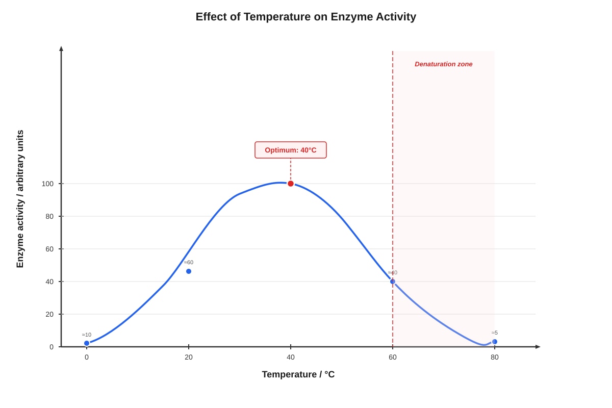
Generated graph for Q4.
At which temperature is the enzyme completely denatured?
A) 40°C
B) 60°C
C) 80°C
D) 0°C
Answer: _________________________
5. Which structure is found in plant cells but NOT in animal cells?
A) Mitochondria
B) Ribosomes
C) Cell wall
D) Nucleus
Answer: _________________________
Section B: Structured Response Questions (Questions 6–12)
Answer all questions in the spaces provided.
6. (a) Name the two types of endoplasmic reticulum found in eukaryotic cells. [2]
(b) State the function of the smooth endoplasmic reticulum. [1]
Total: [3]
7. The diagram shows an animal cell.
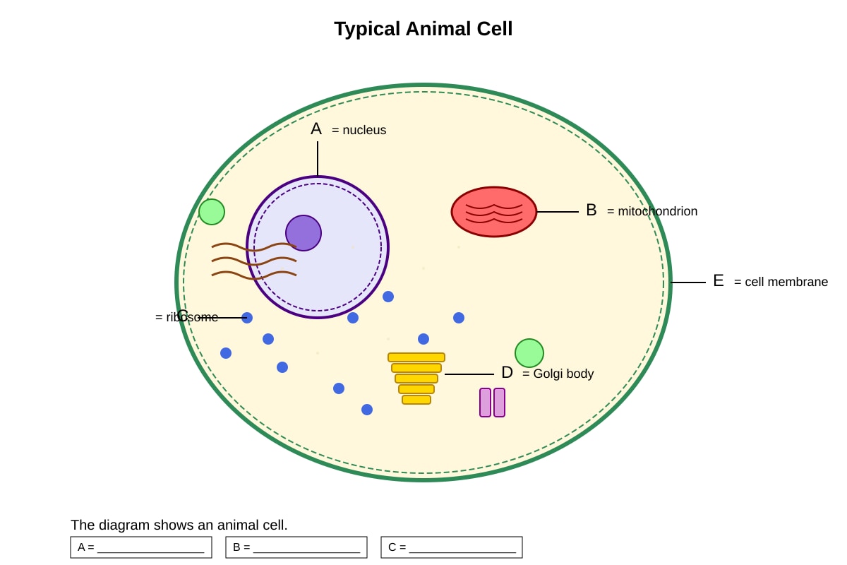
Generated diagram for Q7.
(a) Label structures A, B, and C on the diagram by writing the correct names in the boxes provided. [3]
A = _________________ B = _________________ C = _________________
(b) Structure B is often described as the "powerhouse" of the cell. Explain why this description is appropriate. [2]
Total: [5]
8. A student prepared two cell suspensions: one with intact cells and one with cells that had their cell membranes removed. Both suspensions were placed in distilled water.
(a) Predict and explain what would happen to the intact cells. [2]
(b) Explain why cells without cell membranes would behave differently. [2]
Total: [4]
9. The diagram shows the molecular structure of a triglyceride (fat).
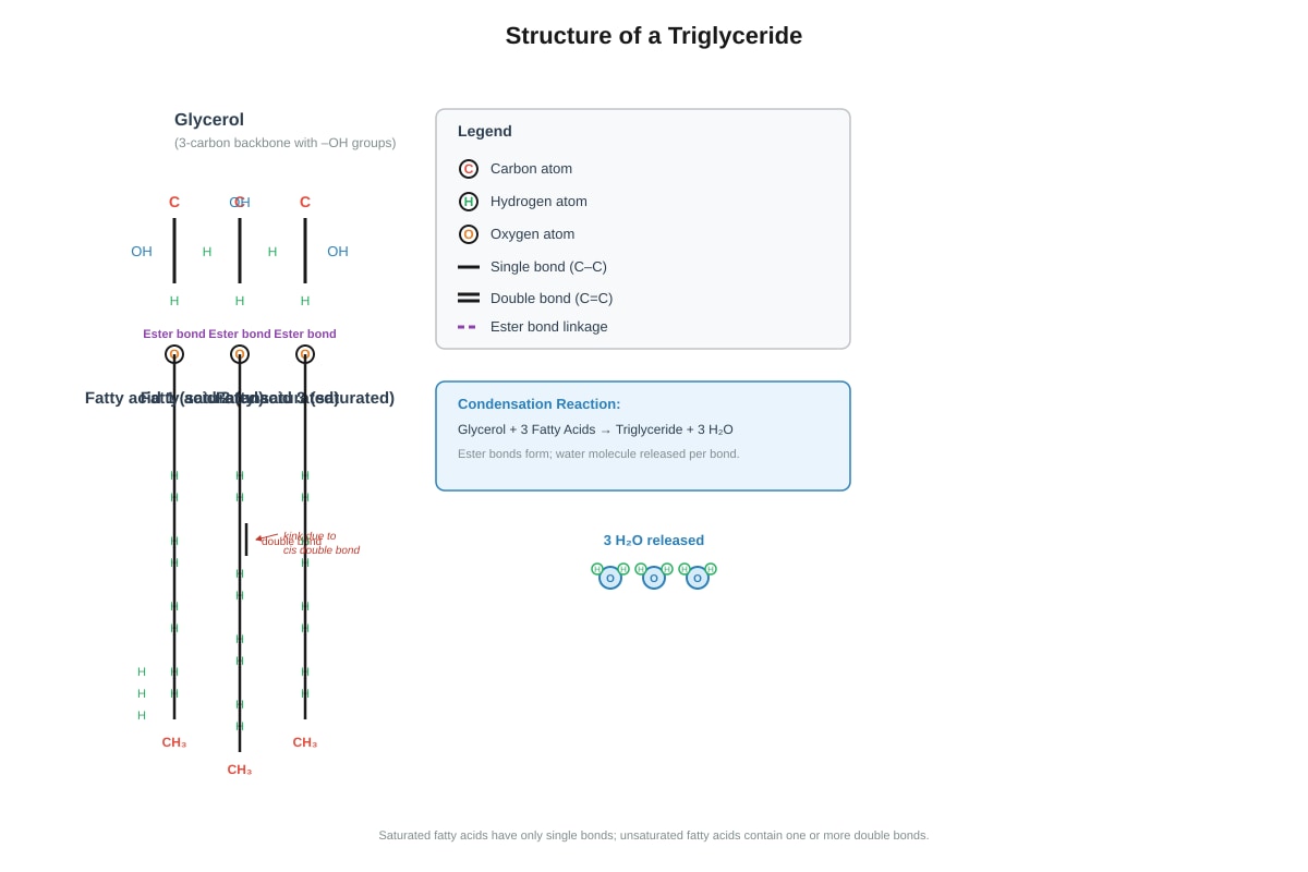
Generated diagram for Q9.
(a) Identify the type of reaction that joins fatty acids to glycerol to form a triglyceride. [1]
(b) State TWO functions of fats in animals. [2]
(c) Explain why fats provide more energy per gram than carbohydrates. [2]
Total: [5]
10. An experiment was conducted to investigate the effect of pH on the activity of the enzyme amylase. The results are shown in the table below.
| pH | Time taken for starch to be completely broken down (minutes) |
|---|---|
| 2 | 20 (no complete breakdown observed) |
| 4 | 15 |
| 7 | 5 |
| 9 | 10 |
| 11 | 18 (no complete breakdown observed) |
(a) Identify the independent variable in this investigation. [1]
(b) State the optimal pH for amylase activity based on these results. [1]
(c) Explain why the enzyme took longer to break down starch at pH 2 and pH 11. [3]
Total: [5]
11. Compare the structure and function of the cell wall and cell membrane in a plant cell. [4]
Total: [4]
12. The diagram shows an experimental setup to demonstrate osmosis.
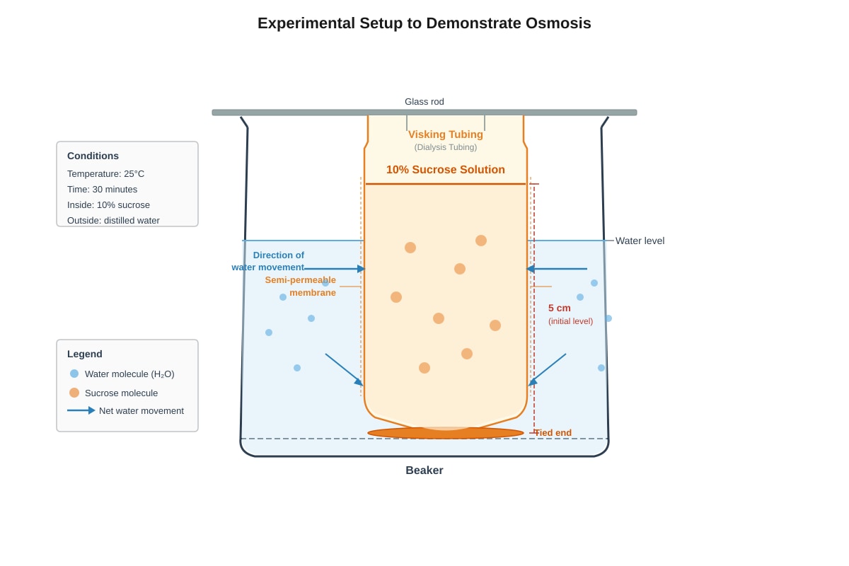
Generated experimental_setup for Q12.
(a) Predict what will happen to the level of liquid inside the Visking tubing after 30 minutes. [1]
(b) Explain your prediction in terms of water potential. [3]
Total: [4]
Section C: Extended Response and Application (Questions 13–20)
13. The electron micrograph below shows part of a secretory cell.
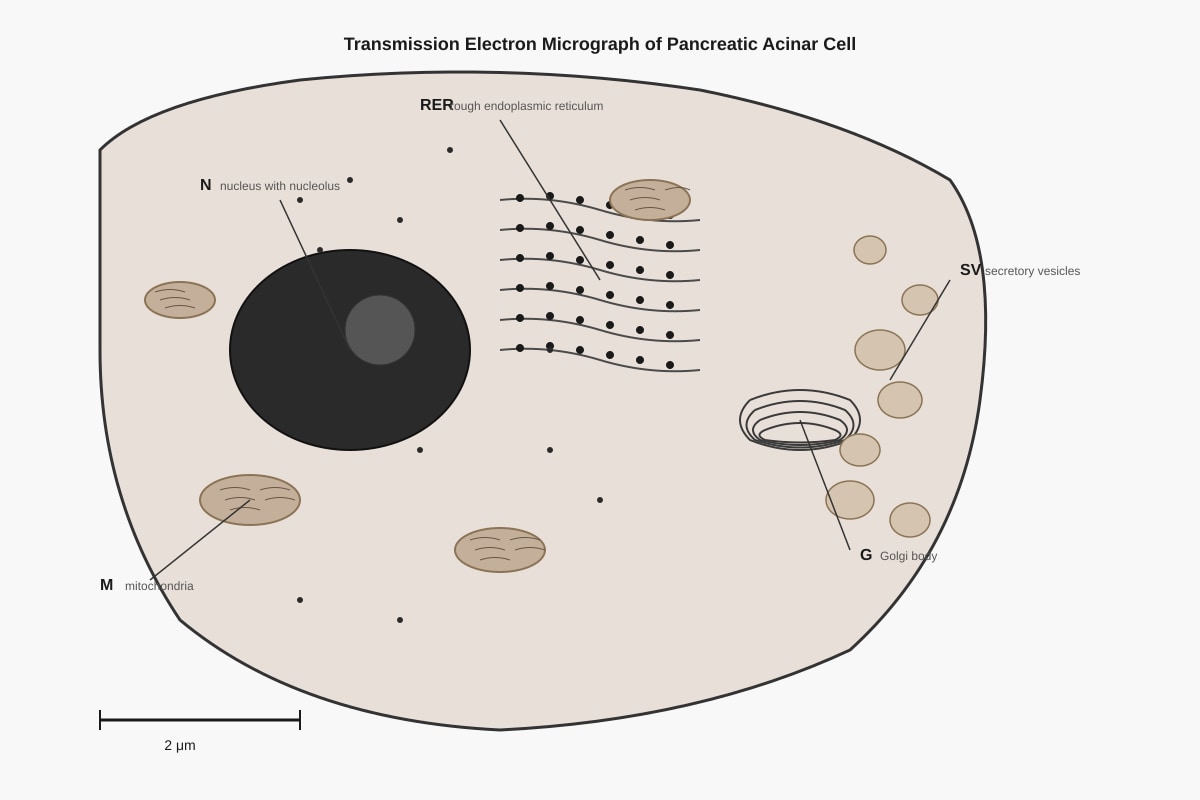
Generated figure for Q13.
(a) Explain the role of the RER and Golgi body in producing and secreting digestive enzymes. [3]
(b) Suggest why this cell contains many mitochondria. [2]
Total: [5]
14. A student was given three unknown solutions: X, Y, and Z. The student performed biochemical tests on each solution. The results are shown below.
| Test | Solution X | Solution Y | Solution Z |
|---|---|---|---|
| Benedict's test (heated) | Brick red precipitate | No change | No change |
| Biuret test | No change | Purple | No change |
| Ethanol emulsion test | No change | No change | White emulsion |
(a) Identify the biomolecule present in each solution. [3]
X: _________________ Y: _________________ Z: _________________
(b) Describe how you would modify the Benedict's test to estimate the concentration of reducing sugar in Solution X. [2]
Total: [5]
15. The graph shows the effect of substrate concentration on the rate of an enzyme-controlled reaction.
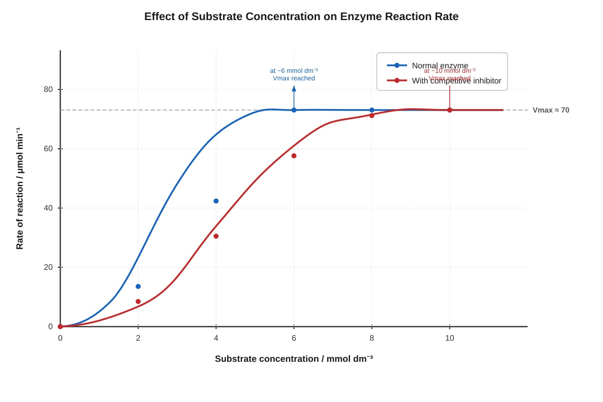
Generated graph for Q15.
(a) Explain why the rate of reaction increases at low substrate concentrations for both curves. [2]
(b) Explain why both curves eventually reach the same maximum rate (Vmax). [2]
(c) A competitive inhibitor binds to the active site of the enzyme. Explain why a higher substrate concentration is needed to reach Vmax in the presence of this inhibitor. [2]
Total: [6]
16. Explain how the properties of phospholipids make them suitable for forming cell membranes. [4]
Total: [4]
17. The table shows some features of DNA and RNA.
| Feature | DNA | RNA |
|---|---|---|
| Number of strands | 2 | 1 |
| Type of sugar | (i) _______ | Ribose |
| Nitrogenous bases | A, T, G, C | (ii) _______ |
(a) Complete the table by filling in the missing information. [2]
(i) _________________ (ii) _________________
(b) Explain why DNA is more stable than RNA and why this is important for its function. [3]
Total: [5]
18. Describe how you would test a leaf for the presence of starch. Include the reason for each step in your method. [5]
Total: [5]
19. The diagram shows a simplified model of enzyme action.
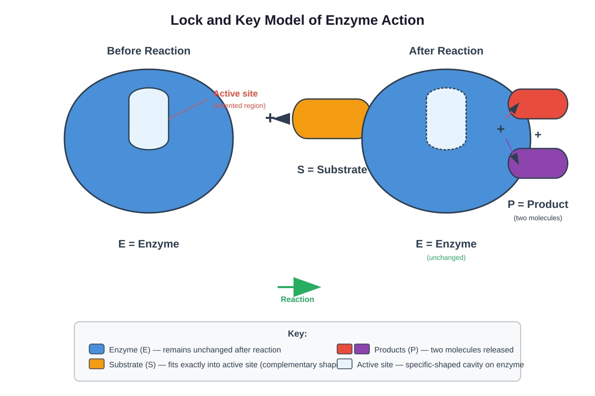
Generated diagram for Q19.
(a) Explain why this model is called the "lock and key" model. [2]
(b) The "induced fit" model is a more modern understanding of enzyme action. State one difference between the induced fit model and the lock and key model shown. [1]
(c) Explain how extreme pH affects enzyme activity, with reference to the active site. [3]
Total: [6]
20. Cyanide is a poison that binds to iron atoms within cytochrome oxidase, an enzyme in mitochondria. This prevents the enzyme from functioning.
(a) Predict the effect of cyanide on the production of ATP in a cell. [1]
(b) Explain your answer with reference to cellular respiration. [3]
(c) Cells in the brain are particularly sensitive to cyanide poisoning. Suggest why this is so. [2]
Total: [6]
END OF QUIZ
Answers
Secondary 3 Biology Quiz - Cells Biomolecules: Answer Key
Total Marks: 45
Section A: Multiple Choice Questions (Questions 1–5)
1. B — Golgi body [1]
Teaching note: The Golgi body (also called Golgi apparatus or Golgi complex) receives proteins from the rough ER, modifies them (e.g., adding carbohydrate groups to make glycoproteins), packages them into vesicles, and sends them to their destinations. The rough ER synthesizes proteins but does not package them for secretion. Ribosomes synthesize proteins but are not membrane-bound organelles for processing. Mitochondria produce ATP through cellular respiration.
2. C — Ribosomes [1]
Teaching note: Radioactive amino acids are the building blocks of proteins. Protein synthesis begins at ribosomes, where amino acids are assembled into polypeptide chains. The ribosomes are therefore the first organelle to incorporate the radioactive label. The rough ER has ribosomes attached to its surface, so it would show radioactivity slightly later as proteins enter its lumen. The Golgi body receives proteins after they have been synthesized and processed in the ER. The nucleus uses nucleotides, not primarily amino acids.
Common mistake: Students confuse the order of the secretory pathway: ribosomes → rough ER → Golgi body → secretory vesicles → cell membrane.
3. D — Fat — glycerol only [1]
Teaching note: Fats (triglycerides) are made of glycerol AND three fatty acids, not glycerol alone. The other pairings are correct: proteins are polymers of amino acids; starch is a polysaccharide of glucose; DNA is a polymer of nucleotides. This question tests understanding of biomolecular structure—knowing that lipids have a distinct structure from the simplistic "glycerol only" description.
4. C — 80°C [1]
Teaching note: At 80°C, the enzyme activity is near zero (approximately 5 arbitrary units), indicating the enzyme is completely denatured. Denaturation occurs when the three-dimensional structure of the enzyme is permanently destroyed by extreme heat, breaking hydrogen bonds and other interactions that maintain the active site's shape. At 40°C, the enzyme is at optimum activity. At 60°C, activity is reduced but not zero—some enzyme molecules still function. At 0°C, activity is low due to reduced kinetic energy, but the enzyme is not denatured; it could regain activity if warmed.
Graph interpretation: The curve shows the typical bell-shaped profile of temperature effects on enzyme activity, with an optimum where molecular motion and enzyme stability are balanced.
5. C — Cell wall [1]
Teaching note: Plant cells have a cell wall made of cellulose outside the cell membrane, providing structural support and protection. Animal cells lack cell walls—they only have a cell membrane. Both plant and animal cells contain mitochondria (for respiration), ribosomes (for protein synthesis), and a nucleus (containing genetic material). The presence of a cell wall is a key distinguishing feature when identifying unknown cells under a microscope.
Section B: Structured Response Questions (Questions 6–12)
6. (a) Rough endoplasmic reticulum (RER) and smooth endoplasmic reticulum (SER) [2] — 1 mark each
(b) Synthesis of lipids / detoxification of drugs and poisons / storage of calcium ions [1] — any one function
Teaching note: The rough ER has ribosomes attached to its surface and is involved in protein synthesis and modification. The smooth ER lacks ribosomes and has a different role profile—it's particularly abundant in cells that synthesize steroid hormones (like testosterone and estrogen) and in liver cells that detoxify harmful substances.
7. (a) A = Nucleus; B = Mitochondrion; C = Ribosome [3] — 1 mark each
(b) The mitochondrion is the site of aerobic respiration [1], where glucose is oxidized to produce ATP (adenosine triphosphate), which is the usable form of energy for cellular processes [1]. [2]
Teaching note: Mitochondria contain their own DNA and ribosomes, supporting the endosymbiotic theory that they evolved from free-living prokaryotes. The inner membrane (cristae) provides a large surface area for the electron transport chain, while the matrix contains enzymes for the Krebs cycle. The "powerhouse" analogy works because mitochondria convert energy from nutrients into ATP, just as a power plant converts fuel into electricity.
8. (a) The intact cells would swell and may burst (lyse) [1]. The cell membrane is partially permeable, allowing water to enter by osmosis [1]. In distilled water (high water potential, low solute concentration), water moves into the cells which have lower water potential due to dissolved cytoplasmic contents. [2]
(b) Cells without cell membranes would not maintain their integrity [1]. The cell contents would disperse into the surrounding water because there is no selectively permeable barrier to contain the cytoplasm and regulate what enters and exits [1]. [2]
Teaching note: Osmosis is the net movement of water molecules from a region of higher water potential to lower water potential across a partially permeable membrane. The cell membrane's selective permeability is essential for maintaining homeostasis. Without it, the organized cellular environment cannot be maintained—this is why cell membrane integrity is a marker of cell viability.
Common mistake: Students confuse "cell wall" and "cell membrane." Plant cells have both; the rigid cell wall prevents bursting under osmotic stress, but animal cells lack this protection and can lyse in hypotonic solutions.
9. (a) Condensation reaction / dehydration synthesis [1]
(b) Any two from: energy storage [1]; insulation (thermal) [1]; protection of organs [1]; hormone production (steroid hormones) [1] [2]
(c) Fats contain more carbon-hydrogen (C-H) bonds per gram than carbohydrates [1]. During respiration, these C-H bonds are oxidized, releasing more energy [1]. OR: Fats are more reduced than carbohydrates, so more oxygen is needed and more energy is released when they are fully oxidized. [2]
Teaching note: The condensation reaction that forms triglycerides releases water molecules—one per ester bond formed (three water molecules total). This is the reverse of hydrolysis. The energy yield from fat oxidation is approximately 38 kJ/g compared to about 17 kJ/g for carbohydrates, making fat a more compact energy store. This is evolutionarily advantageous for animals: migrating birds and hibernating mammals rely heavily on fat reserves.
10. (a) pH [1]
(b) pH 7 [1]
(c) At extreme pH values (pH 2 and pH 11), the hydrogen ion concentration alters the ionization of amino acid side chains [1] in the enzyme's active site. This changes the shape of the active site so the substrate can no longer fit properly [1]. The enzyme is denatured, and the rate of enzyme-substrate complex formation is greatly reduced [1]. [3]
Teaching note: Amylase (salivary and pancreatic) has an optimal pH around 7. In the acidic stomach (pH 2), salivary amylase is inactivated—this is why carbohydrate digestion begins in the mouth and ceases in the stomach. The enzyme pepsin, by contrast, works optimally at pH 2 in the stomach. This illustrates enzyme adaptation to specific environments. The pH affects ionic bonds and hydrogen bonds that maintain the tertiary structure of enzymes.
11. Cell wall: made of cellulose [1]; fully permeable / provides structural support and protection / maintains cell shape / prevents excessive water uptake [1] — any one function with material identified. Cell membrane: made of phospholipid bilayer with embedded proteins [1]; partially/selectively permeable / regulates movement of substances into and out of cell [1]. Both must be compared—structure and function points can be mixed but both aspects must be covered. [4]
Teaching note: The cell wall and cell membrane work together in plant cells. The rigid cell wall provides tensile strength against turgor pressure—the pressure exerted by water pushing against the cell wall when the vacuole is full. Without the cell wall, plant cells would burst in hypotonic solutions like animal cells do. The cell membrane's fluid mosaic structure allows it to be selectively permeable, with transport proteins facilitating movement of ions and large molecules.
12. (a) The level of liquid inside the Visking tubing will rise / increase [1]
(b) The 10% sucrose solution has a lower (more negative) water potential than the distilled water outside [1]. Water moves by osmosis from a region of higher water potential (distilled water) to lower water potential (sucrose solution) through the partially permeable Visking tubing [1]. As water enters the tubing, the volume of liquid increases, causing the level to rise [1]. [3]
Teaching note: This is the classic "osmosis in Visking tubing" demonstration. The sucrose molecules are too large to pass through the pores in the Visking tubing, but water molecules can pass freely. Water potential (Ψ) is the tendency of water to move from one area to another; pure water has Ψ = 0, and adding solute makes Ψ negative. The greater the solute concentration, the more negative the water potential, and the stronger the osmotic gradient.
Common mistake: Students say water moves "to the sugar" or "to where there's more dissolved stuff" without using the term "water potential" or explaining the partially permeable membrane requirement.
Section C: Extended Response and Application (Questions 13–20)
13. (a) Ribosomes on the RER synthesize the digestive enzymes (proteins) [1]. These proteins enter the RER lumen where they are folded and modified [1]. Transport vesicles carry the proteins to the Golgi body, where they are further processed, sorted, and packaged into secretory vesicles that fuse with the cell membrane to release the enzymes [1]. [3]
(b) Protein synthesis, modification, packaging, and secretion are energy-intensive processes requiring ATP [1]. Mitochondria produce ATP through aerobic respiration, so a high mitochondrial density supports the secretory function of this cell [1]. [2]
Teaching note: This question integrates the secretory pathway concept. Pancreatic acinar cells secrete digestive enzymes (proteases, amylase, lipase) into the pancreatic duct. The abundant RER is visible in electron micrographs as dark basophilic regions. The "division of labor" among organelles is key: ribosomes synthesize, ER modifies and transports, Golgi processes and packages, and mitochondria supply energy. This is an example of how cell structure relates to function—one of biology's unifying themes.
14. (a) X = reducing sugar / glucose / maltose [1]; Y = protein [1]; Z = lipid / fat / oil [1] [3]
(b) Prepare serial dilutions of Solution X with known concentrations [1]. Perform Benedict's test on each dilution and compare color intensity; the more concentrated the reducing sugar, the more brick-red precipitate forms, or use a colorimeter for more precise measurement [1]. OR: Compare against standard color chart after consistent heating time and temperature. [2]
Teaching note: The Benedict's test detects reducing sugars (those with free aldehyde groups) by reducing copper(II) sulfate in alkaline conditions to copper(I) oxide, which appears as a brick-red precipitate. Non-reducing sugars like sucrose must first be hydrolyzed to test positive. The Biuret test detects peptide bonds—two or more peptide bonds give a violet/purple color with alkaline copper sulfate. The emulsion test uses ethanol to dissolve lipids; when water is added, a white emulsion forms.
Method note: For semi-quantitative Benedict's testing, control variables are crucial—same volume of test solution, same volume of Benedict's reagent, same heating time and temperature. A colorimeter measures absorbance at the appropriate wavelength for more precise comparison.
15. (a) At low substrate concentrations, there are many available enzyme active sites and few substrate molecules [1]. As substrate concentration increases, more enzyme-substrate complexes form per unit time, increasing the rate of reaction [1]. [2]
(b) At high substrate concentrations, all enzyme active sites are occupied (the enzyme is saturated) [1]. Adding more substrate cannot increase the rate further because there are no free active sites; the limiting factor is now enzyme concentration, not substrate concentration [1]. [2]
(c) A competitive inhibitor competes with substrate for the active site [1]. At low substrate concentrations, inhibitor molecules occupy many active sites. Higher substrate concentration increases the probability that substrate rather than inhibitor binds to the active site [1]. Eventually, with enough substrate, substrate binding outcompetes the inhibitor, and the same Vmax is reached [1]. [2]
Teaching note: This Vmax-and-Km analysis introduces Michaelis-Menten kinetics conceptually without requiring mathematical derivation. Competitive inhibition: Km increases (apparent affinity decreases), Vmax unchanged. Non-competitive inhibition: Vmax decreases, Km unchanged. The key insight is that competitive inhibition is surmountable by excess substrate—this has pharmaceutical relevance. For example, statins competitively inhibit HMG-CoA reductase; the body responds by increasing substrate (HMG-CoA) production, demonstrating competitive dynamics.
16. Phospholipids have a hydrophilic (water-loving) phosphate head and two hydrophobic (water-hating) fatty acid tails [1]. In aqueous environments, they spontaneously form a bilayer with heads facing outwards and tails inwards [1]. This creates a partially permeable membrane that allows lipid-soluble substances to pass while restricting water-soluble substances [1]. The bilayer is fluid, allowing membrane proteins to move and function in transport and signaling [1]. [4]
Teaching note: The amphipathic nature of phospholipids—having both polar and nonpolar regions—is fundamental to membrane structure. The self-assembly property means no energy input is required for membrane formation, which has implications for origin-of-life theories. Fluidity depends on temperature, fatty acid saturation (unsaturated tails create kinks that prevent tight packing), and cholesterol content. This structural foundation explains membrane phenomena from cell signaling to endocytosis.
17. (a) (i) Deoxyribose [1]; (ii) A, U, G, C (uracil instead of thymine) [1] [2]
(b) DNA is double-stranded, with two antiparallel strands held together by hydrogen bonds between complementary bases [1]. This makes the molecule more stable and protects the genetic information from damage and mutations [1]. RNA is single-stranded and more susceptible to degradation, which is appropriate for its temporary roles in transcription and translation [1]. [3]
Teaching note: The sugar difference is subtle but crucial: deoxyribose lacks a hydroxyl (-OH) group at the 2' carbon, having just a hydrogen. This makes DNA less reactive and more chemically stable than RNA. The double helix structure, discovered by Watson and Crick using Rosalind Franklin's X-ray diffraction data, provides redundancy—each strand contains the information to reconstruct the other, enabling accurate replication and repair.
18. Step 1: Boil the leaf in water [1] — to kill the cells and break down the partially permeable membranes, allowing iodine to penetrate / to stop enzyme activity [1]. Step 2: Boil the leaf in ethanol [1] — to remove chlorophyll, which would mask the blue-black color of the starch-iodine complex / chlorophyll is soluble in organic solvents [1]. Step 3: Rinse the leaf in water [1] — to soften the leaf and remove excess ethanol, making it easier to spread iodine solution and preventing ethanol from reacting with iodine [1]. Step 4: Add iodine solution / potassium iodide solution [1] — starch gives a blue-black color with iodine; brown/orange color indicates absence of starch [1]. [5]
Teaching note: This is a standard protocol that tests for multiple practical skills: knowledge of safety (ethanol is flammable—water bath heating, not direct flame), understanding of cell structure (why membranes must be disrupted), and interpretation of biochemical test results. The ethanol step is critical—without it, the green chlorophyll would make the iodine color change impossible to see. The iodine test is specific for starch, not for glucose or other carbohydrates.
Safety emphasis: Ethanol must be heated in a water bath, NOT with a direct Bunsen burner flame, as ethanol vapors are highly flammable.
19. (a) The enzyme's active site has a specific complementary shape to the substrate, like a lock and key fitting together [1]. Only the correct substrate can bind to the active site, in the same way that only the correct key opens a specific lock [1]. [2]
(b) In induced fit, the active site changes shape slightly to accommodate the substrate / the active site is flexible rather than rigid [1]. [1]
(c) Extreme pH alters the ionization of amino acid residues that form the active site [1]. The hydrogen bonding and ionic interactions that maintain the specific three-dimensional shape of the active site are disrupted [1]. The active site loses its complementary shape, so substrate cannot bind effectively, and the enzyme is denatured [1]. [3]
Teaching note: The lock-and-key model (Fischer, 1894) was refined by Koshland's induced fit model (1958), which better explains how some enzymes can bind multiple related substrates and why catalytic activity requires conformational change. pH affects enzyme activity because amino acid side chains have different pKa values—the pH determines whether groups like -COOH/-COO⁻ or -NH₃⁺/-NH₂ predominate, changing charge distribution and thus folding.
20. (a) ATP production would decrease significantly / stop [1]
(b) Cytochrome oxidase is involved in the electron transport chain in mitochondria [1]. It transfers electrons to oxygen, the final electron acceptor, allowing the proton gradient to be maintained for ATP synthesis [1]. Without this enzyme, electron transfer stops, the proton gradient collapses, and oxidative phosphorylation cannot occur, so very little ATP is produced by mitochondria [1]. [3]
(c) Brain cells have a very high metabolic rate and constant energy demand for nerve impulse transmission and maintenance of ion gradients [1]. They do not store glycogen and rely almost entirely on aerobic respiration for ATP; they are therefore highly vulnerable to ATP depletion [1]. [2]
Teaching note: Cyanide (CN⁻) is a potent toxin because it binds with high affinity to the ferric iron (Fe³⁺) in cytochrome a/a₃, blocking cellular respiration at the final step. The electron transport chain backs up, NADH and FADH₂ cannot be reoxidized, and glycolysis must operate anaerobically with lactate fermentation, yielding only 2 ATP per glucose versus ~32 from aerobic respiration. The brain and heart are most affected due to their high oxidative metabolic rates.
Clinical connection: Cyanide poisoning treatment includes nitrites (which oxidize hemoglobin to methemoglobin, which competes with cytochrome oxidase for cyanide binding) and thiosulfate (which converts cyanide to less toxic thiocyanate).
END OF ANSWER KEY
Free quiz and exam paper access
Enter your details to view this paper
Your access is remembered on this device.