AI Generated Exam Paper
Secondary 3 Biology Practice Paper 5
Free Sec 3 Biology Practice Paper 5, Kimi2.6 AI version, with questions, answers, and O Level-style practice for Singapore students.
These static practice materials are generated from the site's syllabus and paper-generation workflow, with source and model context shown so students and parents can evaluate the material before use.
Questions
TuitionGoWhere Practice Paper - Biology Secondary 3
TuitionGoWhere Practice Paper (AI) Version: 5 of 5
| Subject: | Biology |
| Level: | Secondary 3 (Express/G3) |
| Paper: | Practice Paper |
| Duration: | 1 hour 30 minutes |
| Total Marks: | 80 |
| Name: | _________________________ |
| Class: | _________________________ |
| Date: | _________________________ |
Instructions to Candidates
- Answer all questions.
- Write your answers in the spaces provided.
- All working should be shown clearly for questions involving calculations.
- Marks are allocated as shown in brackets [ ] at the end of each question or part question.
- You are advised to spend approximately 20 minutes on Section A, 50 minutes on Section B, and 20 minutes on Section C.
Section A: Multiple Choice [10 marks]
Answer all questions. Each question carries 1 mark.
1. Which structure is present in a palisade mesophyll cell but absent from a root hair cell?
A. Cell wall
B. Chloroplast
C. Vacuole
D. Mitochondrion
Answer: ______
[1]
2. The diagram shows a cell organelle.
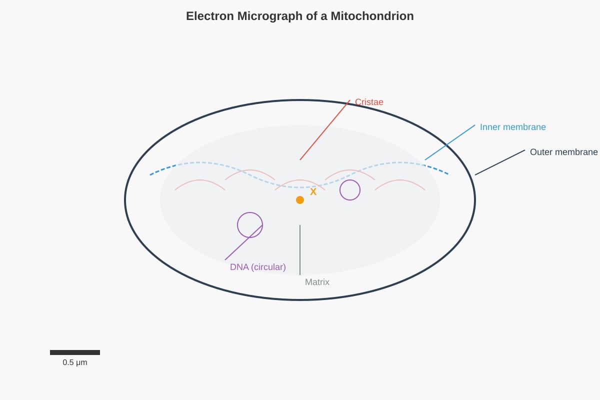
Generated diagram for Q2.
Which process occurs in the region labelled X?
A. Protein synthesis
B. Lipid digestion
C. Krebs cycle reactions
D. Photosynthetic phosphorylation
Answer: ______
[1]
3. A solution of amylase and starch was incubated at 35°C. After 10 minutes, the iodine test gave a yellow-brown colour. One minute later, the Benedict's test was negative. What explains this observation?
A. Amylase was denatured
B. Starch was completely hydrolysed to maltose
C. Maltose had not yet been produced
D. The temperature was below optimum
Answer: ______
[1]
4. Which statement correctly describes the fluid mosaic model of membrane structure?
A. Phospholipids form a single layer with proteins embedded on the surface
B. Proteins are fixed in position and cannot move laterally
C. The hydrophobic tails of phospholipids face outward to interact with water
D. The membrane contains a mosaic of proteins embedded in a fluid phospholipid bilayer
Answer: ______
[1]
5. In an experiment, cells were supplied with radioactive amino acids. Which sequence correctly shows the pathway of radioactivity through organelles in a cell secreting protein?
A. Ribosome → Rough ER → Golgi body → Secretory vesicles
B. Nucleus → Ribosome → Golgi body → Cell membrane
C. Smooth ER → Golgi body → Lysosome → Cell membrane
D. Ribosome → Nucleus → Rough ER → Golgi body
Answer: ______
[1]
6. Which feature of enzymes is explained by the lock and key hypothesis but NOT by the induced fit model?
A. Enzymes are specific in their action
B. Enzymes can be denatured by high temperature
C. The active site changes shape to fit the substrate
D. Enzyme-substrate complexes form temporary bonds
Answer: ______
[1]
7. The graph shows the effect of pH on the activity of three enzymes: pepsin, trypsin, and a plant enzyme from a soil bacterium.
Image pending generation: graph for Q7.
All three enzymes were tested at pH 7. Which statement is correct?
A. Only pepsin would show activity
B. Only the bacterial enzyme would show activity
C. Both trypsin and the bacterial enzyme would show activity
D. All three enzymes would show some activity
Answer: ______
[1]
8. Which column correctly matches the cell structure with its staining properties and function?
| Structure | Staining property | Function | |
|---|---|---|---|
| A | Nucleus | Stains blue with iodine solution | Stores genetic material |
| B | Cytoplasm | Stains pink with eosin | Site of glycolysis and many metabolic reactions |
| C | Cell wall | Stains purple with methylene blue | Provides selective permeability |
| D | Chloroplast | Stains green with iodine solution | Synthesises proteins |
Answer: ______
[1]
9. A student observed that when plant cells were placed in distilled water, they became turgid, but animal cells placed in distilled water burst. What explains this difference?
A. Animal cells lack a cell membrane
B. Plant cells have a cell wall that prevents excessive expansion
C. Animal cells have a higher solute concentration than plant cells
D. Plant cells do not carry out osmosis
Answer: ______
[1]
10. The table shows the results of an investigation into the effect of substrate concentration on enzyme activity.
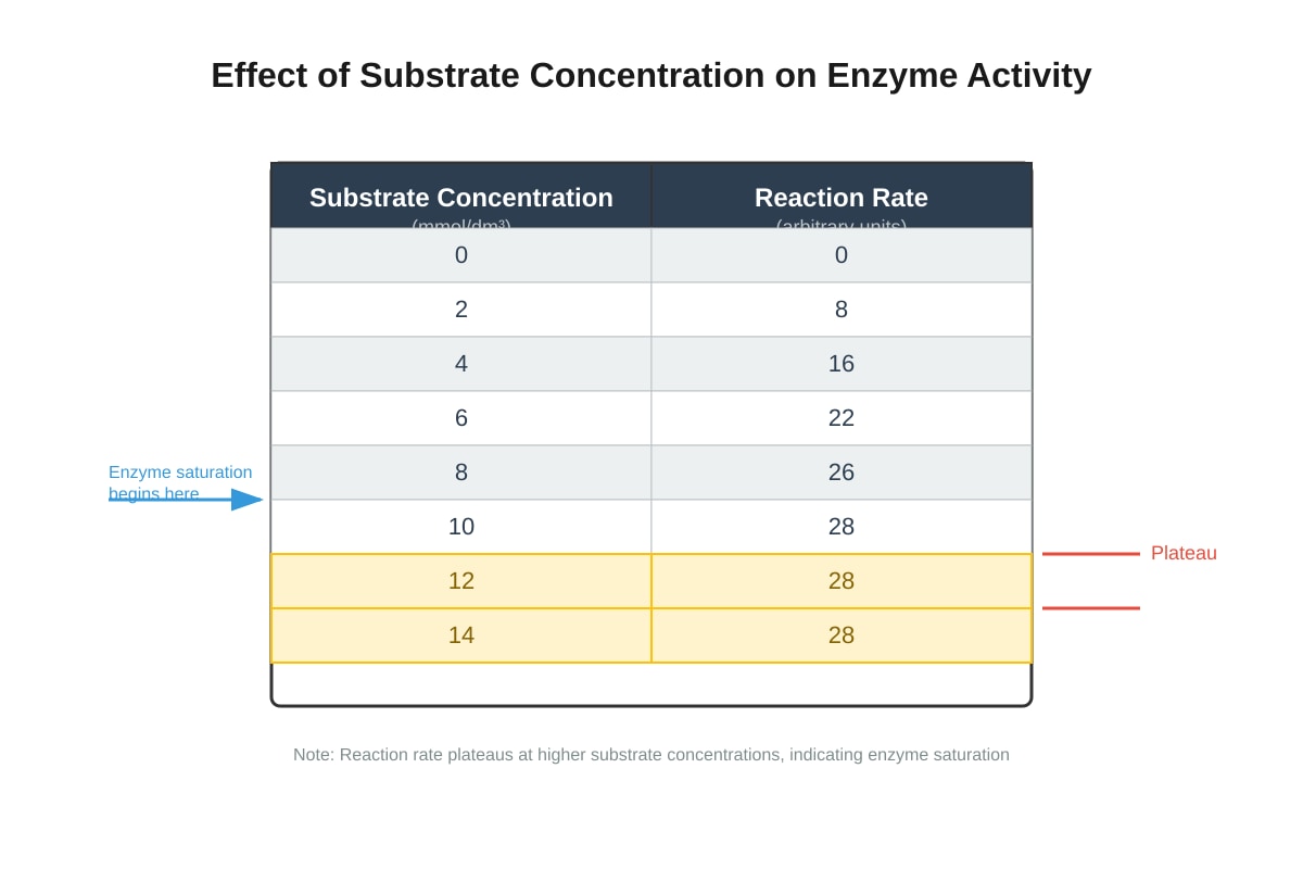
Generated table for Q10.
Between which substrate concentrations does the enzyme become saturated?
A. 0–2 mmol/dm³
B. 2–6 mmol/dm³
C. 6–10 mmol/dm³
D. 10–14 mmol/dm³
Answer: ______
[1]
Section A Total: [10]
Section B: Structured Response [50 marks]
Answer all questions in the spaces provided.
11. The diagram shows an animal cell as seen under an electron microscope.
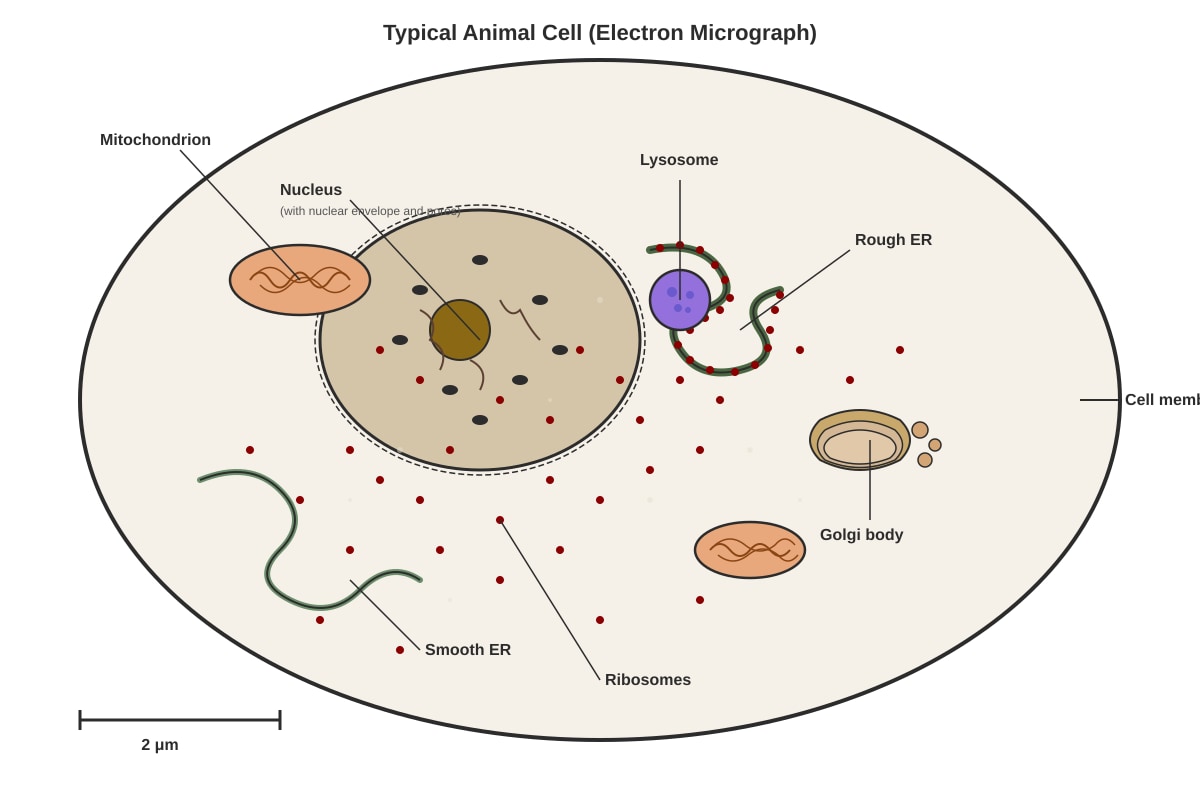
Generated diagram for Q11.
(a) State two structures visible in the diagram that are NOT present in a prokaryotic cell.
[2]
(b) The cell shown is actively synthesising and secreting a protein hormone. Describe the pathway of this protein from synthesis to secretion, naming the organelles involved in the correct sequence.
[3]
(c) Explain why mitochondria are numerous in cells that secrete large amounts of protein.
[2]
12. The photomicrograph shows a plant cell in different solutions.
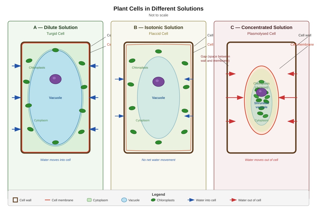
Generated diagram for Q12.
(a) In which solution (A, B, or C) is the cell plasmolysed?
Answer: ______
[1]
(b) Explain fully what happens to the cell in solution C that causes the observed appearance.
[3]
(c) A student placed red blood cells in solution A and observed them under the microscope. Predict what would happen and explain why this differs from the plant cell response.
[3]
13. Enzymes are biological catalysts that speed up chemical reactions in living organisms.
(a) Define the term active site.
[1]
(b) Explain how enzyme activity is affected by high temperature, using ideas about the active site and protein structure.
[3]
(c) The enzyme catalase breaks down hydrogen peroxide:
2H2O2→2H2O+O2
A student investigated the effect of pH on catalase activity using liver extract. The table shows the results.
| pH | Volume of O₂ collected in 2 minutes (cm³) |
|---|---|
| 4 | 2.5 |
| 6 | 8.2 |
| 7 | 12.4 |
| 8 | 9.6 |
| 10 | 3.1 |
(i) State the optimal pH for this enzyme. _______________
[1]
(ii) Calculate the percentage decrease in oxygen production when pH changes from 7 to 10. Show your working.
[2]
(iii) Suggest two variables that must be controlled in this investigation to ensure valid results.
[2]
14. The diagram shows the structure of a cell surface membrane.
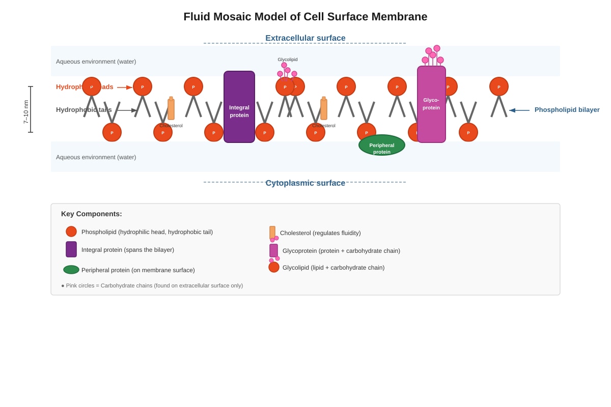
Generated diagram for Q14.
(a) Explain why the phospholipid bilayer forms spontaneously in an aqueous environment.
[2]
(b) State one function of each of the following membrane components:
(i) Glycoproteins: ___________________________________________________
[1]
(ii) Cholesterol: ____________________________________________________
[1]
(c) The cell membrane is described as "selectively permeable." Explain what this means and how the structure of the membrane achieves this property.
[3]
15. Adipose tissue consists of cells specialised for fat storage.
(a) Explain how the structure of adipose cells is adapted for their function.
[2]
(b) Compare and contrast the structure of a typical adipose cell with a palisade mesophyll cell from a leaf. Your answer should include three structural differences linked to their different functions.
[6]
16. The transmission electron micrograph shows part of a chloroplast from a photosynthetic plant cell.
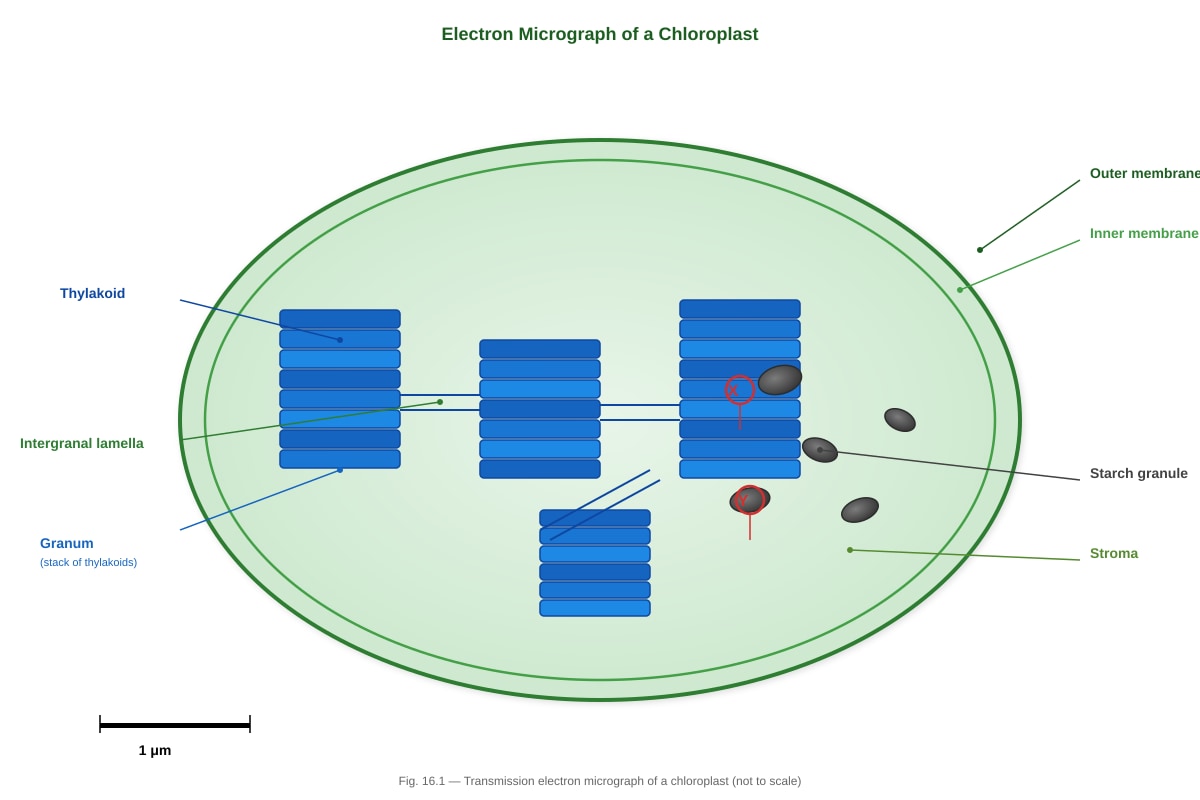
Generated diagram for Q16.
(a) Identify the structures labelled X and Y in the diagram.
X: _________________________________________________________________
Y: _________________________________________________________________
[2]
(b) Explain how the structure of the chloroplast is adapted for the light-dependent reactions of photosynthesis.
[3]
(c) The stroma of isolated chloroplasts was found to contain enzymes for the Calvin cycle but virtually no DNA. In contrast, mitochondria contain both enzymes for the Krebs cycle and their own DNA. Suggest one evolutionary explanation for this difference.
[2]
17. A student carried out an investigation to find the effect of temperature on the activity of amylase. Starch-agar plates were made with wells cut in them. Amylase solution at different temperatures was placed in the wells. After incubation, iodine solution was poured over the plates. Cleared zones indicated where starch had been digested.
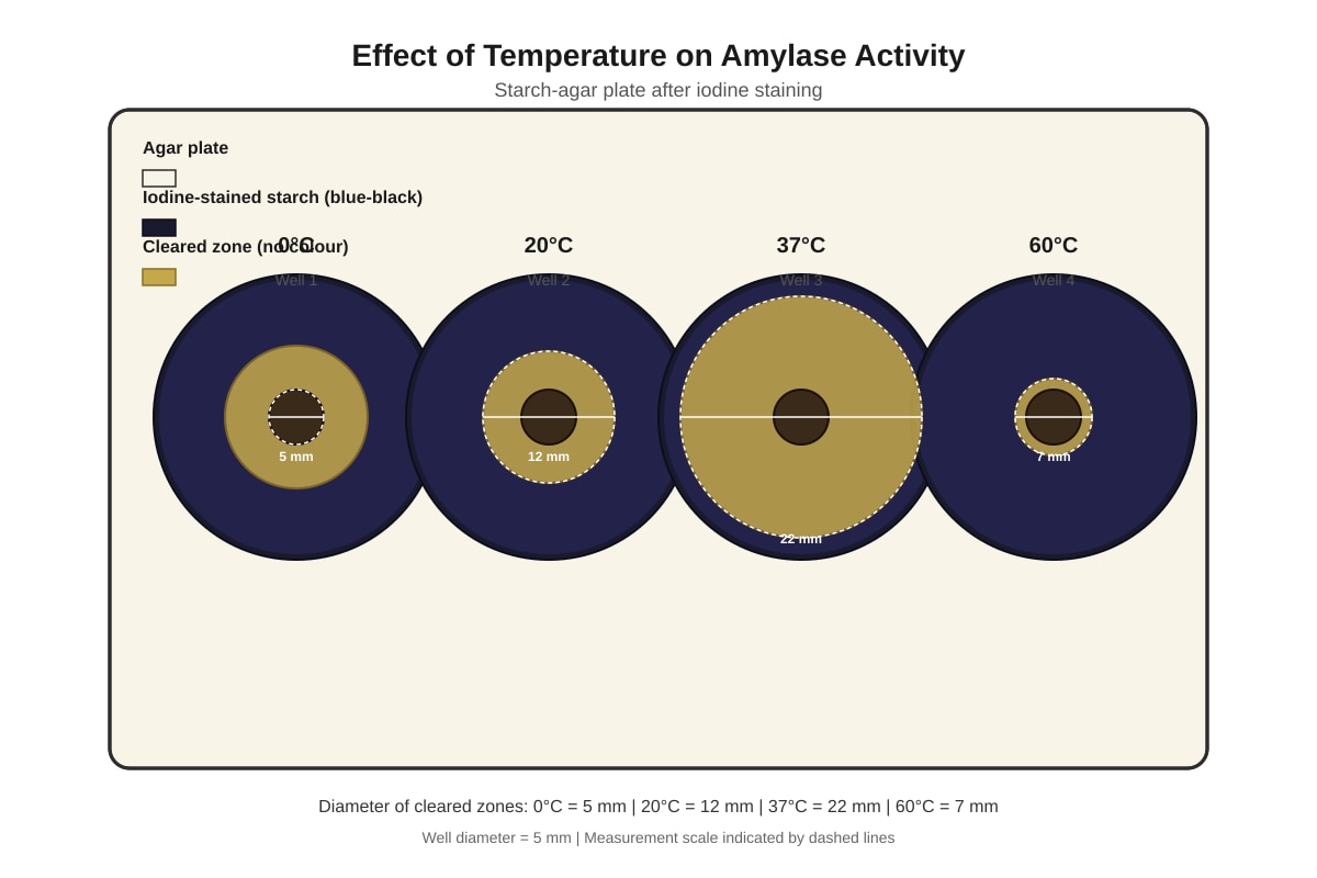
Generated diagram for Q17.
(a) Explain why a cleared zone appears around a well containing active amylase.
[2]
(b) Explain the results at 37°C compared with 0°C.
[3]
(c) Explain why the cleared zone at 60°C is smaller than at 37°C.
[2]
(d) Suggest one modification to this method that would allow the student to calculate the rate of reaction more precisely.
[1]
18. Proteins have diverse functions in living organisms. The table shows information about four proteins.
| Protein | Molecular structure | Function |
|---|---|---|
| Keratin | Fibrous, helix of two polypeptide chains with many disulfide bonds | Structural component of hair, nails, and skin |
| Haemoglobin | Globular, four polypeptide chains each with haem group | Transport of oxygen in blood |
| Insulin | Globular, two polypeptide chains linked by disulfide bonds | Hormone regulating blood glucose |
| Amylase | Globular, single polypeptide with cleft active site | Digestion of starch in mouth and small intestine |
(a) Explain how the structure of keratin is related to its function.
[2]
(b) Insulin is synthesised as a larger single polypeptide called preproinsulin, which is then modified. Explain the importance of this modification in terms of protein transport and secretion.
[3]
(c) Amylase functions at pH 7 in the mouth and pH 7–8 in the small intestine. Explain how a change in pH from the mouth to the stomach affects amylase activity, and why amylase can function again when food enters the small intestine.
[3]
19. The diagram shows the process of cell fractionation used to separate organelles from homogenised liver tissue.
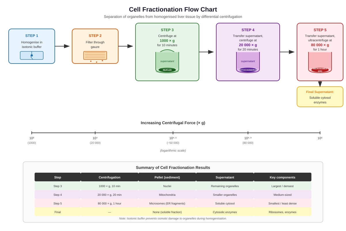
Generated diagram for Q19.
(a) Explain why the buffer used for homogenisation must be:
(i) Isotonic: ________________________________________________________
[1]
(ii) Cold (typically 4°C): ____________________________________________
[1]
(b) Explain how differential centrifugation separates organelles of different sizes.
[2]
(c) The final supernatant was found to contain approximately 50% of the total protein from the original cells. Suggest why this fraction contains such a high proportion of protein, naming the major soluble proteins you would expect to find.
[3]
20. The fluid mosaic model of membrane structure was proposed by Singer and Nicolson in 1972. This model replaced earlier models such as the Davson-Danielli model.
(a) Describe two key features of the fluid mosaic model that were not present in earlier models.
[4]
(b) The discovery of membrane proteins that span the entire membrane (integral proteins) provided evidence against the Davson-Danielli model. Explain why.
[2]
(c) Freeze-fracture electron microscopy provided visual evidence for the fluid mosaic model. Describe what this technique reveals about membrane structure and explain how it supports the model.
[3]
Section B Total: [50]
Section C: Data Response and Analysis [20 marks]
Answer all questions.
21. Investigation of Membrane Permeability
A student investigated the effect of temperature on the permeability of beetroot cell membranes. Beetroot cells contain a red pigment called betacyanin, which is normally contained within the vacuole. If the membrane is damaged, betacyanin leaks out.
The student cut cylinders of beetroot tissue, rinsed them to remove surface pigment, and placed them in tubes of distilled water at different temperatures for 20 minutes. The absorbance of the surrounding liquid was measured using a colorimeter — higher absorbance indicates more pigment leakage.
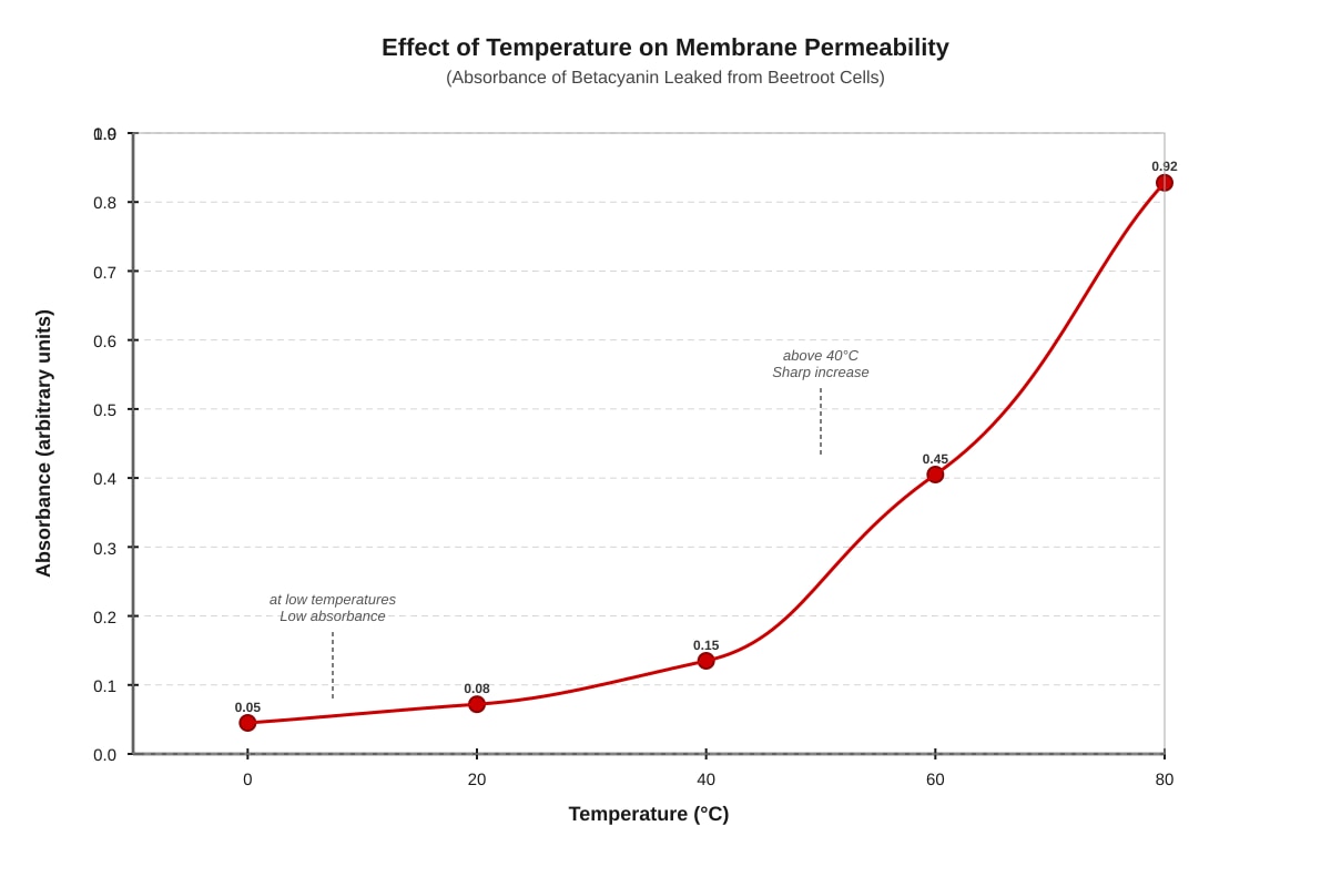
Generated graph for Q21.
(a) Describe the trend shown in the graph.
[2]
(b) Using your knowledge of membrane structure, explain the shape of the graph between 40°C and 80°C.
[4]
(c) Suggest two practical improvements to this investigation, explaining how each would increase the reliability of the results.
[4]
22. Enzyme Inhibition in Pharmaceutical Research
Enzyme inhibitors are important in medicine. The diagram shows two types of inhibition.
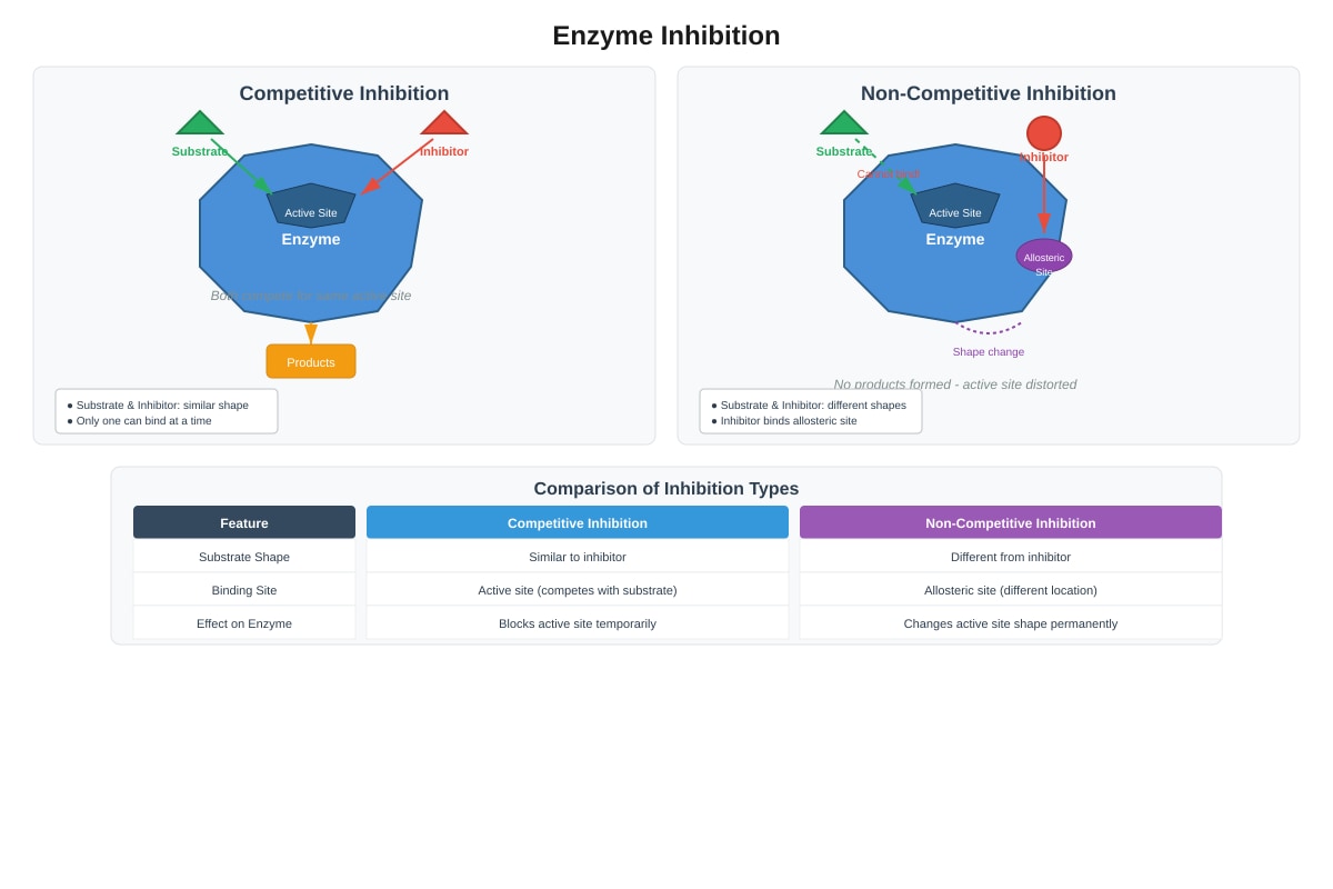
Generated diagram for Q22.
(a) Distinguish between competitive and non-competitive inhibition with reference to the diagrams.
[4]
Statins are drugs used to lower blood cholesterol. Some statins are competitive inhibitors of the enzyme HMG-CoA reductase, which catalyses an early step in cholesterol synthesis.
(b) Explain why taking statins results in lower blood cholesterol levels.
[3]
(c) Another drug, aspirin, irreversibly inhibits the enzyme cyclooxygenase (COX) by permanently modifying the active site. Explain why aspirin must be regularly administered to maintain its therapeutic effect, unlike some other drugs that can be taken less frequently.
[3]
Section C Total: [20]
Paper Total: [80]
END OF PAPER
Answers
TuitionGoWhere Practice Paper Answers - Biology Secondary 3
Version: 5 of 5 Total Marks: 80
Section A: Multiple Choice Answers
| Q | Answer | Explanation |
|---|---|---|
| 1 | B | Chloroplasts are found in photosynthetic plant cells (palisade mesophyll) but not in non-photosynthetic root hair cells. Both cell types have cell walls (plant), vacuoles, and mitochondria. |
| 2 | C | Region X is the matrix of the mitochondrion, where the Krebs cycle (citric acid cycle) occurs. Protein synthesis occurs at ribosomes, lipid digestion in lysosomes, and photosynthetic phosphorylation in chloroplasts. |
| 3 | B | Yellow-brown with iodine indicates no starch present; complete hydrolysis to maltose has occurred. Benedict's test is negative because maltose is a reducing sugar but needs heating — the negative result after one minute suggests the test wasn't performed correctly or the timing was too early, but the key point is starch is gone. More precisely: the iodine result proves starch hydrolysis; Benedict's needs boiling so "one minute later" implies no heating was done. The best explanation is that starch was hydrolysed to maltose (which would give positive Benedict's if heated). |
| 4 | D | The fluid mosaic model describes proteins embedded in a fluid phospholipid bilayer. Option A is wrong (bilayer, not single layer); B is wrong (proteins can move); C is wrong (hydrophobic tails face inward, away from water). |
| 5 | A | Radioactive amino acids are incorporated into protein at ribosomes, enter RER for folding, pass to Golgi for modification and packaging, then leave in secretory vesicles. The nucleus is not on the secretion pathway. |
| 6 | C | The induced fit model specifically proposes that the active site changes shape to fit the substrate — this is the key difference from lock and key, which assumes a rigid active site. Both models explain specificity (A), denaturation (B), and temporary bonding (D). |
| 7 | C | At pH 7: pepsin is inactive (optimum pH 2, inactive by pH 6–7); trypsin shows some activity (optimum pH 8, still active at pH 7); bacterial enzyme shows moderate activity (active across pH 7–12 with optimum at 10). |
| 8 | B | Eosin stains cytoplasm pink; cytoplasm is the site of glycolysis and many reactions. Nucleus stains with methylene blue/acetic orcein, not iodine. Cell wall doesn't stain with methylene blue (it's not living). Chloroplasts are naturally green — iodine stains starch blue-black inside them but the chloroplast itself is green due to chlorophyll. Option B is correct. |
| 9 | B | Plant cells have a rigid cell wall that prevents excessive water uptake and bursting; animal cells lack this protection and undergo cytolysis in distilled water. Both have cell membranes; osmosis occurs in plant cells; animal cell solute concentration is irrelevant to the structural difference. |
| 10 | D | The enzyme is saturated when further increases in substrate concentration do not increase reaction rate. From 10–14 mmol/dm³, the rate remains constant at 28 units, indicating all active sites are occupied. |
Section A Total: [10]
Section B: Structured Response Answers
11.
(a) [2 marks]
- Membrane-bound nucleus / nuclear envelope [1]
- Mitochondria [1]
- Endoplasmic reticulum (rough or smooth) [1]
- Golgi body / Golgi apparatus [1]
- Lysosomes [1]
(Any two correct; accept also: nuclear pores, 70S vs 80S ribosomes if visible, though typical EM distinction is membrane-bound organelles)
(b) [3 marks]
- Ribosomes: protein synthesis using mRNA template [1]
- Rough endoplasmic reticulum: folding and initial modification of polypeptide; transport in vesicles [1]
- Golgi body / apparatus: further modification, sorting, and packaging into secretory vesicles [1]
- Secretory vesicles fuse with cell membrane to release hormone by exocytosis [1]
(Maximum 3 marks; must have correct sequence)
(c) [2 marks]
- Protein synthesis and secretion are energy-requiring processes (ATP needed) [1]
- Mitochondria are sites of aerobic respiration, producing large amounts of ATP [1]
- More mitochondria provide sufficient ATP to power ribosome function, vesicle transport, and exocytosis [1]
(Any 2 points)
12.
(a) [1 mark]
- C [1]
(b) [3 marks]
- In solution C (concentrated solution / hypertonic), water potential is lower outside the cell than inside [1]
- Water leaves the cell by osmosis (down water potential gradient, from high to low water potential) [1]
- Cytoplasm and vacuole lose water, shrink, and pull away from cell wall (plasmolysis) [1]
- Cell membrane no longer pushed against cell wall; chloroplasts cluster in center [1]
(Maximum 3 marks)
(c) [3 marks]
- Red blood cells would burst / undergo haemolysis / cytolysis [1]
- Animal cells lack cell wall, so no protective structure against water entry [1]
- Distilled water is hypotonic compared to cytoplasm, so water enters continuously by osmosis [1]
- Plant cell wall is rigid and provides resistance to expansion, preventing bursting [1]
(First 3 valid points; must include contrast with plant cell)
13.
(a) [1 mark]
- The region of the enzyme where the substrate binds / a specific pocket or cleft in the tertiary structure [1]
(b) [3 marks]
- High temperature increases kinetic energy of molecules, increasing collision frequency [1]
- Above optimum temperature, bonds holding enzyme in shape (hydrogen bonds, ionic bonds) vibrate and break [1]
- Tertiary structure / 3D shape of enzyme is altered / denatured [1]
- Active site shape changes permanently, substrate can no longer fit / enzyme-substrate complexes cannot form [1]
(Maximum 3 marks; must include denaturation and active site change)
(c)(i) [1 mark]
- pH 7 [1]
(c)(ii) [2 marks]
- Change in oxygen: 28 → 3.1 = decrease of 25.3 cm³ [1]
- Percentage decrease = (25.3 / 28) × 100 = 90.4% (accept 90–91%) [1]
(c)(iii) [2 marks]
- Concentration / volume of hydrogen peroxide (substrate) [1]
- Concentration / volume of liver extract (enzyme source) [1]
- Temperature of incubation [1]
- Time of collection / same apparatus [1]
- Same batch of liver / same pH of buffer used [1]
(Any two with brief explanation of why control is needed)
14.
(a) [2 marks]
- Phospholipids have hydrophilic (water-loving) phosphate heads and hydrophobic (water-fearing) fatty acid tails [1]
- In aqueous environment, heads face water on both sides while tails are shielded in the membrane interior, minimising contact with water / creating energetically favourable arrangement [1]
(b)(i) [1 mark]
- Cell recognition / antigen markers / cell adhesion / receptors for hormones and neurotransmitters [1]
(b)(ii) [1 mark]
- Stabilises membrane / maintains fluidity / prevents phospholipids packing too closely at low temperatures / prevents excessive fluidity at high temperatures [1]
(c) [3 marks]
- Selectively permeable means some substances can cross while others are restricted / controlled entry and exit of substances [1]
- Small non-polar molecules (O₂, CO₂) and small polar uncharged molecules (water, urea) diffuse through lipid bilayer [1]
- Large, polar, or charged molecules need protein channels or carriers [1]
- Specific proteins allow selectivity — only molecules matching channel size or carrier specificity can pass / active transport moves substances against concentration gradients [1]
(Maximum 3 marks; must explain both lipid barrier and protein-mediated transport)
15.
(a) [2 marks]
- Large central lipid droplet pushes cytoplasm and nucleus to periphery, maximising storage volume [1]
- Thin cell membrane / absence of large cytoplasmic organelles reduces metabolic demands, allowing more space for fat [1]
(b) [6 marks]
| Palisade mesophyll cell | Adipose cell | Linked to function |
|---|---|---|
| Cell wall present, cell membrane inside | No cell wall, only cell membrane [1] | Adipose needs flexibility for fat expansion; palisade needs rigidity for structural support [1] |
| Many chloroplasts present | No chloroplasts [1] | Palisade photosynthesises; adipose stores energy as chemical potential in lipids [1] |
| Large vacuole for turgor / no fat droplet | Large central lipid droplet [1] | Palisade maintains turgidity and positions chloroplasts for light capture; adipose maximises lipid storage [1] |
| Cytoplasm present with many organelles | Thin layer of cytoplasm at edge [1] | Palisade actively synthesises sugars; adipose minimises metabolic machinery for storage efficiency [1] |
| No large fat droplet (starch stored temporarily in chloroplasts) | Dominant lipid droplet occupies most volume [1] | Palisade produces but exports sugars; adipose specialises in long-term energy reserve [1] |
(Maximum 6 marks; need 3 valid differences with structures and functions linked)
16.
(a) [2 marks]
- X: Granum / stack of thylakoids [1]
- Y: Stroma [1] (Accept: thylakoid for X if clearly pointing to single disc; stroma must be ground substance)
(b) [3 marks]
- Thylakoid membranes / grana contain chlorophyll and electron transport chains for light harvesting [1]
- Large surface area of thylakoid membranes provides extensive area for photosystems and electron carriers [1]
- Stacked grana allow interconnected membrane system for efficient electron and proton transfer [1]
- Surrounding stroma contains enzymes for ATP production via chemiosmosis and the Calvin cycle [1]
(Maximum 3 marks; must link structure to light-dependent reactions specifically)
(c) [2 marks]
- Mitochondria evolved from free-living prokaryotes (endosymbiotic theory) and retained their own DNA for some proteins [1]
- Chloroplasts also evolved from endosymbionts but have transferred most genes to nucleus / lost most original DNA over evolution [1]
- Chloroplasts are more dependent on nuclear-encoded proteins imported from cytoplasm / different evolutionary reduction of genome [1]
(Any valid evolutionary explanation; accept endosymbiotic theory discussion)
17.
(a) [2 marks]
- Amylase diffuses from well into surrounding agar [1]
- Amylase hydrolyses / digests starch to maltose; starch absent so iodine no longer gives blue-black colour [1]
- Cleared zone indicates region where starch has been broken down [1]
(Maximum 2 marks)
(b) [3 marks]
- 37°C is near optimum temperature for human salivary / pancreatic amylase [1]
- Kinetic energy of enzyme and substrate molecules is higher at 37°C than 0°C [1]
- More frequent successful collisions / more enzyme-substrate complexes form per unit time [1]
- Rate of starch digestion is faster, so cleared zone is larger [1]
(Maximum 3 marks; must compare temperatures and explain rate difference)
(c) [2 marks]
- 60°C is above optimum temperature for amylase [1]
- Enzyme begins to denature / tertiary structure disrupted / active site shape altered [1]
- Fewer effective collisions / enzyme-substrate complexes cannot form efficiently [1]
- Some remaining activity as not all molecules denatured immediately, giving small zone [1]
(Maximum 2 marks)
(d) [1 mark]
- Measure the diameter / area of cleared zones at regular time intervals [1]
- Use a colorimeter to measure starch disappearance / reducing sugar appearance [1]
- Maintain constant temperature with water bath for each test [1]
(Any valid improvement for precision)
18.
(a) [2 marks]
- Fibrous structure with cross-linked polypeptides (disulfide bonds) provides strength and insolubility [1]
- Coiled arrangement allows some elasticity while resisting stretching / mechanical stress [1]
- Keratin is insoluble in water, making it durable for protective functions (hair, nails, skin) [1]
(Maximum 2 marks)
(b) [3 marks]
- Preproinsulin contains a signal sequence directing it to RER [1]
- Signal sequence removed to form proinsulin; C-peptide removed during processing in Golgi [1]
- Final active insulin (two chains) is smaller, allowing packaging into secretory vesicles [1]
- Folding and disulfide bond formation in ER/Golgi ensures correct tertiary structure for binding to receptors [1]
(Maximum 3 marks; must explain why modification is necessary for transport and secretion)
(c) [3 marks]
- Stomach pH is approximately 1.5–2 (highly acidic) due to hydrochloric acid [1]
- Amylase has optimum pH 7; at stomach pH the enzyme is denatured / active site charges altered [1]
- Amylase cannot bind starch / enzyme-substrate complexes do not form in stomach [1]
- In small intestine, pH rises to 7–8 due to bicarbonate from pancreas; if amylase secreted from pancreas, it can function again / salivary amylase may have some residual activity if pH neutralises [1]
(Must explain inactivation in stomach and reactivation/continuation in small intestine)
19.
(a)(i) [1 mark]
- Prevents osmotic water gain or loss that would burst or shrink organelles / maintains organelle integrity and shape [1]
(a)(ii) [1 mark]
- Reduces enzyme activity / metabolic reactions that could degrade organelles / prevents denaturation of proteins [1]
(b) [2 marks]
- Different organelles have different sizes and therefore different sedimentation rates [1]
- Larger / denser organelles sediment at lower centrifugal force; smaller ones need higher force [1]
- Sequential centrifugation at increasing speeds pellets organelles from largest to smallest [1]
(Maximum 2 marks)
(c) [3 marks]
- Cytosol contains many soluble enzymes involved in glycolysis and other metabolic pathways [1]
- No membrane to pellet during centrifugation, so remains in supernatant [1]
- Expected proteins: enzymes of glycolysis (phosphofructokinase, pyruvate kinase), transaminases, dehydrogenases [1]
- High protein concentration because cytosol is major metabolic compartment / many reactions occur there [1]
(Maximum 3 marks; must explain why high proportion and name examples)
20.
(a) [4 marks]
- Membrane proteins are embedded within the lipid bilayer, not just on the surface [1]
- Earlier Davson-Danielli model had proteins as separate layers on either side [1]
- Phospholipid bilayer is fluid — lipids and proteins can move laterally [1]
- Earlier models depicted rigid sandwich structure [1]
- Proteins are a mosaic — varied types with different functions, distributed asymmetrically [1]
- Hydrophobic and hydrophilic regions of proteins match their positions in/through the membrane [1]
(Maximum 4 marks; need two features with contrast to earlier model)
(b) [2 marks]
- Davson-Danielli proposed continuous protein layers on membrane surfaces [1]
- Integral (transmembrane) proteins span the membrane, with regions exposed on both sides and within the lipid core [1]
- This is incompatible with surface-only protein layers — proteins must penetrate through the hydrophobic core [1]
(Maximum 2 marks)
(c) [3 marks]
- Freeze-fracture splits the membrane along the hydrophobic core between phospholipidleaflets [1]
- Reveals internal view showing proteins embedded throughout the lipid matrix as bumps/particles [1]
- Distribution of proteins is irregular mosaic pattern, not continuous layers [1]
- Particle movement at different temperatures demonstrates membrane fluidity [1]
(Maximum 3 marks)
Section B Total: [50]
Section C: Data Response and Analysis Answers
21.
(a) [2 marks]
- Low absorbance at 0–40°C with slight gradual increase [1]
- Sharp / steep increase in absorbance between 40°C and 60°C [1]
- Highest absorbance / near-maximum leakage at 80°C [1]
(Maximum 2 marks; need description of overall trend with key temperature points)
(b) [4 marks]
- Phospholipid bilayer has hydrophobic fatty acid tails in membrane interior [1]
- Above 40°C, thermal energy increases phospholipid movement / membrane becomes more fluid [1]
- At 60°C and above, increased kinetic energy disrupts bonds holding membrane structure [1]
- Membrane proteins denature / channel proteins change shape, creating gaps [1]
- Betacyanin leaks through damaged membrane; extensive leakage at 80°C indicates complete loss of membrane integrity [1]
(Maximum 4 marks; must explain membrane structure changes causing increased permeability)
(c) [4 marks]
Improvement 1: [2 marks]
- Use a water bath or thermostatically controlled heater to maintain precise temperatures [1]
- Reason: Reduces temperature fluctuation, ensures all samples at exact target temperature, improves reliability / reproducibility [1]
Improvement 2: [2 marks]
- Use replicates at each temperature (e.g., 5 cylinders per temperature) and calculate mean absorbance [1]
- Reason: Identifies and reduces effect of anomalous results / biological variation between beetroot samples; statistical validity improves [1]
(Other valid improvements: standardise cylinder size with cork borer; use buffer instead of distilled water to control pH; standardise rinsing time; use colorimeter calibration; blind measurement to reduce bias)
22.
(a) [4 marks]
| Competitive inhibition | Non-competitive inhibition |
|---|---|
| Inhibitor has similar shape to substrate / competes for same active site [1] | Inhibitor has different shape / binds at different site (allosteric site) [1] |
| Inhibitor and substrate cannot both bind simultaneously [1] | Substrate can still bind active site but binding is ineffective / product not formed [1] |
| Inhibition is overcome by increasing substrate concentration [1] | Inhibition cannot be overcome by excess substrate [1] |
| No change to enzyme shape / active site remains normal [1] | Binding causes conformational change / alters active site shape permanently or temporarily [1] |
(Maximum 4 marks; need clear distinction using diagram information)
(b) [3 marks]
- HMG-CoA reductase catalyses formation of mevalonate, a precursor for cholesterol synthesis [1]
- Statins occupy / compete with substrate for the active site of this enzyme [1]
- Reduced enzyme activity means less mevalonate produced, therefore less cholesterol synthesised [1]
- Blood cholesterol levels decrease as liver takes in more LDL from blood to compensate / de novo synthesis is blocked [1]
(Maximum 3 marks)
(c) [3 marks]
- Irreversible inhibition permanently modifies the active site / forms covalent bonds with enzyme [1]
- Inhibited enzyme molecules cannot function again; they must be replaced by new enzyme synthesis [1]
- Enzymes are continually broken down and resynthesised in cells / have finite lifespan [1]
- Regular aspirin administration needed to inhibit newly synthesised COX enzymes / maintain therapeutic enzyme inhibition as old enzymes degrade [1]
(Maximum 3 marks; must explain irreversibility and need for repeated dosing)
Section C Total: [20]
Grand Total: [80]
END OF ANSWER KEY
Free quiz and exam paper access
Enter your details to view this paper
Your access is remembered on this device.