AI Generated Exam Paper
Secondary 3 Biology Practice Paper 2
Free Sec 3 Biology Practice Paper 2, Kimi2.6 AI version, with questions, answers, and O Level-style practice for Singapore students.
These static practice materials are generated from the site's syllabus and paper-generation workflow, with source and model context shown so students and parents can evaluate the material before use.
Questions
TuitionGoWhere Practice Paper - Biology Secondary 3
TuitionGoWhere Practice Paper (AI)
| Subject: | Biology |
| Level: | Secondary 3 (G3/Express) |
| Paper: | Practice Paper — Cells and Biomolecules |
| Version: | 2 of 5 |
| Duration: | 1 hour 15 minutes |
| Total Marks: | 60 |
| Name: | _________________________________ |
| Class: | _________________________________ |
| Date: | _________________________________ |
Instructions
- Answer all questions.
- Write your answers in the spaces provided.
- For multiple choice questions, circle the correct answer.
- For structured questions, show your working and reasoning clearly.
- Marks are indicated in brackets [ ] at the end of each question or part.
- Calculators are not required unless specified.
Section A: Multiple Choice [10 marks]
Answer all questions. Circle the correct answer. Each question carries 1 mark.
1. Which of the following organelles is responsible for producing ATP through aerobic respiration?
A) Golgi body
B) Mitochondrion
C) Rough endoplasmic reticulum
D) Lysosome
Answer: _________________ [1]
2. A scientist supplies a population of actively dividing culture cells with radioactive uracil. Which cellular component will first show increased radioactivity?
A) Ribosome
B) Nucleus
C) Cell membrane
D) Cytoplasm
Answer: _________________ [1]
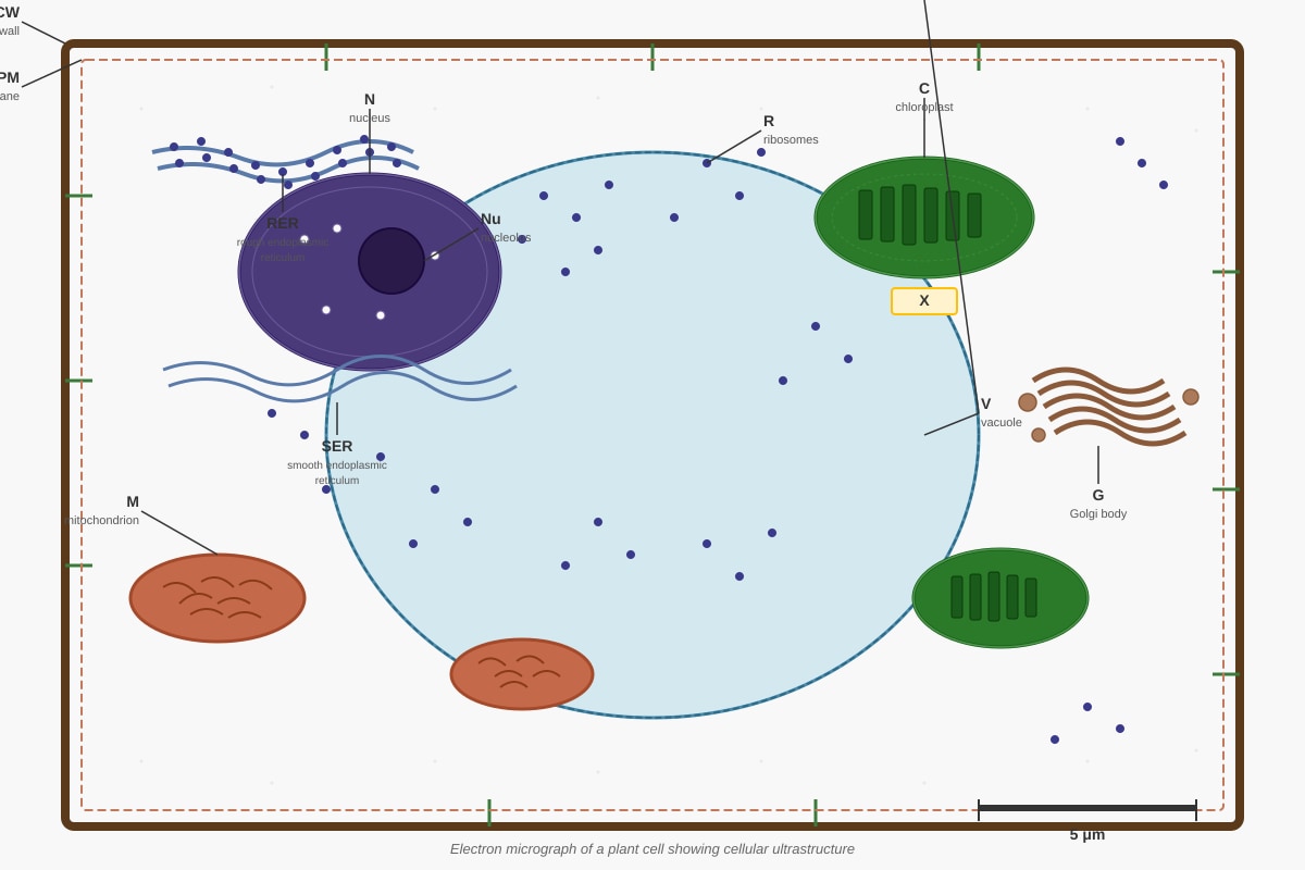
Generated diagram for Q3.
3. Refer to the electron micrograph of a plant cell above. Structure X is labelled but not identified. Structure X shows flattened membranous sacs stacked into grana and is surrounded by a double membrane. Name structure X and state one function specific to plant cells.
Name: _________________________________ [1]
Function: ________________________________________________________________ [1]
4. Which biomolecule test would produce a purple colour in the presence of short peptide chains?
A) Benedict's test
B) Biuret test
C) Iodine test
D) Emulsion test
Answer: _________________ [1]
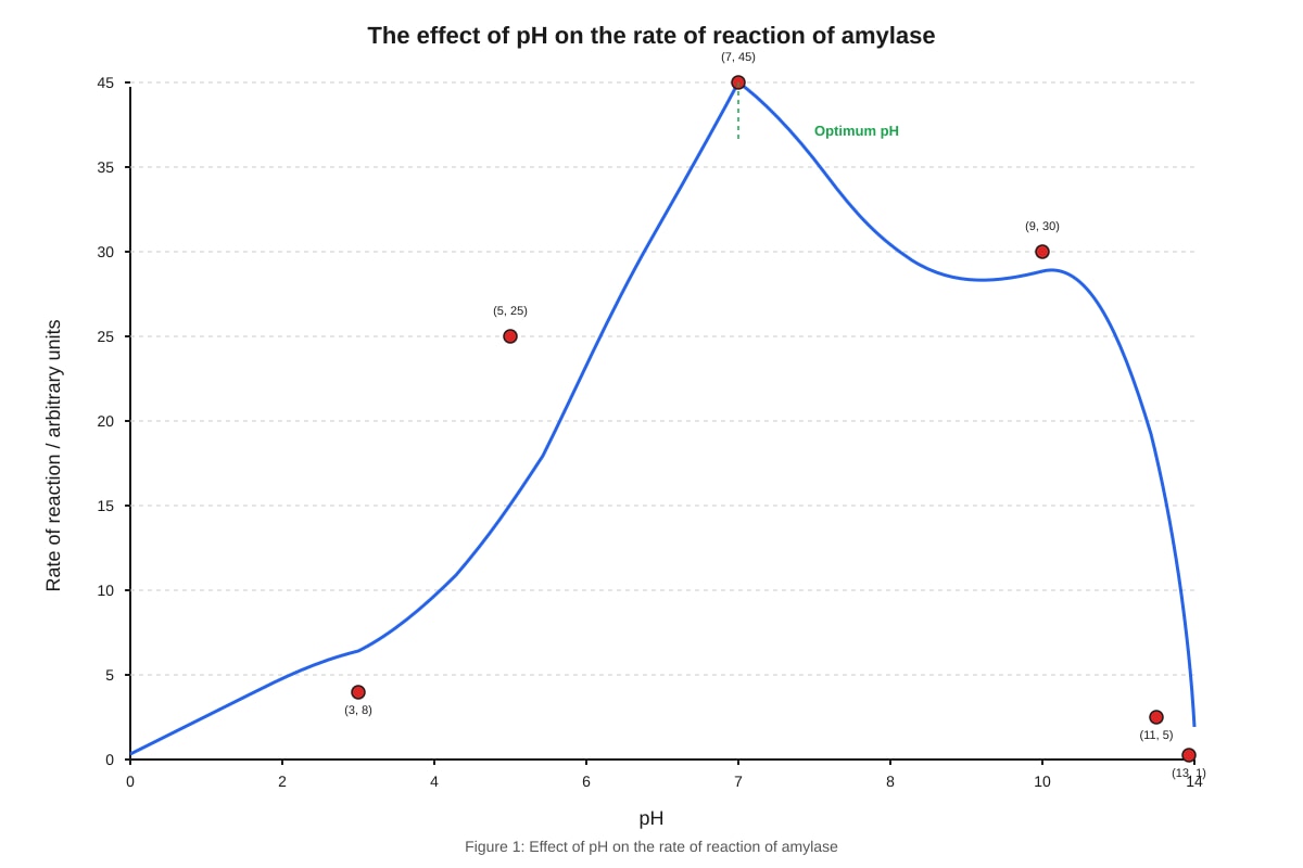
Generated graph for Q5.
5. The graph shows the effect of pH on amylase activity. At which pH value does amylase show approximately 67% of its maximum activity?
A) pH 5
B) pH 6
C) pH 8
D) pH 9
Answer: _________________ [1]
6. Which statement correctly describes the fluid mosaic model of membrane structure?
A) Phospholipids form a bilayer with hydrophilic heads facing inward and hydrophobic tails facing outward
B) Proteins are fixed in position and do not move within the membrane
C) Cholesterol molecules increase membrane fluidity at low temperatures by preventing tight packing
D) Carbohydrate chains are attached to the inner surface of the membrane for cell recognition
Answer: _________________ [1]
7. During protein synthesis, a secretory protein follows a specific pathway through organelles. Which sequence correctly traces this pathway from synthesis to secretion?
A) Ribosome → Smooth ER → Golgi body → secretory vesicle → cell membrane
B) Ribosome → Rough ER → Golgi body → lysosome → cell membrane
C) Ribosome → Rough ER → Golgi body → secretory vesicle → cell membrane
D) Nucleus → Rough ER → Golgi body → secretory vesicle → cell membrane
Answer: _________________ [1]
8. Which of the following increases the surface area to volume ratio of a cell?
A) Increasing cell diameter while maintaining spherical shape
B) Flattening the cell into a disc-like shape
C) Increasing the number of mitochondria
D) Developing a thicker cell wall
Answer: _________________ [1]
9. The enzyme catalase breaks down hydrogen peroxide. A student investigates catalase activity using potato discs of different surface areas but constant total volume. What is the independent variable in this investigation?
A) Volume of oxygen produced
B) Temperature of the water bath
C) Surface area of potato tissue
D) Concentration of hydrogen peroxide
Answer: _________________ [1]
10. Which observation from a microscope would indicate that a cell is undergoing cytokinesis?
A) Chromosomes aligned at the equatorial plate
B) Nuclear envelope reforming around separated chromosomes
C) Cleavage furrow visible at the cell periphery
D) Chromatin condensing into visible chromosomes
Answer: _________________ [1]
Section A Total: _____ / 10
Section B: Short Structured Questions [24 marks]
Answer all questions.
11. The table below shows features of different types of cells in multicellular organisms.
| Cell type | Presence of nucleus | Presence of cell wall | Presence of centrioles | Presence of chloroplasts |
|---|---|---|---|---|
| P | Yes | No | Yes | No |
| Q | Yes | Yes | No | Yes |
| R | No | Yes | No | No |
(a) Identify cell type P and suggest one specialized function this cell type might have in an organism. [2]
(b) Cell type Q is found in leaf tissue. Explain how one structural feature of cell type Q relates to its primary function in photosynthesis. [2]
(c) Cell type R is found in the vascular tissue of plants. Suggest the identity of cell type R and explain why the absence of a nucleus is advantageous for its function. [2]
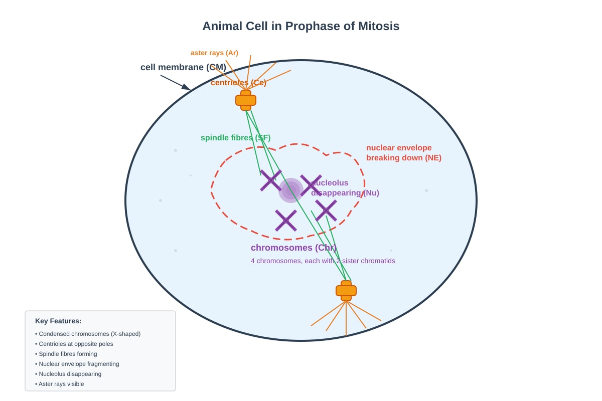
Generated diagram for Q12.
12. The diagram shows an animal cell in a particular stage of cell division.
(a) Name the stage of mitosis shown and describe two visible features that identify this stage. [3]
(b) Explain why it is important that the nuclear envelope breaks down during this stage. [2]
(c) After this stage, the cell shown will complete mitosis and undergo cytokinesis. Calculate how many chromosomes would be present in each daughter cell if the diploid number of this organism is 12. Explain your reasoning. [2]
13. An investigation was carried out to study the effect of temperature on the activity of the enzyme lipase. Lipase breaks down lipids into fatty acids and glycerol. The pH of the reaction mixture was monitored using a pH probe, as the production of fatty acids causes a decrease in pH.
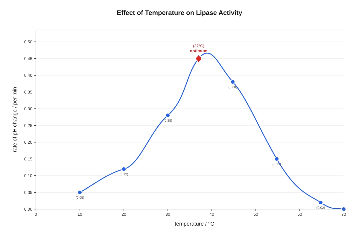
Generated graph for Q13.
(a) Describe the trend shown in the graph between 20°C and 55°C. [2]
(b) With reference to enzyme structure, explain why the rate of reaction decreases above 45°C. [3]
(c) The investigation was repeated at pH 2 instead of the optimal pH for lipase. Predict how the curve would differ from the original and explain your answer. [2]
14. Phospholipids are essential components of all cell membranes.
(a) Draw a simple diagram to show how phospholipids are arranged in a cell membrane. Label the hydrophilic and hydrophobic regions. [2]
(b) Explain how the properties of phospholipids contribute to the selective permeability of the cell membrane. [3]
15. The endosymbiotic theory proposes that mitochondria and chloroplasts evolved from free-living prokaryotic organisms.
(a) State two structural features that mitochondria share with free-living prokaryotes. [2]
(b) Explain why the presence of DNA in mitochondria supports the endosymbiotic theory. [2]
Section B Total: _____ / 24
Section C: Data Response and Extended Answer [26 marks]
Answer all questions.
16. The water potential of plant cells is an important factor in maintaining cell shape and facilitating transport. The table below shows measurements from three adjacent cells (X, Y, and Z) in a plant tissue.
| Cell | Solute potential / MPa | Pressure potential / MPa |
|---|---|---|
| X | -0.5 | +0.2 |
| Y | -0.8 | +0.1 |
| Z | -0.3 | +0.3 |
(a) Calculate the water potential of each cell. Show your working. [3]
(b) Using your calculated values, predict the direction of net water movement between the cells and explain your reasoning. [3]
(c) Explain what would happen to cell Y if it were placed in pure water. Include reference to water potential terms in your answer. [3]
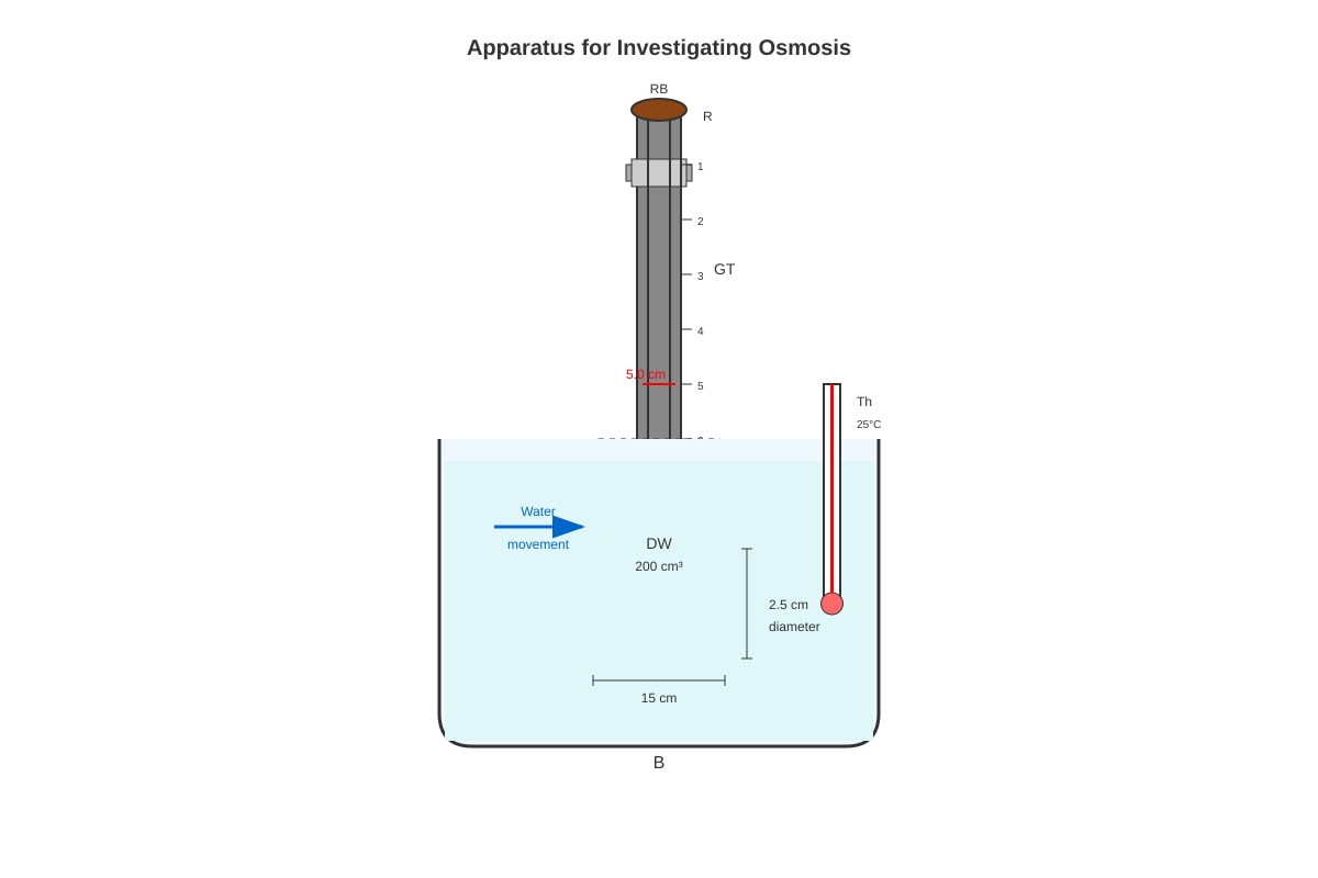
Generated experimental_setup for Q17.
17. The diagram shows apparatus used to investigate osmosis. A Visking tubing bag containing 20% glucose solution was placed in a beaker of distilled water. The initial water level in the glass tubing was recorded.
(a) Predict what will happen to the water level in the glass tubing over the next 30 minutes. Explain your answer in terms of water potential. [3]
(b) After 30 minutes, the student adds more glucose to the solution inside the Visking tubing to make the concentration 40%. Explain how this would affect the rate of water movement and why. [2]
(c) List two variables that should be kept constant to ensure valid results, and explain how each could be controlled. [2]
18. Enzymes are used in many industrial processes. The table compares the use of free enzymes versus immobilized enzymes in starch processing.
| Feature | Free enzymes | Immobilized enzymes |
|---|---|---|
| Reusability | Single use only | Can be reused multiple times |
| Product contamination | Enzyme present in product | Enzyme remains in reactor |
| Optimal temperature stability | Activity decreases rapidly above 60°C | More stable at higher temperatures |
| Cost per batch | Higher | Lower over multiple batches |
| Initial setup cost | Low | High |
(a) Using information from the table, explain why immobilized enzymes are more suitable for large-scale industrial starch processing. [3]
(b) Suggest and explain one environmental advantage of using immobilized enzymes instead of free enzymes. [2]
19. The discovery of the structure of DNA was a major milestone in biology. The diagram below shows part of a DNA molecule.
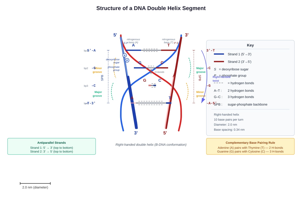
Generated diagram for Q19.
(a) Name the type of chemical bond that joins: (i) adjacent nucleotides in a single strand: _________________________________ [1] (ii) complementary bases on opposite strands: _________________________________ [1]
(b) Explain why the structure shown allows DNA to be replicated accurately. [3]
(c) A DNA molecule contains 32% guanine. Calculate the percentage of thymine in this molecule. Show your working. [2]
20. Stem cells have the potential to develop into various specialized cell types. The diagram shows how a fertilized egg cell gives rise to different cell types in an animal.
Image pending generation: diagram for Q20.
(a) Define the term "differentiation" and explain why it is essential for the development of a multicellular organism. [3]
(b) Compare embryonic stem cells and tissue stem cells with respect to: (i) their potency (range of cell types they can form): [2]
(ii) their location in the body: [2]
(c) Research is being conducted to use stem cells to treat type 1 diabetes, where pancreatic β-cells are destroyed. Evaluate the potential benefits and ethical concerns of using embryonic stem cells for this treatment. [4]
Section C Total: _____ / 26
Paper Total: _____ / 60
END OF PAPER
Answers
TuitionGoWhere Practice Paper Answers - Biology Secondary 3
Version: 2 of 5
Topic: Cells and Biomolecules
Total Marks: 60
Section A: Multiple Choice [10 marks]
1. B) Mitochondrion
Explanation: The mitochondrion is the site of aerobic respiration, where ATP is produced through the Krebs cycle and oxidative phosphorylation. The inner mitochondrial membrane contains electron transport chains and ATP synthase. The Golgi body modifies and packages proteins; the rough endoplasmic reticulum synthesizes proteins; lysosomes contain digestive enzymes. [1 mark]
2. B) Nucleus
Explanation: Uracil is a nitrogenous base found only in RNA, not DNA. The nucleus is where transcription occurs—DNA is used as a template to synthesize RNA molecules. Ribosomes contain rRNA but do not synthesize RNA; the cell membrane and cytoplasm are not primary sites of RNA synthesis. Radioactive uracil would be incorporated into newly made RNA in the nucleus first. [1 mark]
3.
- Name: Chloroplast [1]
- Function: Site of photosynthesis / contains chlorophyll to absorb light energy / produces glucose from carbon dioxide and water [1]
Explanation for visual: The double membrane, stacked internal membranes (grana/thylakoids), and location in a plant cell identify this as a chloroplast. The grana maximize surface area for light absorption. Accept: "stores starch" or "site of photophosphorylation."
4. B) Biuret test
Explanation: The Biuret test detects peptide bonds, producing a purple colour in the presence of proteins and shorter polypeptide chains. Benedict's test detects reducing sugars (brick red precipitate); iodine test detects starch (blue-black); emulsion test detects lipids (white emulsion). [1 mark]
5. C) pH 8
Explanation: Maximum activity = 45 units at pH 7. Sixty-seven percent of 45 = 0.67 × 45 ≈ 30 units. From the graph, the rate at pH 9 is 30 units, which is closest to 67% of maximum. At pH 8, the rate would be approximately 38-40 units (higher than 67%); at pH 5, rate is 25 units (closer to 55%); at pH 6, rate would be approximately 40 units. Note: pH 9 gives exactly 30 units = 66.7%, which is approximately 67%. [1 mark]
6. C) Cholesterol molecules increase membrane fluidity at low temperatures by preventing tight packing
Explanation: Cholesterol is a buffer—in cold, it separates phospholipids to maintain fluidity; in heat, it restricts movement. A is incorrect: hydrophilic heads face outward, hydrophobic tails inward. B is incorrect: proteins can move (fluid mosaic). D is incorrect: carbohydrates are on the outer surface for cell recognition. [1 mark]
7. C) Ribosome → Rough ER → Golgi body → secretory vesicle → cell membrane
Explanation: Secretory proteins are synthesized on ribosomes attached to rough ER, folded and modified in the ER lumen, transported in vesicles to the Golgi body for further processing, then packaged into secretory vesicles that fuse with the cell membrane. The smooth ER lacks ribosomes and is not involved in protein synthesis. Lysosomes are for intracellular digestion, not secretion. The nucleus contains DNA but proteins are not synthesized there. [1 mark]
8. B) Flattening the cell into a disc-like shape
Explanation: Surface area to volume ratio increases when surface area increases relative to volume. A sphere has the minimum surface area for a given volume; flattening increases surface area without changing volume. Increasing diameter decreases SA:V ratio (volume grows faster than surface area). More mitochondria or thicker cell walls do not change the cell's dimensions. [1 mark]
9. C) Surface area of potato tissue
Explanation: The independent variable is the factor deliberately changed by the investigation. The student varies surface area while keeping total volume constant. Volume of oxygen is the dependent variable (measured). Temperature and hydrogen peroxide concentration should be controlled variables (kept constant). [1 mark]
10. C) Cleavage furrow visible at the cell periphery
Explanation: Cytokinesis is division of cytoplasm. In animal cells, a cleavage furrow forms at late anaphase/telophase and pinches the cell in two. A describes metaphase; B describes telophase (nuclear division ending); D describes prophase. C is the defining feature of cytokinesis itself. [1 mark]
Section B: Short Structured Questions [24 marks]
11. (a) Cell type P: Animal cell [1]
Specialized function example: Nerve cell/neuron — transmits electrical impulses; OR Sperm cell — fertilization; OR Muscle cell — contraction; OR White blood cell — phagocytosis/immune defence [1]
Marking note: Any reasonable specialized animal cell with matching function accepted.
11. (b) Structural feature: Cell wall (made of cellulose) / Large permanent vacuole / Chloroplasts [1]
Explanation: The cell wall provides structural support and maintains cell shape against turgor pressure, preventing the cell from bursting when water enters. The large vacuole maintains turgidity and stores cell sap. Chloroplasts contain chlorophyll and are the site of photosynthesis. [1]
Accept any one feature with correct linked explanation. Must link to photosynthesis function.
11. (c) Cell type R: Sieve tube element (in phloem) OR Xylem vessel element [1]
Explanation for sieve tube element: Absence of nucleus allows more space for translocation of organic solutes (mainly sucrose), and the companion cell adjacent to it carries out metabolic functions. The cross-walls (sieve plates) allow easier flow.
Explanation for xylem vessel: Absence of nucleus allows hollow, continuous tube for water transport; lignified walls provide strength and waterproofing for long-distance water conduction without living cell contents. [1]
12. (a) Stage: Prophase [1]
Visible features (any two):
- Chromosomes condense and become visible as thread-like structures (then X-shaped with two chromatids) [1]
- Nuclear envelope breaks down/disappears [1]
- Nucleolus disappears [1]
- Centrioles move to opposite poles with spindle fibres forming between them [1]
- Chromosomes begin to attach to spindle fibres [1]
Need 2 features for 2 marks, plus correct stage identification.
12. (b) The nuclear envelope breaks down to allow spindle fibres to attach to chromosomes [1]. The spindle fibres need direct access to kinetochores on chromosomes to move and separate them accurately to opposite poles during metaphase and anaphase [1]. Without nuclear envelope breakdown, the spindle apparatus cannot interact with chromosomes.
12. (c) 6 chromosomes in each daughter cell [1]
Working/Reasoning: In mitosis, daughter cells are genetically identical to the parent cell. The parent cell shown is diploid (2n = 12). During prophase, each chromosome has replicated to form two sister chromatids, but the chromosome number is still counted as 12 (based on centromeres). After cytokinesis, each daughter cell receives a complete set: 12 ÷ 2 = 6 chromatids? No—correction: each daughter cell receives 12 chromatids that become 12 chromosomes, but the question states diploid number is 12, so each daughter has 2n = 12 chromosomes.
Wait—let me recalculate: The diagram shows 4 chromosomes in prophase, each with 2 chromatids = 8 chromatids. If diploid number is 12, then 2n = 12 chromosomes. Each daughter cell after mitosis receives n chromosomes? No—mitosis maintains diploid number.
Correct answer: 12 chromosomes [1]
Explanation: Mitosis produces two genetically identical daughter cells with the same chromosome number as the parent cell [1]. The parent cell is diploid (2n = 12), so each daughter cell will also be diploid with 12 chromosomes. (In prophase, chromosomes have replicated, but sister chromatids remain attached at the centromere; after anaphase, chromatids separate to become daughter chromosomes, and each daughter cell receives 12 chromosomes.)
Common error: Students may think 12 ÷ 2 = 6, confusing mitosis with meiosis.
13. (a) Trend between 20°C and 55°C:
- From 20°C to 37°C (optimum): Rate increases (from 0.12 to 0.45) — steep rise, rate approximately doubles between 30°C and 37°C [1]
- From 37°C to 55°C: Rate decreases (from 0.45 to 0.15) — gradual decline as temperature exceeds optimum [1]
Must describe both parts of trend for full marks.
13. (b) Above 45°C, the rate decreases due to enzyme denaturation [1].
Explanation: Enzymes are proteins with a specific tertiary structure maintained by hydrogen bonds, ionic bonds, and disulfide bridges [1]. As temperature increases beyond optimum, thermal energy disrupts these bonds, causing the active site to change shape. The substrate (lipid) no longer fits the deformed active site — the enzyme-substrate complex cannot form [1]. This denaturation is typically irreversible.
13. (c) Prediction: The optimum temperature would shift or the curve would show lower overall rates, with reduced peak activity [1].
Explanation: Lipase has an optimal pH around 7-8 (neutral to slightly alkaline). At pH 2 (strongly acidic), excess H⁺ ions disrupt ionic bonds and alter the charge distribution in the active site and protein structure. The enzyme is denatured or its conformation is altered, reducing catalytic efficiency even before temperature effects are considered. The shape of the curve might be similar but with much lower y-values throughout [1].
14. (a)
Diagram should show:
- Phospholipid bilayer with hydrophilic phosphate heads facing outward (toward aqueous environments) and hydrophobic fatty acid tails facing inward, tail-to-tail [1]
- Correct labels: hydrophilic heads and hydrophobic tails indicated [1]
   [aqueous]  O====      O====  [aqueous]   ← hydrophilic heads
                  ||||      ||||
                  ||||      ||||                 ← hydrophobic tails
                  ||||      ||||
   [aqueous]  O====      O====  [aqueous]
                    hydrophobic core
14. (b) Selective permeability explanation:
- The hydrophobic core (fatty acid tails) repels polar molecules and ions, preventing their free passage [1]
- Small, non-polar molecules (O₂, CO₂, lipid-soluble substances) can dissolve in and diffuse through the lipid bilayer [1]
- Polar molecules and ions (glucose, amino acids, Na⁺, K⁺) cannot pass freely and require channel proteins or carrier proteins — this allows the cell to regulate what enters and exits [1]
The phospholipid arrangement creates a barrier that is permeable to some substances but not others — the basis of selective permeability.
15. (a) Any two from:
- Circular DNA (not linear chromosomes in a nucleus) [1]
- 70S ribosomes (smaller than eukaryotic 80S ribosomes) [1]
- Double membrane — outer from host cell engulfment, inner from original prokaryote [1]
- Reproduce by binary fission independently of host cell division [1]
- Similar size to prokaryotic cells [1]
15. (b) Mitochondria contain their own DNA (mtDNA), which is circular and resembles bacterial DNA in structure and gene organization [1]. This suggests mitochondria were once independent free-living bacteria that were engulfed by a larger host cell. Over evolutionary time, they became endosymbiotic — the host provided nutrients, and the prokaryote provided ATP through aerobic respiration [1]. The retention of independent DNA supports their bacterial origin.
Section C: Data Response and Extended Answer [26 marks]
16. (a) Water potential (Ψ) = Solute potential (Ψs) + Pressure potential (Ψp) [1]
Cell X: Ψ = -0.5 + 0.2 = -0.3 MPa [1] Cell Y: Ψ = -0.8 + 0.1 = -0.7 MPa [1] Cell Z: Ψ = -0.3 + 0.3 = 0.0 MPa [1]
Show working clearly. Accept if all three correct with formula stated.
16. (b) Net water movement: From Z → X → Y / From Z to X to Y / From highest to lowest water potential [1]
Reasoning: Water moves by osmosis from a region of higher (less negative) water potential to lower (more negative) water potential [1]. Cell Z has the highest water potential (0.0 MPa), cell X is intermediate (-0.3 MPa), and cell Y is lowest (-0.7 MPa). The gradient drives net water movement down the water potential gradient until equilibrium or until pressure changes alter the gradient [1].
16. (c) In pure water (Ψ = 0 MPa), cell Y has Ψ = -0.7 MPa, which is lower than pure water [1]. Therefore, water will enter cell Y by osmosis [1]. The cell will become turgid as the vacuole expands and pressure potential increases. If the cell wall is strong enough, the pressure potential will rise until Ψcell = 0, at which point net water movement ceases. Without a cell wall (animal cell), the cell would burst/lyse [1].
17. (a) Prediction: The water level in the glass tubing will rise over 30 minutes [1].
Explanation: The glucose solution inside the Visking tubing has a lower (more negative) water potential than the distilled water in the beaker (Ψ = 0). Water moves by osmosis from the beaker into the Visking tubing down the water potential gradient [1]. As water enters, the volume inside increases, forcing the liquid column up the narrow glass tubing, causing the water level to rise [1].
17. (b) The rate of water movement will increase [1].
Explanation: Increasing glucose concentration from 20% to 40% makes the solute potential more negative, thereby lowering the water potential inside the Visking tubing. This creates a steeper water potential gradient between the tubing contents and the distilled water outside. A larger gradient provides greater driving force for osmosis, increasing the rate of water entry. [1]
17. (c) Any two valid variables with control methods:
| Variable | How controlled |
|---|---|
| Temperature | Use a water bath maintained at constant temperature (e.g., 25°C); monitor with thermometer [1] |
| Volume of glucose solution / distilled water | Measure with graduated cylinder/pipette; keep volumes constant [1] |
| Concentration of glucose solution initially | Prepare solutions carefully with weighing and dilution [1] |
| Size/dimensions of Visking tubing | Cut to same length and width; use same batch [1] |
| Time of observation | Use stopwatch; record at regular intervals [1] |
Need two variables with matching controls for 2 marks.
18. (a) Immobilized enzymes are more suitable because:
- Reusability: Can be used for multiple batches, reducing enzyme cost per unit of product [1]
- No product contamination: Enzyme remains in reactor, so product is purer without need for expensive enzyme removal steps [1]
- Thermal stability: Higher temperature stability allows faster reaction rates without rapid loss of activity, increasing throughput and efficiency [1]
- Lower cost per batch: Despite high initial setup cost, long-term operational costs are reduced for large-scale continuous processing [1]
Any three well-explained points for 3 marks.
18. (b) Environmental advantage: Reduced waste / less enzyme disposal needed [1]
Explanation: Free enzymes are discarded with each batch, requiring disposal that may have environmental impacts. Immobilized enzymes are retained in the reactor, producing less waste material over the lifetime of processing. Additionally, the higher efficiency reduces energy and resource consumption per unit of product, lowering the overall environmental footprint [1].
19. (a) (i) Phosphodiester bond (between sugar of one nucleotide and phosphate of next) [1]
(ii) Hydrogen bond (between complementary bases — A-T with 2 hydrogen bonds, G-C with 3 hydrogen bonds) [1]
19. (b) Accurate replication is possible because:
- Complementary base pairing: A always pairs with T, and G always pairs with C [1]
- Antiparallel strands: The sequence of one strand determines the sequence of the other, providing a template for synthesis [1]
- Semi-conservative replication: Each daughter DNA molecule contains one original strand and one new strand, ensuring fidelity is preserved [1]
The specific hydrogen bonding pattern and consistent base-pairing rules mean errors are rare, and when they occur, proofreading mechanisms can correct them.
19. (c) Thymine = 18% [1]
Working:
- If guanine (G) = 32%, then cytosine (C) = 32% (Chargaff's rules: G = C) [1]
- G + C = 32% + 32% = 64%
- Therefore A + T = 100% - 64% = 36%
- Since A = T, then T = 36% ÷ 2 = 18% [1]
Method must be shown for full marks.
20. (a) Differentiation: The process by which a less specialized cell becomes a more specialized cell type with a specific structure and function [1].
Essential for multicellular organisms because:
- Allows division of labour — different cells perform different functions efficiently [1]
- Enables complex tissue and organ formation — specialized cells group together to form tissues with specific functions (e.g., muscle tissue for contraction, nerve tissue for conduction) [1]
- Increases efficiency — specialized cells are optimized for their role rather than performing all functions moderately
20. (b) (i) Embryonic stem cells: Totipotent or pluripotent — can form all cell types in the body (embryonic and extra-embryonic tissues if totipotent, or all three germ layers if pluripotent) [1]
Tissue stem cells: Multipotent or unipotent — can form limited range of cell types specific to their tissue of origin (e.g., haematopoietic stem cells form blood cells; neural stem cells form neurons and glia) [1]
20. (b) (ii) Embryonic stem cells: Found in the inner cell mass of the blastocyst (early embryo, ~5-7 days post-fertilization) [1]
Tissue stem cells: Found in specific tissues throughout the body — e.g., bone marrow, skin basal layer, intestinal crypts, brain subventricular zone [1]
20. (c) Potential benefits (up to 2 marks):
- Embryonic stem cells are pluripotent, so they can be differentiated into pancreatic β-cells to replace those destroyed in type 1 diabetes [1]
- Could provide a renewable source of transplantable cells, reducing reliance on donated organs and immunosuppressive drugs if patient-specific cells are used [1]
- Could cure the underlying cause rather than just managing symptoms (insulin injections)
Ethical concerns (up to 2 marks):
- Embryos are destroyed to obtain inner cell mass cells — some believe this destroys potential human life and is morally equivalent to killing a person [1]
- Slippery slope: Could lead to reproductive cloning or creation of embryos for tissue farming [1]
- Alternative: Induced pluripotent stem cells (iPSCs) — reprogram adult cells to pluripotency without embryo destruction, addressing ethical concerns but with technical challenges
Need balanced evaluation for full 4 marks. Accept other valid ethical arguments.
Mark Summary
| Section | Marks |
|---|---|
| Section A | 10 |
| Section B | 24 |
| Section C | 26 |
| Total | 60 |
Free quiz and exam paper access
Enter your details to view this paper
Your access is remembered on this device.