From Real Exams Exam Paper
Secondary 3 Biology Semestral Assessment 2 (End of Year) Paper 4
Free Sec 3 Biology SA2 Paper 4, Kimi2.6 Exam version, with questions, answers, and O Level-style practice for Singapore students.
These static practice materials are generated from the site's syllabus and paper-generation workflow, with source and model context shown so students and parents can evaluate the material before use.
Questions
TuitionGoWhere Practice Paper - Secondary 3 Biology
TuitionGoWhere Exam Practice (AI)
| Subject: | Biology |
| Level: | Secondary 3 (Express/G3) |
| Paper: | SA2 Practice Paper - Version 4 of 5 |
| Duration: | 1 hour 15 minutes |
| Total Marks: | 60 |
Name: _________________________ Class: ___________ Date: ___________
Instructions
- Answer ALL questions.
- Write your answers in the spaces provided.
- For multiple choice questions, circle the correct answer.
- All working for calculations should be shown.
- Marks are allocated for clear and accurate labelled diagrams where required.
Section A: Multiple Choice (Questions 1–10)
Choose the correct answer for each question. Each question carries 2 marks. Section Total: 20 marks
1. Which structure is found in a plant cell but NOT in an animal cell?
| A | Centrioles |
|---|---|
| B | Mitochondria |
| C | Cell wall |
| D | Nucleus |
Answer: _______________ (2 marks)
2. A student observed a cell under the microscope and noted the following features: a large central vacuole, chloroplasts, and a regular shape. Which type of cell was most likely observed?
| A | White blood cell |
|---|---|
| B | Muscle cell |
| C | Onion epidermal cell |
| D | Palisade mesophyll cell |
Answer: _______________ (2 marks)
3. The diagram below shows an animal cell.
Generated diagram for Q3.
Which labelled structure is responsible for modifying, sorting, and packaging proteins for secretion?
Answer: _______________ (2 marks)
4. Which biomolecule is the primary structural component of cell membranes?
| A | Starch |
|---|---|
| B | Phospholipid |
| C | Glycogen |
| D | Cellulose |
Answer: _______________ (2 marks)
5. An actively growing cell is supplied with radioactive amino acids. Which cell component would FIRST show an increase in radioactivity?
| A | Golgi body |
|---|---|
| B | Rough endoplasmic reticulum |
| C | Mitochondria |
| D | Nucleus |
Answer: _______________ (2 marks)
6. The graph below shows the effect of pH on the activity of two enzymes, P and Q.
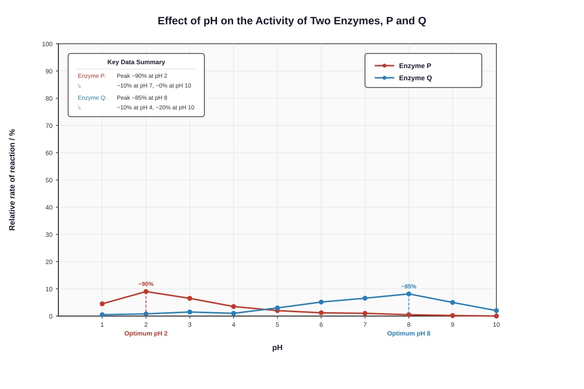
Generated graph for Q6.
Which statement correctly describes the enzymes?
| A | Both enzymes work best in acidic conditions |
|---|---|
| B | Enzyme P is likely to be pepsin and enzyme Q is likely to be trypsin |
| C | Enzyme Q has a higher rate of reaction than enzyme P at all pH values |
| D | Both enzymes are denatured at pH 7 |
Answer: _______________ (2 marks)
7. Which of the following is a correct statement about diffusion?
| A | Diffusion requires energy from ATP |
|---|---|
| B | Diffusion occurs only in gases |
| C | Diffusion is the net movement of particles from a region of higher concentration to lower concentration |
| D | Diffusion requires a partially permeable membrane |
Answer: _______________ (2 marks)
8. The diagram shows the structure of a protein molecule.
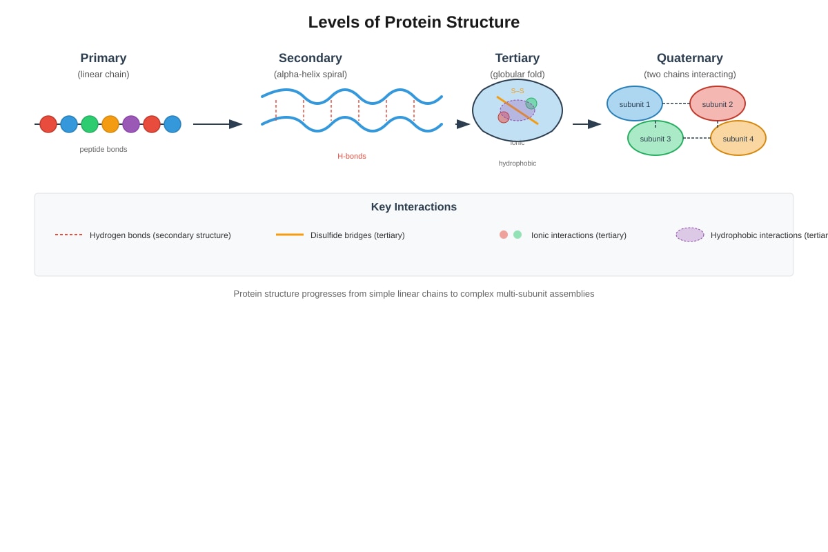
Generated diagram for Q8.
Which level of protein structure is maintained primarily by hydrogen bonds between peptide groups?
| A | Primary structure |
|---|---|
| B | Secondary structure |
| C | Tertiary structure |
| D | Quaternary structure |
Answer: _______________ (2 marks)
9. A red blood cell is placed in distilled water. What would be observed after 30 minutes?
| A | The cell becomes flaccid |
|---|---|
| B | The cell shrinks and becomes crenated |
| C | The cell swells and may burst |
| D | No visible change occurs |
Answer: _______________ (2 marks)
10. Which of the following correctly matches a cell structure with its function?
| Structure | Function | |
|---|---|---|
| A | Ribosome | Synthesis of lipids |
| B | Lysosome | Intracellular digestion |
| C | Smooth endoplasmic reticulum | Protein synthesis |
| D | Cell wall | Selective control of substances entering and leaving cell |
Answer: _______________ (2 marks)
Section B: Structured Response (Questions 11–16)
Answer all questions in the spaces provided. Section Total: 24 marks
11. The diagram below shows a plant cell as seen under a light microscope.
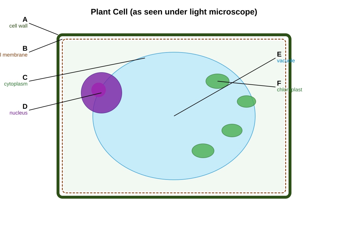
Generated diagram for Q11.
(a) State the names of structures labelled A and F.
A: _________________________________ (1 mark)
F: _________________________________ (1 mark)
(b) Explain how structure A helps maintain the shape of the plant cell. (2 marks)
(c) Structure E plays an important role in maintaining cell turgidity. Explain what would happen to the plant cell if it was placed in a concentrated salt solution and why this occurs. (3 marks)
[Total: 7 marks]
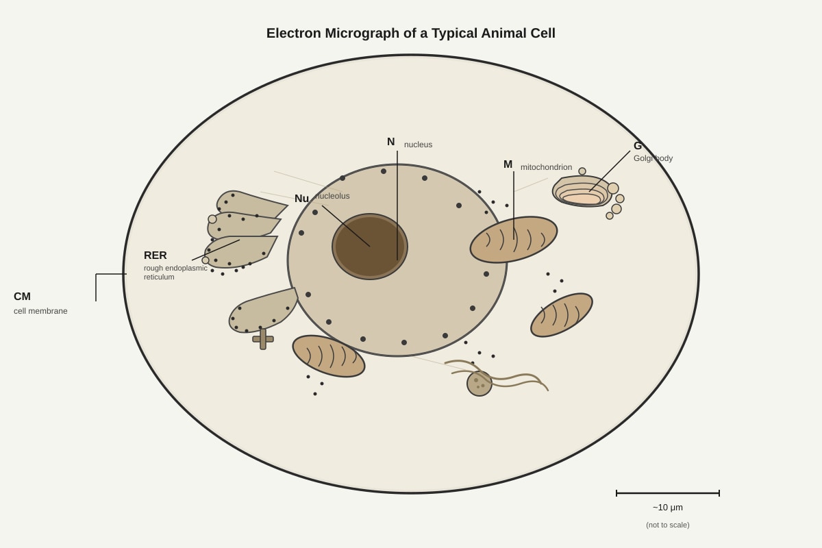
12. The electron micrograph below shows part of an animal cell.
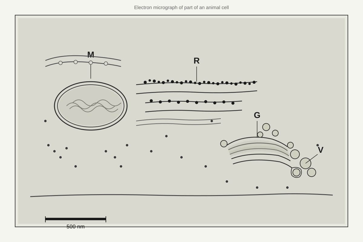
Generated diagram for Q12.
(a) Identify the organelle labelled M and describe two features visible in the diagram that support your identification. (3 marks)
(b) Explain the functional relationship between the rough endoplasmic reticulum (R), the Golgi body (G), and the secretory vesicles (V) in the production and release of proteins from the cell. (4 marks)
[Total: 7 marks]
13. An experiment was conducted to investigate the effect of temperature on the activity of the enzyme amylase. The results are shown in the table below.
| Temperature / °C | Time taken for starch to be completely broken down / minutes |
|---|---|
| 10 | 12 |
| 20 | 8 |
| 30 | 4 |
| 40 | 2 |
| 50 | 5 |
| 60 | 15 |
| 70 | no reaction after 30 minutes |
(a) Calculate the rate of reaction at 40°C. Show your working. (2 marks)
(b) Explain why the reaction time was shortest at 40°C. (2 marks)
(c) Explain why there was no reaction at 70°C even after 30 minutes. (2 marks)
[Total: 6 marks]
14. The diagram below shows the fluid mosaic model of a cell membrane.
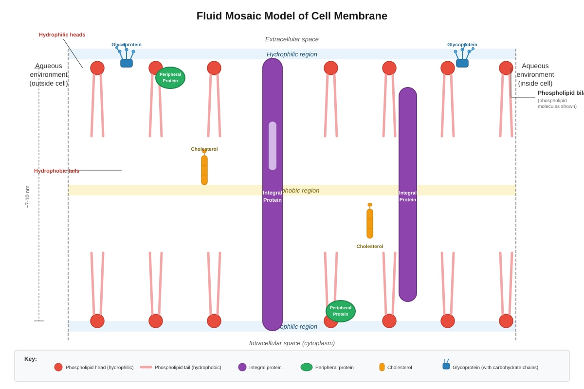
Generated diagram for Q14.
(a) Describe the arrangement of phospholipid molecules in the membrane and explain why they are arranged in this way. (3 marks)
(b) State two functions of membrane proteins and give one example of each. (2 marks)
[Total: 5 marks]
15. The diagram shows an experimental set-up used to demonstrate osmosis.
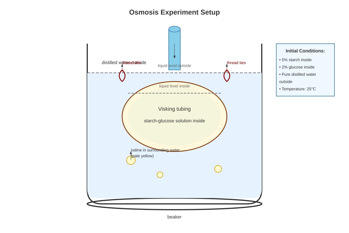
Generated experimental_setup for Q15.
(a) The Visking tubing acts as a partially permeable membrane. Explain what is meant by "partially permeable." (2 marks)
(b) After 30 minutes, the water in the beaker was tested with Benedict's solution and heated. An orange-red precipitate was observed. Explain this result with reference to the properties of the Visking tubing and the molecules involved. (3 marks)
[Total: 5 marks]
16. The table below compares features of prokaryotic and eukaryotic cells.
| Feature | Prokaryotic cell | Eukaryotic cell |
|---|---|---|
| Nucleus | No true nucleus; DNA in nucleoid | _______________ |
| Ribosomes | Smaller (70S) | _______________ |
| Membrane-bound organelles | Absent | _______________ |
| Cell wall | Present (contains peptidoglycan) | _______________ |
| Example | _______________ | Amoeba |
(a) Complete the table by filling in the missing information. (4 marks)
(b) State two advantages of compartmentalisation in eukaryotic cells. (2 marks)
[Total: 6 marks]
Section C: Data Analysis and Extended Response (Questions 17–20)
Answer all questions in the spaces provided. Section Total: 16 marks
17. The graph below shows the absorption spectra of three different photosynthetic pigments found in plant cells.
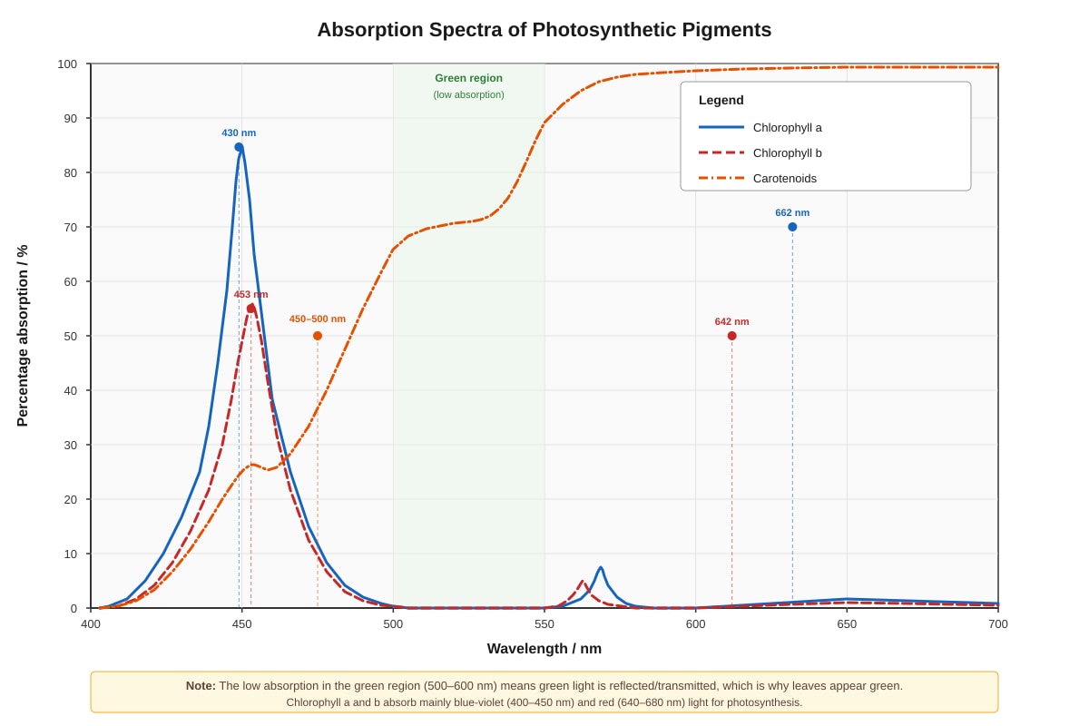
Generated graph for Q17.
(a) State the wavelength at which chlorophyll a shows maximum absorption. (1 mark)
(b) Explain why leaves appear green to the human eye, using evidence from the graph. (2 marks)
(c) A plant is grown under green light only. Predict and explain what would happen to the growth rate of this plant compared to one grown under white light. (3 marks)
[Total: 6 marks]
18. The diagram shows an experiment to investigate the effect of concentration on the rate of diffusion.
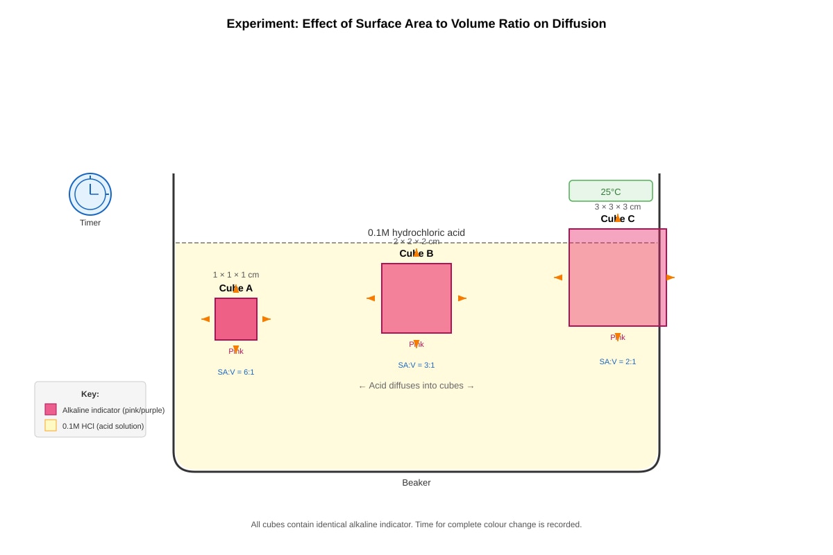
Generated experimental_setup for Q18.
The cubes contain an alkaline indicator and are placed in dilute hydrochloric acid. The time taken for the acid to completely neutralise the indicator (as shown by complete colour change) is recorded.
| Cube | Dimensions / cm | Total surface area / cm² | Volume / cm³ | Surface area to volume ratio | Time for complete colour change / s |
|---|---|---|---|---|---|
| A | 1 × 1 × 1 | 6 | 1 | 6:1 | 45 |
| B | 2 × 2 × 2 | __ | 8 | __ | 180 |
| C | 3 × 3 × 3 | __ | __ | 2:1 | __ |
(a) Complete the missing values in the table. Show your working. (3 marks)
(b) Explain why the complete colour change takes longer for larger cubes, using the concept of surface area to volume ratio. (3 marks)
[Total: 6 marks]
19. Proteins are essential biomolecules with diverse functions in living organisms.
(a) Describe the structural difference between a condensation reaction and a hydrolysis reaction in the context of protein formation and breakdown. (2 marks)
(b) Explain how the structure of an enzyme relates to its specificity in catalysing reactions. (4 marks)
[Total: 6 marks]
20. The following passage describes the development of a new drug to lower blood cholesterol levels.
Statins are drugs that inhibit the enzyme HMG-CoA reductase, which catalyses an early step in cholesterol synthesis in liver cells. The original statins were derived from fungal products. Researchers have now developed synthetic statins that are more effective at lower doses. These drugs must be absorbed from the digestive system, enter liver cells, and bind specifically to the active site of HMG-CoA reductase.
Using your knowledge of cell membranes, enzymes, and biomolecules:
(a) Explain why the synthetic statins need to be able to cross cell membranes to be effective. (2 marks)
(b) The original fungal-derived statins and the new synthetic statins both inhibit the same enzyme but the synthetic version works at lower concentrations. Using your knowledge of enzyme action and molecular structure, suggest why the synthetic statin might be more effective. (4 marks)
(c) Some patients cannot take statins due to side effects. Describe one alternative approach to lowering blood cholesterol that does not involve inhibiting this enzyme. (2 marks)
[Total: 8 marks]
END OF PAPER
Total Marks: 60
Answers
TuitionGoWhere Practice Paper - Secondary 3 Biology
SA2 Practice Paper - Version 4 of 5 — Answer Key
Total Marks: 60
Section A: Multiple Choice (Questions 1–10)
Each question: 2 marks
| Question | Answer | Explanation |
|---|---|---|
| 1 | C | Cell wall — Plant cells possess a cell wall made of cellulose outside the cell membrane, providing structural support. Animal cells lack cell walls. Centrioles (A) are found in animal cells but not most plant cells. Mitochondria (B) and nucleus (D) are present in both. |
| 2 | D | Palisade mesophyll cell — This is a plant cell with chloroplasts (for photosynthesis) and a large vacuole. Onion epidermal cells (C) lack chloroplasts. White blood cells (A) and muscle cells (B) are animal cells with no cell wall, vacuole, or chloroplasts. |
| 3 | G (Golgi body) | The Golgi body modifies, sorts, and packages proteins received from the rough ER into vesicles for transport. The RER (rough ER) synthesises proteins; mitochondria produce ATP; the nucleus stores genetic information. |
| 4 | B | Phospholipid — Cell membranes are composed of a phospholipid bilayer. The hydrophilic phosphate heads face outward and hydrophobic fatty acid tails face inward. Starch (A), glycogen (C), and cellulose (D) are carbohydrates, not membrane structural components. |
| 5 | B | Rough endoplasmic reticulum — Radioactive amino acids are incorporated into proteins at ribosomes on the RER. The protein then moves to the Golgi body for processing. Thus the RER shows radioactivity FIRST. |
| 6 | B | Enzyme P is likely pepsin; enzyme Q is likely trypsin — Pepsin works in the stomach at pH ~2 (acidic). Trypsin works in the small intestine at pH ~8 (alkaline). Both have different pH optima; neither is denatured at pH 7 (both retain some activity). |
| 7 | C | Diffusion is net movement from higher to lower concentration — Diffusion is passive (no ATP needed, A incorrect), occurs in gases AND liquids (B incorrect), and does NOT require a membrane (D describes osmosis or facilitated diffusion). |
| 8 | B | Secondary structure — Alpha-helices and beta-pleated sheets are maintained by hydrogen bonds between the amide hydrogen and carbonyl oxygen of peptide groups. Primary structure uses peptide bonds; tertiary uses various interactions including disulfide bonds and ionic interactions. |
| 9 | C | The cell swells and may burst — Distilled water is hypotonic to the cytoplasm. Water enters by osmosis, causing the cell to swell. Animal cells lack cell walls, so they may burst (lyse). Plant cells would become turgid but not burst. |
| 10 | B | Lysosome: intracellular digestion — Lysosomes contain hydrolytic enzymes. Ribosomes synthesise proteins (not lipids, A wrong). Smooth ER synthesises lipids (not proteins, C wrong). Cell wall is fully permeable; the cell membrane controls entry/exit (D wrong). |
Section A Total: 20 marks
Section B: Structured Response (Questions 11–16)
Question 11 [Total: 7 marks]
(a) State the names of structures labelled A and F. (2 marks)
- A: Cell wall (1 mark)
- F: Chloroplast (1 mark)
Marking note: Accept phonetic spellings if clear. "Cell wall" must distinguish from cell membrane.
(b) Explain how structure A helps maintain the shape of the plant cell. (2 marks)
- The cell wall is rigid and surrounds the cell membrane, providing mechanical support (1 mark)
- It is made of cellulose which has high tensile strength; when the cell is turgid (vacuole full of water pushing against cell wall), the combination of wall rigidity and turgor pressure maintains the firm shape (1 mark)
Common mistake: Students confuse cell wall with cell membrane. The wall is fully permeable and rigid; the membrane is selectively permeable and flexible.
(c) Structure E plays an important role in maintaining cell turgidity. Explain what would happen to the plant cell if it was placed in a concentrated salt solution and why this occurs. (3 marks)
- The cell would become flaccid and may undergo plasmolysis (1 mark)
- A concentrated salt solution is hypertonic (higher solute concentration) compared to the cell sap (1 mark)
- Water leaves the vacuole and cytoplasm by osmosis (net movement of water from higher water potential to lower water potential) through the partially permeable membrane (1 mark)
- The cell membrane pulls away from the cell wall as the vacuole shrinks
Key terms to award: hypertonic, osmosis, water potential, flaccid/plasmolysis
Question 12 [Total: 7 marks]
(a) Identify the organelle labelled M and describe two features visible in the diagram that support your identification. (3 marks)
- Mitochondrion (1 mark)
- Feature 1: Double membrane (outer smooth membrane and inner membrane folded into cristae) — increases surface area (1 mark)
- Feature 2: Cristae — the folded inner membrane projections visible in the diagram (1 mark)
Alternative: Matrix (the inner space) containing enzymes for Krebs cycle — but only if clearly described from image features.
(b) Explain the functional relationship between RER, Golgi body, and secretory vesicles in protein production and release. (4 marks)
| Mark point | Explanation |
|---|---|
| 1 mark | Ribosomes on RER synthesise proteins using mRNA templates; proteins enter the lumen of RER for folding and initial modification |
| 1 mark | Transport vesicles bud off from RER and carry proteins to the Golgi body |
| 1 mark | In the Golgi body, proteins are further modified (e.g., glycosylation — adding carbohydrate chains), sorted, and packaged into secretory vesicles |
| 1 mark | Secretory vesicles move to and fuse with the cell membrane, releasing proteins outside by exocytosis |
This is the secretory pathway — sequential processing ensures correct protein structure and destination.
Question 13 [Total: 6 marks]
(a) Calculate the rate of reaction at 40°C. Show your working. (2 marks)
Rate=time1=2 minutes1=0.5 min−1
Or: \frac{60}{2} = 30 \text{ arbitrary units per minute (or % per minute)}
- Correct formula/reciprocal relationship stated (1 mark)
- Correct answer with units: 0.5 min⁻¹ or equivalent (1 mark)
Accept: 30 % min⁻¹, 0.0083 s⁻¹, etc. if working shown
(b) Explain why the reaction time was shortest at 40°C. (2 marks)
- 40°C is the optimum temperature for amylase activity (1 mark)
- At this temperature, enzyme and substrate molecules have sufficient kinetic energy to collide frequently, AND the enzyme maintains its tertiary structure/active site shape for effective substrate binding (1 mark)
Common error: Saying "enzyme works fastest" without explaining WHY — need to mention both kinetic energy AND maintained structure.
(c) Explain why there was no reaction at 70°C even after 30 minutes. (2 marks)
- At 70°C, the enzyme is denatured (1 mark)
- The tertiary structure is disrupted; the active site changes shape permanently and can no longer bind to the starch substrate (1 mark)
- This is irreversible; cooling will not restore activity
Question 14 [Total: 5 marks]
(a) Describe the arrangement of phospholipid molecules and explain why. (3 marks)
- Arrangement: Phospholipids form a bilayer with hydrophilic phosphate heads facing the aqueous environments on both sides, and hydrophobic fatty acid tails pointing inward, shielded from water (1 mark)
- Reason for arrangement: The aqueous cytoplasm and external environment mean the hydrophilic heads interact with water; the hydrophobic tails are repelled by water and cluster together (1 mark)
- This creates a ** stable barrier** that controls what enters and leaves the cell — hydrophobic core prevents free passage of charged/polar substances (1 mark)
(b) State two functions of membrane proteins and give one example of each. (2 marks)
| Function | Example |
|---|---|
| Transport/carrier proteins — move specific substances across membrane | Glucose transporters (GLUT proteins) in cell membranes |
| Receptor proteins — bind signalling molecules | Insulin receptors on liver/muscle cells |
| Enzymes — catalyse reactions at membrane surface | Digestive enzymes on microvilli of intestinal cells |
| Cell recognition/antigens — identify self/non-self | Glycoproteins (MHC proteins) for immune recognition |
- Any two functions with valid examples (1 mark each = 2 marks)
Question 15 [Total: 5 marks]
(a) Explain what is meant by "partially permeable." (2 marks)
- A partially permeable membrane allows certain molecules to pass through while preventing or restricting others (1 mark)
- It permits small molecules (like water, glucose) to diffuse through but blocks larger molecules (like starch) (1 mark)
Related terms: selectively permeable, differentially permeable — accept these as equivalent.
(b) Explain the orange-red precipitate after heating with Benedict's solution. (3 marks)
- Benedict's test indicates reducing sugars; orange-red colour confirms glucose has diffused out of the Visking tubing (1 mark)
- Glucose molecules are small enough to pass through the pores in the partially permeable Visking tubing membrane (1 mark)
- Starch molecules are too large to pass through, so no starch is detected outside (1 mark)
- The glucose concentration gradient (high inside, zero outside initially) drives net movement by diffusion
Expected final state: Glucose present inside and outside (equilibrated); starch remains inside.
Question 16 [Total: 6 marks]
(a) Complete the table. (4 marks)
| Feature | Prokaryotic cell | Eukaryotic cell |
|---|---|---|
| Nucleus | No true nucleus; DNA in nucleoid | True nucleus with nuclear envelope |
| Ribosomes | Smaller (70S) | Larger (80S) |
| Membrane-bound organelles | Absent | Present (e.g., mitochondria, Golgi body, ER) |
| Cell wall | Present (contains peptidoglycan) | Absent in animals; present in plants/fungi (cellulose/chitin) |
| Example | Bacterium / Cyanobacterium | Amoeba |
- 4 correct entries (1 mark each) = 4 marks
(b) State two advantages of compartmentalisation in eukaryotic cells. (2 marks)
-
Different chemical environments: Each organelle can maintain optimal pH, enzymes, and substrates for specific reactions (e.g., acidic pH in lysosomes for hydrolysis) without interfering with other cellular processes (1 mark)
-
Increased efficiency of metabolic reactions: Concentrating enzymes and substrates in specific compartments (e.g., mitochondria for respiration, chloroplasts for photosynthesis) increases reaction rates and allows simultaneous but incompatible processes (e.g., protein synthesis on RER and protein digestion in lysosomes) (1 mark)
Alternative: Protection of cell from harmful reactions (e.g., lysosomal enzymes contained); DNA separated from cytoplasm for controlled gene expression.
Section B Total: 24 marks
Section C: Data Analysis and Extended Response (Questions 17–20)
Question 17 [Total: 6 marks]
(a) State the wavelength at which chlorophyll a shows maximum absorption. (1 mark)
- 430 nm (or approximately 430–435 nm) — the first/main peak in the blue-violet region (1 mark)
- Accept 662 nm as secondary peak if specified, but 430 nm has higher absorption
(b) Explain why leaves appear green using evidence from the graph. (2 marks)
- All three pigments show low percentage absorption in the green region (~500–600 nm) (1 mark)
- Green light is reflected or transmitted rather than absorbed; this reflected green light reaches our eyes, making leaves appear green (1 mark)
Chlorophyll a and b both have absorption minima around 550 nm; carotenoids absorb poorly above 500 nm.
(c) Predict and explain growth under green light only compared to white light. (3 marks)
| Mark point | Content |
|---|---|
| 1 mark (prediction) | Plant under green light would show slower growth / reduced growth / stunted growth compared to white light |
| 1 mark (explanation from data) | Green light is poorly absorbed by chlorophyll a, chlorophyll b, and carotenoids (as shown by low % absorption, ~10–20% at 550 nm vs. 60–75% at optimum wavelengths) |
| 1 mark (consequence) | Less light energy is captured for photosynthesis; less glucose produced; therefore less energy and carbon skeletons for growth, cell division, and metabolic processes |
White light contains all wavelengths, including the blue-violet and red regions where chlorophylls absorb maximally.
Question 18 [Total: 6 marks]
(a) Complete the table. Show your working. (3 marks)
Cube B:
- Surface area = 6×(2)2=6×4=24 cm²
- SA:V ratio = 24:8=3:1
Cube C:
- Surface area = 6×(3)2=6×9=54 cm²
- Volume = 33=27 cm³
| Cube | Dimensions / cm | Total surface area / cm² | Volume / cm³ | Surface area to volume ratio | Time for complete colour change / s |
|---|---|---|---|---|---|
| A | 1 × 1 × 1 | 6 | 1 | 6:1 | 45 |
| B | 2 × 2 × 2 | 24 | 8 | 3:1 | 180 |
| C | 3 × 3 × 3 | 54 | 27 | 2:1 | 405 (or proportional: 45 × 9 = 405, or 180 × 2.25 = 405) |
- Surface area calculations correct (1 mark)
- Volume and SA:V ratio correct (1 mark)
- Extrapolated time for C based on pattern (proportional to volume, or inverse to SA:V) (1 mark)
Pattern: Time increases with volume; Cube A (1 cm³, ratio 6:1) → 45s; Cube B (8 cm³, ratio 3:1) → 180s (4×); Cube C (27 cm³, ratio 2:1) → 405s or 540s depending on model used. Accept reasonable extrapolation with justification.
(b) Explain why larger cubes take longer, using SA:V ratio. (3 marks)
| Mark point | Content |
|---|---|
| 1 mark | Larger volume means more indicator molecules need to be neutralised by acid; more substance to be affected |
| 1 mark | As size increases, SA:V ratio decreases (from 6:1 to 3:1 to 2:1); proportionally less surface area available for diffusion per unit volume |
| 1 mark | Diffusion distance increases — acid must penetrate further to reach centre; hence rate of complete neutralisation slows as cube size increases |
Key concept: Efficiency of exchange processes depends on SA:V ratio. Small cells/organisms have higher SA:V ratios = more efficient diffusion.
Question 19 [Total: 6 marks]
(a) Describe structural difference between condensation and hydrolysis. (2 marks)
| Condensation | Hydrolysis |
|---|---|
| Builds larger molecules from smaller subunits | Breaks down larger molecules into smaller subunits |
| Removes water — H from one subunit, OH from another, forming a peptide bond (in proteins) or glycosidic bond (in carbohydrates) | Adds water — H and OH split across the bond, breaking the peptide/glycosidic bond |
- One mark for each correct structural description (2 marks)
In proteins: condensation joins amino acids → peptide bond + H₂O lost. Hydrolysis breaks peptide bond → amino acids + H₂O used.
(b) Explain how enzyme structure relates to specificity. (4 marks)
| Mark point | Content |
|---|---|
| 1 mark | Enzymes are proteins with a specific tertiary structure; this creates a unique 3-dimensional shape |
| 1 mark | The active site is a specific region with particular arrangement of amino acid R-groups, forming a complementary shape to the substrate |
| 1 mark | This is the lock and key (or induced fit) model — only substrate(s) with matching shape/fit can bind |
| 1 mark | Specific amino acid side chains in the active site form temporary bonds (hydrogen bonds, ionic interactions) with the substrate, stabilising the transition state and lowering activation energy — different substrates cannot form these interactions |
Named models accepted; emphasis should be on structural complementarity and specific molecular interactions.
Question 20 [Total: 8 marks]
(a) Explain why statins must cross cell membranes to be effective. (2 marks)
- HMG-CoA reductase is an intracellular enzyme located inside liver cells (cytoplasm) (1 mark)
- The drug must cross the cell membrane to reach the enzyme's active site and inhibit it; if it remains outside, it cannot access its target (1 mark)
Membranes act as barriers — only lipid-soluble or appropriately transported substances can enter.
(b) Suggest why synthetic statin works at lower concentrations. (4 marks)
| Mark point | Content |
|---|---|
| 1 mark | Synthetic statin likely has a chemical structure more similar/complementary to the enzyme's active site |
| 1 mark | Better fit to active site — higher binding affinity; forms more non-covalent interactions (hydrogen bonds, ionic bonds, hydrophobic interactions) |
| 1 mark | Under induced fit model, the synthetic statin may induce a more favourable conformational change, stabilising the inactive enzyme form |
| 1 mark | Higher affinity means lower Kₘ (lower concentration needed for half-maximal inhibition); the drug competes more effectively with the natural substrate for the active site |
Binding affinity relates to structure — medicinal chemistry optimises molecular shape and charge distribution for target specificity and efficacy.
(c) Describe one alternative approach to lowering blood cholesterol. (2 marks)
| Approach | Explanation |
|---|---|
| Dietary modification | Reduce intake of saturated fats and cholesterol; increase dietary fibre (soluble fibre binds bile salts/cholesterol in gut, increasing excretion) |
| Exercise | Increases LDL receptor expression in liver cells, enhancing cholesterol removal from blood |
| Bile acid sequestrants | Drugs that bind bile acids in intestine, preventing reabsorption; liver must use more cholesterol to make new bile acids |
| Statins from natural sources | Red yeast rice contains monacolin K (natural statin-like compound) — though this is still enzyme inhibition |
- Valid alternative with reasonable mechanism (2 marks)
Section C Total: 16 marks
GRAND TOTAL: 60 MARKS
Mark Distribution Summary
| Section | Marks |
|---|---|
| A (MCQ 1–10) | 20 |
| B (Structured 11–16) | 24 |
| C (Extended 17–20) | 16 |
| Total | 60 |
Duration Check
- MCQ: ~15 minutes (1.5 min/question)
- Section B: ~30 minutes (5 min/question)
- Section C: ~25 minutes (6–8 min/question)
- Review buffer: ~5 minutes
- Total: 75 minutes ✓
Free quiz and exam paper access
Enter your details to view this paper
Your access is remembered on this device.