From Real Exams Exam Paper
Secondary 3 Biology Semestral Assessment 2 (End of Year) Paper 3
Free Sec 3 Biology SA2 Paper 3, Kimi2.6 Exam version, with questions, answers, and O Level-style practice for Singapore students.
These static practice materials are generated from the site's syllabus and paper-generation workflow, with source and model context shown so students and parents can evaluate the material before use.
Questions
TuitionGoWhere Practice Paper - Biology Secondary 3
TuitionGoWhere Secondary School (AI)
| Subject: | Biology |
| Level: | Secondary 3 (G3/Express) |
| Paper: | SA2 Practice Paper |
| Duration: | 1 hour 15 minutes |
| Total Marks: | 60 |
| Version: | 3 of 5 |
Name: _________________________________ Class: _________________ Date: _________________
INSTRUCTIONS TO CANDIDATES
- Write your name, class, and date in the spaces provided.
- Answer ALL questions.
- Write your answers in the spaces provided.
- All essential working in calculations must be shown.
- Marks are allocated as shown in brackets [ ] at the end of each question or part question.
- The use of an approved calculator is allowed.
SECTION A: Multiple Choice [10 marks]
Answer ALL questions. Each question carries 1 mark. Choose the letter corresponding to the best answer and write it in the space provided.
1 An electron micrograph shows a cell with extensive membrane-bound organelles. The cell is actively secreting digestive enzymes. Which organelle would first show an increase in radioactivity if radioactive amino acids were supplied?
A Nucleus
B Golgi body
C Rough endoplasmic reticulum
D Mitochondria
Answer: _________________ [1]
2 The diagram below shows a simplified view of enzyme action.
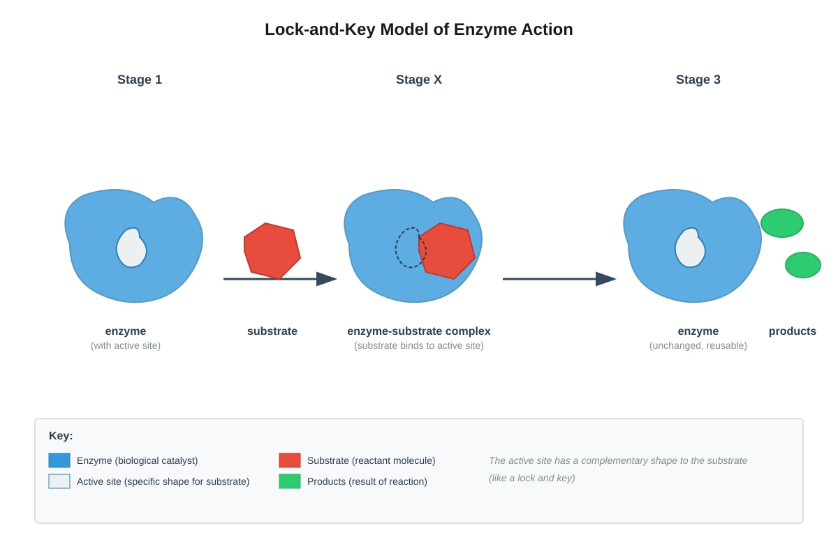
Generated diagram for Q2.
Which statement correctly describes what is happening at stage X?
A The substrate is being broken down into smaller molecules
B The enzyme is being permanently changed in shape
C The products are being released from the active site
D The substrate is binding to the active site of the enzyme
Answer: _________________ [1]
3 A student investigates the effect of temperature on the rate of amylase activity. The results are shown below.
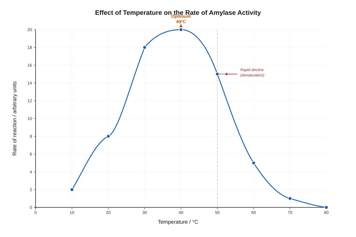
Generated graph for Q3.
At temperatures above 50°C, the rate of reaction decreases sharply because:
A the substrate molecules move more slowly
B the enzyme becomes denatured
C the enzyme is used up in the reaction
D the products inhibit the enzyme
Answer: _________________ [1]
4 Which of the following molecules is a disaccharide?
A Glucose
B Fructose
C Sucrose
D Glycogen
Answer: _________________ [1]
5 A plant cell is placed in a concentrated salt solution. Which of the following correctly describes what would happen?
| Water movement | Final state of cell | |
|---|---|---|
| A | Into the cell | Turgid |
| B | Out of the cell | Flaccid |
| C | Into the cell | Flaccid |
| D | Out of the cell | Turgid |
Answer: _________________ [1]
6 Which cell structure is responsible for producing ATP during aerobic respiration?
A Chloroplast
B Golgi body
C Mitochondrion
D Ribosome
Answer: _________________ [1]
7 The table shows the composition of four unknown solutions.
| Solution | Reducing sugar | Non-reducing sugar | Protein | Starch |
|---|---|---|---|---|
| W | Present | Absent | Absent | Present |
| X | Present | Present | Absent | Absent |
| Y | Absent | Absent | Present | Absent |
| Z | Absent | Present | Present | Present |
Which solution(s) would give a positive result with Benedict's solution after heating?
A W and X only
B X and Y only
C W, X, and Z
D All of W, X, Y, and Z
Answer: _________________ [1]
8 During protein synthesis, which sequence correctly traces the path of a newly formed protein from synthesis to secretion?
A Ribosome → Cytoplasm → Nucleus → Cell membrane
B Ribosome → Rough ER → Golgi body → Vesicle → Cell membrane
C Nucleus → Ribosome → Golgi body → Cytoplasm → Cell membrane
D Rough ER → Ribosome → Golgi body → Lysosome → Cell membrane
Answer: _________________ [1]
9 Which of the following is NOT a function of lipids in organisms?
A Storage of energy
B Formation of cell membranes
C Insulation and protection
D Catalysis of metabolic reactions
Answer: _________________ [1]
10 A cell has the following features: no cell wall, no chloroplasts, contains centrioles, small or no vacuoles. This cell is most likely from:
A a leaf mesophyll layer
B a fungal hypha
C a human cheek epithelium
D a bacterial colony
Answer: _________________ [1]
SECTION A TOTAL: [10]
SECTION B: Structured Questions [30 marks]
Answer ALL questions. Write your answers in the spaces provided.
11 The diagram below shows an animal cell as seen under an electron microscope.
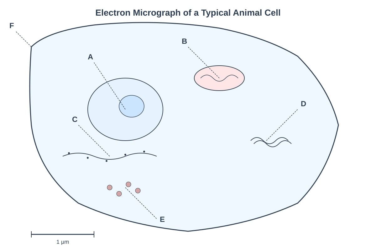
Generated diagram for Q11.
(a) Identify the organelles labelled A, C, and D. [3]
A: _________________________________________________________________
C: _________________________________________________________________
D: _________________________________________________________________
(b) Explain how the structure of organelle B is adapted to its function. [2]
(c) Describe the role of organelle E in protein synthesis. [2]
[7]
12 An investigation was carried out to study the effect of pH on the activity of catalase in potato tissue. Catalase breaks down hydrogen peroxide into water and oxygen. The rate of reaction was measured by recording the volume of oxygen produced over time at different pH values.
(a) Complete the table by writing the word equation for this reaction. [1]
Hydrogen peroxide → _______________________ + _______________________
(b) Suggest two variables that should be kept constant in this investigation. [2]
(c) The results of the investigation are shown in the graph below.
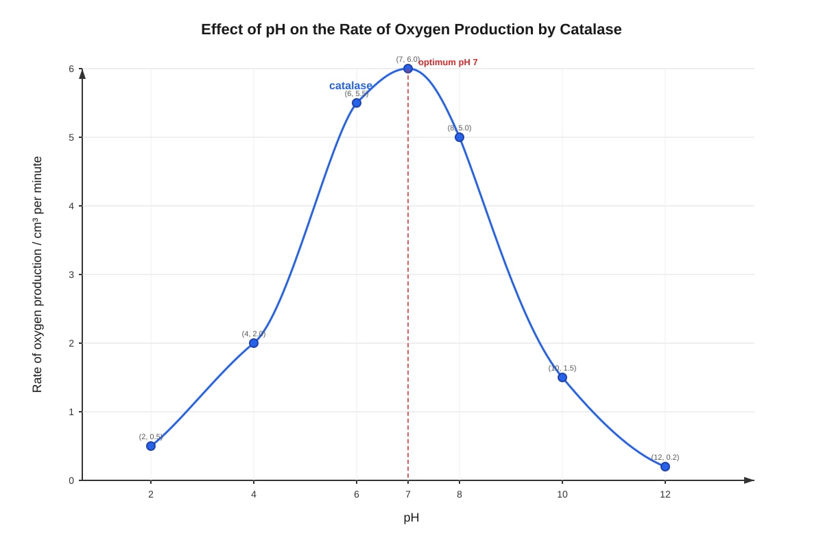
Generated graph for Q12.
Using the graph, determine:
(i) The optimum pH for catalase activity. [1]
(ii) The rate of reaction at pH 5. [1]
(iii) The rate of reaction at pH 9. [1]
(d) Explain why the rate of reaction is very low at pH 2 and pH 12. [3]
[9]
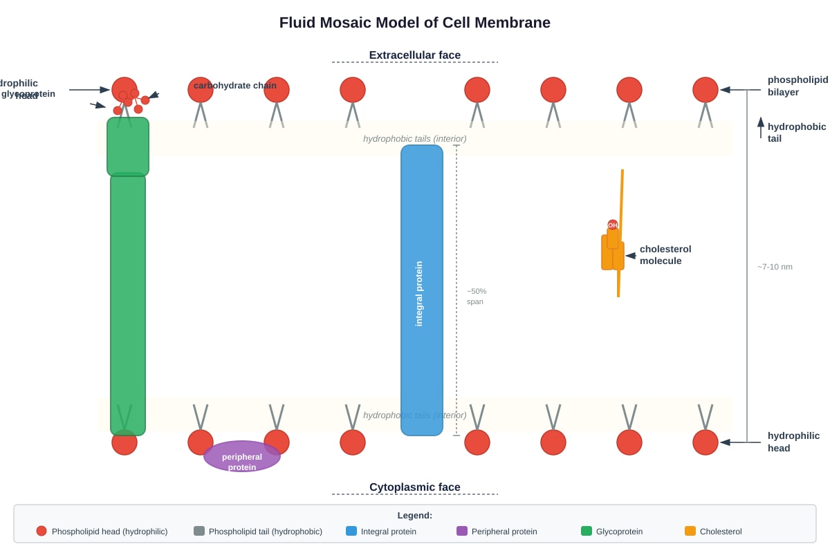
13 The diagram below shows the fluid mosaic model of a cell membrane.
Generated diagram for Q13.
(a) State two functions of the proteins found in the cell membrane. [2]
(b) Explain how the structure of phospholipids makes them suitable for forming the cell membrane. [3]
(c) Cholesterol is found in animal cell membranes but is absent in most plant cell membranes. Suggest one reason why plant cells do not require cholesterol in their membranes. [2]
[7]
14 The table below shows the results of food tests carried out on three unknown food samples P, Q, and R.
| Food Test | Solution P | Solution Q | Solution R |
|---|---|---|---|
| Benedict's test (heated) | Brick red precipitate | No change | No change |
| Biuret test | No change | Purple colour | No change |
| Iodine test | No change | No change | Blue-black colour |
| Emulsion test | White emulsion | No change | No change |
(a) Identify the type of biomolecule present in each solution. [3]
P: _________________________________________________________________
Q: _________________________________________________________________
R: _________________________________________________________________
(b) Solution P was then boiled before the Benedict's test was repeated. The result was negative. Explain this observation. [2]
[5]
15 The diagram shows two cells, X and Y, of different surface area to volume ratios.
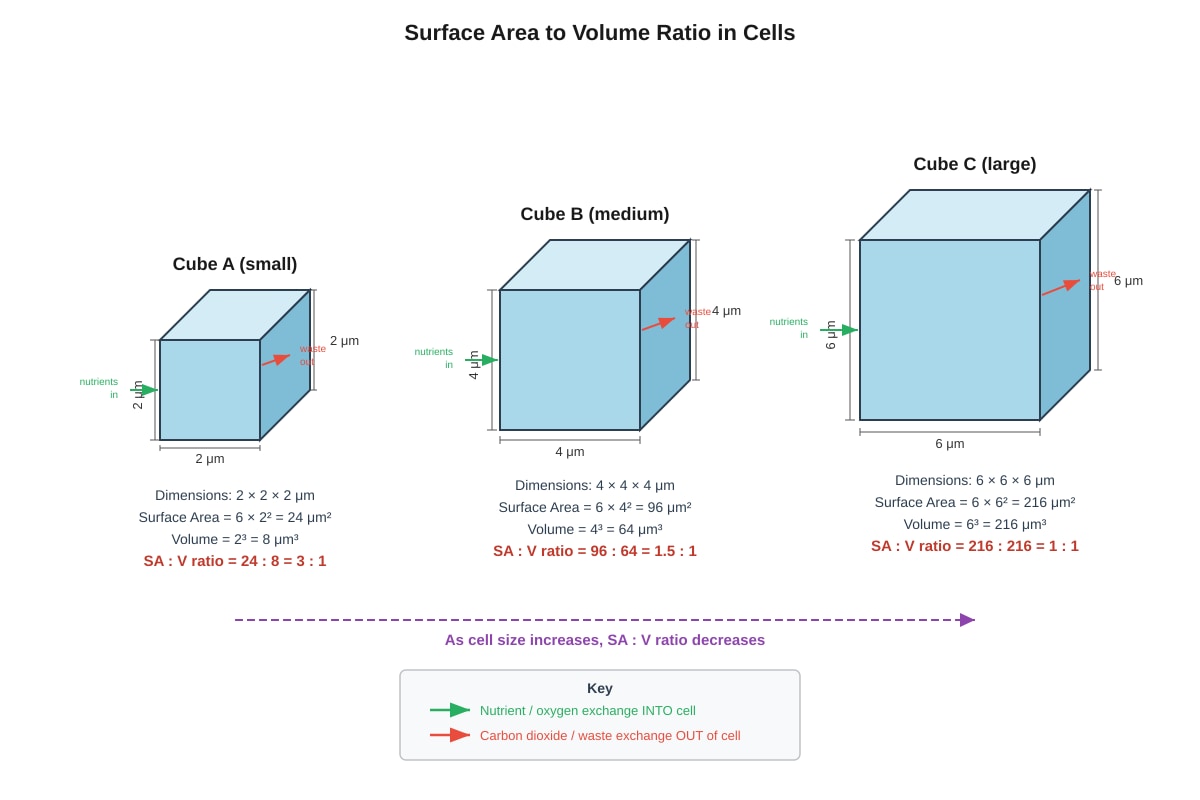
Generated diagram for Q15.
(a) Using the information provided, explain why cells cannot grow indefinitely large. [3]
(b) Explain two adaptations that multicellular organisms have evolved to overcome the limitations of surface area to volume ratio. [4]
[7]
SECTION B TOTAL: [30]
SECTION C: Data Analysis and Extended Response [20 marks]
Answer ALL questions. Write your answers in the spaces provided.
16 The passage below describes an investigation into membrane permeability.
Red blood cells were placed in solutions of different concentrations of sodium chloride. The cells were examined under a microscope after 10 minutes. The results are shown in the table.
| Sodium chloride concentration / % | Appearance of red blood cells | Percentage of cells that appear damaged |
|---|---|---|
| 0.0 (distilled water) | Swollen, some burst | 98% |
| 0.5 | Slightly swollen | 15% |
| 0.9 | Normal biconcave shape | 0% |
| 1.5 | Shrunken, spiky appearance | 5% |
| 2.0 | Very shrunken, crenated | 25% |
| 5.0 | Severely crenated, clumped | 85% |
(a) Explain why red blood cells placed in distilled water swell and burst. [3]
(b) Explain the appearance of red blood cells in the 1.5% sodium chloride solution. [2]
(c) Using your knowledge of osmosis, predict what would happen to plant cells placed in each of the following solutions. Explain your answers.
(i) 0.0% sodium chloride solution [2]
(ii) 5.0% sodium chloride solution [2]
(d) The 0.9% sodium chloride solution is described as isotonic to red blood cells. Explain what is meant by "isotonic" and why this concentration is important in medical practices such as blood transfusions. [3]
[12]
17 Enzymes are biological catalysts that are essential for life processes. The graph below shows the energy changes during a chemical reaction with and without an enzyme.
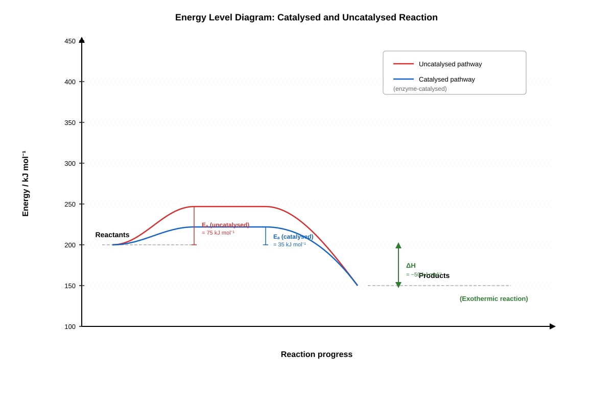
Generated graph for Q17.
(a) Define the term "activation energy". [2]
(b) Using the graph, explain how enzymes increase the rate of reaction without being used up in the process. [4]
(c) The enzyme carbonic anhydrase catalyses the reaction between carbon dioxide and water to form carbonic acid. This enzyme has one of the highest known turnover numbers, converting approximately 600,000 molecules of substrate per second.
(i) Calculate how many molecules of carbonic acid would be produced in 5 minutes by one molecule of carbonic anhydrase. Show your working. [2]
(ii) Suggest two advantages of having such a high turnover number for an enzyme involved in gas transport in the blood. [2]
[10]
18 The electron micrograph below shows part of a chloroplast from a plant cell.
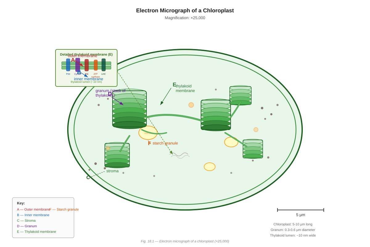
Generated diagram for Q18.
(a) Identify structures D and E, and state the function of each in photosynthesis. [4]
D: _________________________________________________________________
Function: ___________________________________________________________
E: _________________________________________________________________
Function: ___________________________________________________________
(b) Explain how the internal structure of the chloroplast is adapted to maximise the efficiency of photosynthesis. [4]
(c) The plant was kept in darkness for 48 hours before being exposed to light for 2 hours. A section of the leaf was then examined. Predict what would be observed in structure F after this treatment, and explain your answer. [2]
[10]
19 The table below compares some features of DNA and RNA.
| Feature | DNA | RNA |
|---|---|---|
| Number of strands | Usually single | |
| Type of sugar | Deoxyribose | |
| Nitrogenous bases | Adenine, guanine, cytosine, thymine | |
| Location in cell | Mainly nucleus |
(a) Complete the table by filling in the missing information. [4]
(b) DNA replication must occur before cell division. Describe the process of DNA replication, including the role of enzymes. [6]
[10]
20 The diagram below represents a eukaryotic cell cycle.
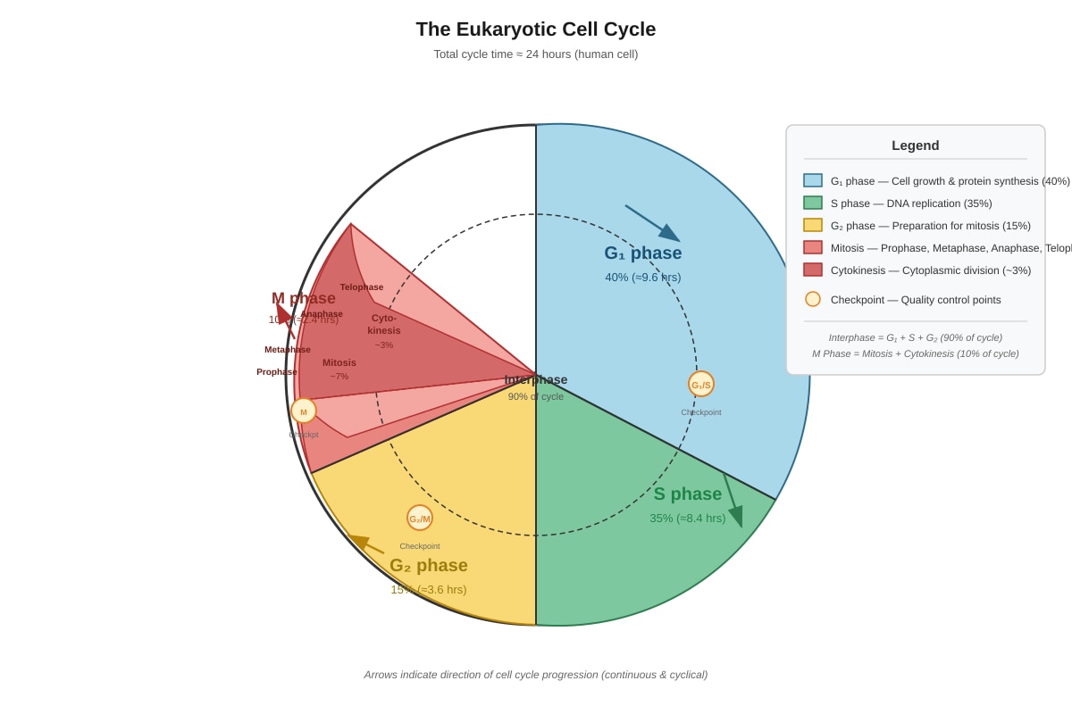
Generated diagram for Q20.
(a) State the name of the phase in which DNA replication occurs. [1]
(b) Describe TWO events that occur during prophase of mitosis. [2]
(c) Explain why it is important that the S phase is separated from the actual division of the cell (M phase) by G1 and G2 phases. [3]
(d) Cancer cells often have defective checkpoints that allow them to progress through the cell cycle more rapidly. Explain how understanding the normal cell cycle can help in the development of cancer treatments. [4]
[10]
SECTION C TOTAL: [20]
PAPER TOTAL: [60]
END OF PAPER
Answers
TuitionGoWhere Practice Paper - Biology Secondary 3
ANSWER KEY and MARKING SCHEME
Version 3 of 5
SECTION A: Multiple Choice [10 marks]
| Question | Answer | Explanation |
|---|---|---|
| 1 | C | Rough endoplasmic reticulum (RER). Radioactive amino acids are the building blocks of proteins. Protein synthesis begins at ribosomes on the RER. The RER is the first organelle to receive these amino acids for protein assembly. Common mistake: Choosing Golgi body (D) — the Golgi modifies and packages proteins AFTER they leave the RER. |
| 2 | D | The substrate is binding to the active site of the enzyme. Stage X represents the formation of the enzyme-substrate complex, where the substrate fits into the complementary active site (lock-and-key model). The enzyme's shape is temporary and returns to original after product release. |
| 3 | B | The enzyme becomes denatured. Above the optimum temperature (shown as ~40°C), the thermal energy disrupts the hydrogen bonds and other interactions that maintain the enzyme's tertiary structure. The active site changes shape permanently, so substrate can no longer bind. Note: enzymes are not "used up" (C is wrong); they are catalysts. |
| 4 | C | Sucrose is a disaccharide composed of glucose + fructose. Glucose (A) and fructose (B) are monosaccharides. Glycogen (D) is a polysaccharide. |
| 5 | B | Water moves out of the cell; the cell becomes flaccid. A concentrated salt solution is hypertonic to the cell cytoplasm. Water moves by osmosis from high water potential (inside cell) to low water potential (outside). The vacuole shrinks, and the cell membrane pulls away from the cell wall in plant cells plasmolysis. |
| 6 | C | Mitochondrion. This organelle carries out aerobic respiration (Krebs cycle and electron transport chain) to produce ATP. Chloroplasts (A) produce ATP only for photosynthesis, not general cell use. |
| 7 | A | W and X only. Benedict's test detects reducing sugars directly. Non-reducing sugars must first be hydrolyzed (acid + heat) before they give a positive Benedict's test. Solution W contains reducing sugar; Solution X contains reducing sugar. Solutions Y (protein only) and Z (non-reducing sugar + starch, but no reducing sugar present) would not give immediate positive Benedict's test. |
| 8 | B | Ribosome → Rough ER → Golgi body → Vesicle → Cell membrane. This is the secretory pathway. Proteins synthesized at ribosomes enter the RER lumen for folding, travel to the Golgi for modification and sorting, then packaged into vesicles that fuse with the plasma membrane for secretion. |
| 9 | D | Catalysis of metabolic reactions — this is the function of enzymes (proteins), not lipids. Lipids store energy (as triglycerides, A), form membranes (as phospholipids, B), and provide insulation/padding (C). |
| 10 | C | Human cheek epithelium — animal cells lack cell walls and chloroplasts, and contain centrioles for cell division. Plant cells (A) have both cell walls and chloroplasts. Fungi (B) have cell walls. Bacteria (D) are prokaryotes with different structures entirely. |
SECTION B: Structured Questions [30 marks]
11 Total: [7]
(a) [3 marks — 1 each]
- A: Nucleus (with nucleolus) — contains genetic material (DNA), controls cell activities
- C: Rough endoplasmic reticulum (RER) — site of protein synthesis and folding; "rough" due to ribosomes on surface
- D: Golgi body/apparatus — modifies, sorts, and packages proteins and lipids for secretion or delivery to other organelles
(b) [2 marks]
Mitochondrion B has highly folded inner membrane (cristae) [1], which greatly increases the surface area available for the electron transport chain and ATP synthase enzymes [1]. The matrix contains enzymes for the Krebs cycle and mitochondrial DNA/ribosomes for synthesizing some of its own proteins [1] — any 2 points.
(c) [2 marks]
Ribosomes (E) are the site of protein synthesis [1]. They read the mRNA sequence and catalyze the assembly of amino acids into polypeptide chains according to the genetic code [1]. Free ribosomes make proteins for use within the cell; bound ribosomes make proteins for secretion or membrane insertion.
12 Total: [9]
(a) [1 mark]
Hydrogen peroxide → water + oxygen
(b) [2 marks — 1 each]
Any two from:
- Temperature — must be constant as it affects enzyme activity and kinetic energy
- Concentration of hydrogen peroxide (substrate) — must be same to compare pH effect fairly
- Volume/mass of potato tissue (enzyme source) — must be same to ensure same enzyme concentration
- Surface area of potato — affects access of enzyme to substrate
(c)
(i) [1 mark] pH 7 (accept pH 6.5–7.5 from graph reading)
(ii) [1 mark] Approximately 2.0 cm³ per minute (accept 1.8–2.2 from graph)
(iii) [1 mark] Approximately 5.0 cm³ per minute (accept 4.8–5.2 from graph)
(d) [3 marks]
At pH 2 and pH 12, the solution is highly acidic or highly alkaline [1]. These extremes of pH cause denaturation of the catalase enzyme [1]. The hydrogen ions (H⁺ at low pH) or hydroxide ions (OH⁻ at high pH) interfere with ionic bonds and hydrogen bonds that maintain the enzyme's specific tertiary structure [1]. The active site changes shape permanently, so substrate (hydrogen peroxide) can no longer bind effectively [1] — any 3 points.
13 Total: [7]
(a) [2 marks — 1 each]
Any two from:
- Transport proteins — carrier proteins and channel proteins for facilitated diffusion and active transport of ions/molecules
- Receptor proteins — bind signaling molecules (hormones, neurotransmitters) for cell communication
- Enzymatic proteins — catalyze reactions at the membrane surface
- Cell recognition/adhesion — glycoproteins involved in immune recognition and tissue formation
- Structural support — anchor cytoskeleton and extracellular matrix
(b) [3 marks]
Phospholipids have a hydrophilic (water-loving) phosphate head and hydrophobic (water-fearing) fatty acid tails [1]. In an aqueous environment, they arrange themselves into a bilayer with heads facing outward and tails sandwiched inward [1]. This creates a partially permeable barrier that allows small non-polar molecules to pass while restricting polar/charged substances [1], essential for controlling what enters and exits the cell.
(c) [2 marks]
Plant cells have cell walls made of cellulose that provide structural rigidity and protection [1], reducing the need for membrane fluidity regulation. Additionally, plant cells can adjust turgor pressure through vacuole water content to maintain membrane integrity across temperature ranges [1].
14 Total: [5]
(a) [3 marks — 1 each]
- P: Reducing sugar (positive Benedict's test; brick red precipitate on heating)
- Q: Protein (positive Biuret test; purple colour with copper sulfate in alkaline conditions)
- R: Starch (positive iodine test; blue-black colour)
(b) [2 marks]
The reducing sugar in P was originally part of a larger carbohydrate structure or the solution contained a non-reducing sugar that was hydrolyzed by heating [1]. Alternatively, if P was a mixture or the reducing sugar was present in a bound form, boiling may have caused Maillard browning or caramelization that masked the result, or more likely: the sample contained sucrose (non-reducing sugar) with trace reducing sugar impurity that was destroyed or the test conditions were altered [1].
Better explanation: The original solution P was likely a disaccharide like sucrose that had been partially hydrolyzed; upon boiling without acid, any existing reducing sugar may have undergone degradation, or the test was not performed correctly (needed acid hydrolysis first to detect non-reducing sugars).
Accept: Solution P contained glucose and fructose that reacted in the first test; upon boiling the solution, the reducing sugars were oxidized by atmospheric oxygen or polymerized, making them unavailable for the Benedict's test [1].
15 Total: [7]
(a) [3 marks]
As cells grow larger, their volume increases faster than surface area (SA:V ratio decreases) [1]. This means diffusion distances increase and the surface area available for exchange becomes insufficient relative to the metabolic needs of the increased volume [1]. Waste products accumulate and nutrients cannot reach the centre fast enough, limiting metabolic efficiency [1].
(b) [4 marks — 2 each]
-
Specialized exchange surfaces — e.g., lungs with alveoli (thin walls, large surface area, moist), intestinal villi, or root hair cells. These maximize surface area while allowing efficient diffusion/osmosis across membranes [2 marks: structure + function explanation].
-
Transport/circulatory systems — e.g., blood circulatory system in animals or vascular tissues (xylem and phloem) in plants. These actively move substances over longer distances, compensating for large body size where diffusion alone is inadequate [2 marks: system + explanation].
Alternative: Flat, thin, or elongated body shapes (e.g., tapeworms); cellular organization into thin layers/sheets.
SECTION C: Data Analysis and Extended Response [20 marks]
16 Total: [12]
(a) [3 marks]
Distilled water is hypotonic to red blood cell cytoplasm (lower solute concentration, higher water potential outside) [1]. Water moves into the cells by osmosis down the water potential gradient [1]. The cell membrane cannot withstand the increasing pressure (no cell wall in animal cells), so cells swell and burst (haemolysis) [1].
(b) [2 marks]
The 1.5% solution is hypertonic to red blood cells (higher solute concentration, lower water potential outside) [1]. Water moves out of the cells by osmosis, causing the cells to shrink and develop spiky projections (crenation) as membrane pulls inward [1].
(c)
(i) [2 marks] Turgid / swollen [1]. The plant cell has a cell wall that prevents bursting. Water enters by osmosis, the vacuole expands, and the cell pushes against the cell wall becoming turgid (rigid and firm), which is the healthy state for plant cells [1].
(ii) [2 marks] Plasmolysed / flaccid [1]. Water leaves the cell by osmosis. The cell membrane pulls away from the cell wall as the cytoplasm and vacuole shrink. The cell becomes flaccid; if severe, plasmolysis occurs where protoplast completely detaches from wall [1].
(d) [3 marks]
Isotonic means the solution has the same water potential/solute concentration as the cell cytoplasm, resulting in no net water movement [1]. This maintains red blood cells in their normal biconcave shape, ensuring efficient gas transport and preventing cell damage [1]. In medical practice, intravenous fluids and blood products must be isotonic to prevent haemolysis (if hypotonic) or crenation (if hypertonic), which would destroy blood cells and harm the patient [1].
17 Total: [10]
(a) [2 marks]
Activation energy is the minimum energy required for a chemical reaction to start [1]. It is the energy needed to break existing bonds in reactant molecules and form the transition state, enabling the reaction to proceed to products [1].
(b) [4 marks]
Enzymes lower the activation energy required for the reaction by providing an alternative reaction pathway [1], shown by the lower peak on the catalysed curve [1]. They do this by binding substrates at the active site, where amino acid side chains stabilize the transition state and orient substrates for reaction [1]. The enzyme itself returns to its original form unchanged after product release, so one enzyme molecule can catalyze many reaction cycles — it is not consumed [1].
(c)
(i) [2 marks]
Working: 600,000 molecules per second × 60 seconds × 5 minutes [1] = 600,000 × 300 = 180,000,000 molecules (or 1.8 × 10⁸) [1]
(ii) [2 marks — 1 each]
- Rapid removal of carbon dioxide from tissues, maintaining steep diffusion gradients for efficient gas exchange between blood and cells [1]
- Prevents excessive acidification of blood — quick conversion to carbonic acid allows prompt transport and subsequent dissociation to bicarbonate, maintaining blood pH homeostasis [1]
Alternative: Allows efficient gas transport despite low CO₂ solubility in plasma; enables rapid response to changing metabolic demands during exercise.
18 Total: [10]
(a) [4 marks — 1 identification + 1 function each]
-
D: Granum/stack of thylakoids [1]
- Function: Site of light-dependent reactions in photosynthesis [1]; contains chlorophyll and electron transport chain proteins to capture light energy and produce ATP and NADPH
-
E: Thylakoid membrane [1]
- Function: Contains photosystems, electron carriers, and ATP synthase [1]; provides large surface area for embedding protein complexes involved in photophosphorylation and electron transport
(b) [4 marks]
- Double membrane envelope — controls entry/exit of substances; maintains internal environment optimized for photosynthesis [1]
- Thylakoid membrane system (grana and lamellae) — provides extensive surface area for light-harvesting complexes and electron transport chains [1]; stacked grana allow efficient absorption at different light intensities and wavelengths [1]
- Stroma — contains enzymes for Calvin cycle (light-independent reactions), DNA, ribosomes for protein synthesis, and starch granules for glucose storage [1]; fluid medium allows enzyme-substrate interactions
- Chlorophyll arrangement in photosystems — maximizes light capture and allows resonance energy transfer to reaction centres [1] — any 4 well-developed points
(c) [2 marks]
Large starch granules would be visible in F [1]. The 48-hour darkness period depleted starch stores (starch broken down to glucose for respiration/transport). The subsequent 2 hours of light allowed photosynthesis to occur, producing glucose that was converted to starch for storage in the chloroplast stroma [1].
19 Total: [10]
(a) [4 marks — 1 each row]
| Feature | DNA | RNA |
|---|---|---|
| Number of strands | Two (double helix) | Usually single |
| Type of sugar | Deoxyribose | Ribose |
| Nitrogenous bases | Adenine, guanine, cytosine, thymine | Adenine, guanine, cytosine, uracil |
| Location in cell | Mainly nucleus | Nucleus, cytoplasm, ribosomes |
(b) [6 marks]
DNA replication is semi-conservative and occurs during S phase of the cell cycle [1 mark for overview].
Process:
- Helicase enzyme unwinds the double helix and breaks hydrogen bonds between base pairs, separating the two strands at the replication fork [1]
- DNA polymerase reads the template strand in the 3' to 5' direction and adds complementary nucleotides in the 5' to 3' direction [1]. It proofreads and corrects errors [1]
- Primase synthesizes a short RNA primer to initiate DNA synthesis [1]
- Ligase joins Okazaki fragments on the lagging strand [1]
- Each new DNA molecule contains one original (parental) strand and one newly synthesized strand [1]
20 Total: [10]
(a) [1 mark] S phase (synthesis phase)
(b) [2 marks — 1 each]
Any two from:
- Chromatin condenses into visible chromosomes — DNA wraps around proteins and coils tightly to facilitate movement
- Nuclear envelope breaks down — allows spindle microtubules to access chromosomes
- Centrioles move to opposite poles and spindle fibres begin to form
- Chromosomes become visible as two sister chromatids joined at centromere
(c) [3 marks]
G1 and G2 phases provide time for cell growth and preparation [1]. G1 allows the cell to synthesize proteins and organelles needed for DNA replication and cell division [1]. G2 allows for further protein synthesis, particularly of spindle proteins, and provides time to check for DNA damage or replication errors before committing to mitosis [1]. This ensures the cell is fully prepared and that DNA integrity is maintained.
(d) [4 marks]
- Cancer cells have mutations in genes encoding checkpoint proteins (e.g., p53 tumor suppressor, cyclins, CDKs) [1]
- Understanding normal cell cycle identifies molecular targets — drugs can be designed to restore or enhance checkpoint function [1]
- Specific inhibitors can block cyclin-CDK complexes or target signaling pathways that drive uncontrolled proliferation [1]
- Microtubule inhibitors (e.g., taxanes) prevent spindle formation, arresting cells in metaphase and triggering apoptosis [1]
Alternative points: Radiation/chemotherapy targets rapidly dividing cells by damaging DNA during S phase; understanding repair mechanisms helps design combination therapies.
MARK SUMMARY
| Section | Marks |
|---|---|
| A | 10 |
| B | 30 |
| C | 20 |
| TOTAL | 60 |
END OF ANSWER KEY
Free quiz and exam paper access
Enter your details to view this paper
Your access is remembered on this device.