AI Generated Quiz
Secondary 1 Science Life Sciences Quiz
Free Sec 1 Science Life Sciences quiz, Kimi2.6 AI version, with questions, answers, and syllabus-aligned practice for Singapore students.
These static practice materials are generated from the site's syllabus and paper-generation workflow, with source and model context shown so students and parents can evaluate the material before use.
Questions
Secondary 1 Science Quiz - Life Sciences
Name: _______________________________ Class: __________ Date: __________
Score: ______ / 50 marks
Duration: 45 minutes
Instructions:
- Answer all questions.
- Write your answers in the spaces provided.
- For multiple choice questions, circle the correct answer.
- Read each question carefully before answering.
Section A: Multiple Choice Questions (Questions 1–10)
Choose the correct answer for each question. Each question carries 2 marks.
Total: 20 marks
1. Which of the following is the basic unit of life?
A) Tissue
B) Organ
C) Cell
D) Organelle
Answer: _________________________________________________
2. The cell wall is found in which type of cells?
A) Animal cells only
B) Plant cells only
C) Both animal and plant cells
D) Neither animal nor plant cells
Answer: _________________________________________________
3. Which organelle is responsible for photosynthesis in plant cells?
A) Mitochondrion
B) Nucleus
C) Chloroplast
D) Vacuole
Answer: _________________________________________________
4. During an experiment, a student observes a specimen under a microscope. The image appears upside down and reversed. Which part of the microscope causes this?
A) Eyepiece lens
B) Objective lens
C) Mirror
D) The combination of lenses in the optical path
Answer: _________________________________________________
5. Which of the following is a characteristic of all living things?
A) Ability to fly
B) Ability to reproduce
C) Ability to photosynthesise
D) Ability to move from place to place
Answer: _________________________________________________
6. The diagram below shows a plant cell.
Image pending generation: diagram for Q6.
Structure X is the large fluid-filled space that helps maintain the shape of the cell. What is structure X?
A) Nucleus
B) Mitochondrion
C) Vacuole
D) Chloroplast
Answer: _________________________________________________
7. Which cell structures contain genetic material?
A) Nucleus and mitochondria
B) Nucleus and vacuole
C) Mitochondria and chloroplasts only
D) Ribosomes and nucleus
Answer: _________________________________________________
8. A student prepares a wet mount of an onion epidermal cell. She places a drop of iodine solution on the specimen before covering it with a coverslip. What is the purpose of the iodine solution?
A) To kill the cell
B) To stain the cell and make structures more visible
C) To preserve the cell permanently
D) To dissolve the cell wall
Answer: _________________________________________________
9. Which statement correctly compares animal and plant cells?
A) Both have chloroplasts, but only plant cells have mitochondria.
B) Both have cell walls, but only animal cells have centrioles.
C) Both have mitochondria, but only plant cells have large central vacuoles and chloroplasts.
D) Both have nuclei, but only animal cells have cytoplasm.
Answer: _________________________________________________
10. The magnification of a specimen viewed under a microscope is calculated as:
A) Eyepiece magnification + Objective magnification
B) Eyepiece magnification × Objective magnification
C) Objective magnification ÷ Eyepiece magnification
D) Eyepiece magnification − Objective magnification
Answer: _________________________________________________
Section B: Short Answer Questions (Questions 11–15)
Answer each question in the space provided. Show all working where required.
Total: 15 marks
11. State three differences between animal cells and plant cells. [3 marks]
12. (a) Name the two main types of cells based on the complexity of their structure. [1 mark]
(b) Give one example of an organism made up of each type of cell you named in part (a). [2 marks]
13. A student views a specimen using a light microscope with an eyepiece lens of ×10 and an objective lens of ×40.
(a) Calculate the total magnification of the specimen. [2 marks]
(b) The student then changes the objective lens to ×10. Without changing the eyepiece, what is the new magnification? [1 mark]
14. The diagram below shows a specialised animal cell.
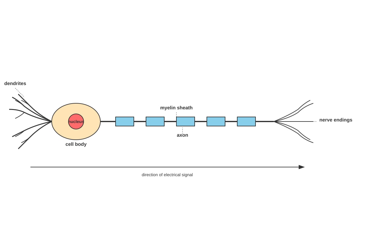
Generated diagram for Q14.
(a) Identify the type of cell shown in the diagram. [1 mark]
(b) Explain how the structure of this cell is adapted to its function of transmitting electrical signals. [2 marks]
15. Complete the table below by stating the function of each cell structure. [3 marks]
| Cell Structure | Function |
|---|---|
| Nucleus | _________________________________ |
| Mitochondrion | _________________________________ |
| Cell membrane | _________________________________ |
Section C: Structured Questions (Questions 16–20)
Answer all questions. These questions require more detailed explanations and application of knowledge.
Total: 15 marks
16. The diagram below shows an experimental setup to investigate the effect of temperature on the movement of red pigment in water.
Image pending generation: experimental_setup for Q16.
(a) Predict in which beaker the red colour will spread faster. Explain your answer with reference to the movement of particles. [3 marks]
17. Meera prepares a slide of human cheek cells. She follows these steps:
- Step 1: Gently scrape the inside of her cheek with a clean spatula
- Step 2: Smear the cells on a clean glass slide
- Step 3: Add a drop of methylene blue stain
- Step 4: Cover with a coverslip
(a) State one safety precaution Meera should take during this procedure. [1 mark]
(b) Explain why methylene blue stain is used in this procedure. [2 marks]
18. The table below shows the sizes of various specimens and the magnification needed to view them.
| Specimen | Actual Size | Magnification Needed |
|---|---|---|
| Human red blood cell | 0.007 mm | ×2000 |
| Onion epidermal cell | 0.25 mm | ×100 |
| Pollen grain | 0.05 mm | ×400 |
(a) One student claims that the onion cell is the largest specimen in the table. Another student disagrees, saying the red blood cell viewed at ×2000 would appear largest. Who is correct? Explain your reasoning with a calculation. [3 marks]
(b) Why do we need to use a microscope to view these specimens even though some are visible to the naked eye? [2 marks]
19. The diagram below shows two different types of cells from a leaf.
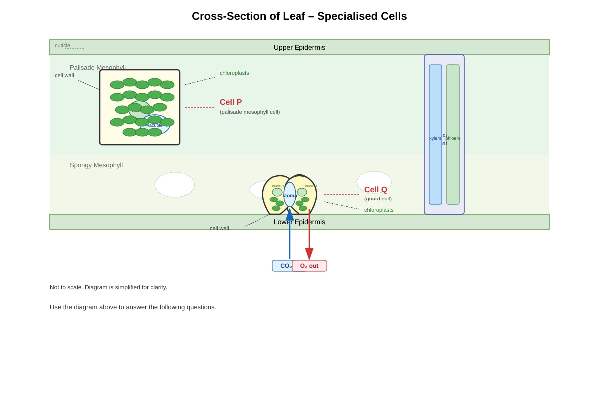
Generated diagram for Q19.
(a) Cell P is a palisade mesophyll cell. Explain why having many chloroplasts is important for this cell's function. [2 marks]
(b) Cell Q is a guard cell. Describe how the structure shown helps the leaf carry out gas exchange. [2 marks]
20. A biologist studies a unicellular organism called Paramecium under a microscope. The organism is 0.25 mm long and moves using hair-like structures.
(a) Name the hair-like structures that Paramecium uses for movement. [1 mark]
(b) Explain why Paramecium, despite being only one cell, is considered a living organism. [3 marks]
— END OF QUIZ —
Answers
Secondary 1 Science Quiz - Life Sciences (Answer Key)
Total Marks: 50
Section A: Multiple Choice Questions (Questions 1–10)
2 marks each
| Question | Answer | Explanation |
|---|---|---|
| 1 | C | The cell is the basic unit of life. All living organisms are made of cells, and cells carry out all the functions of life. Tissues are groups of cells, organs are made of tissues, and organelles are structures within cells. |
| 2 | B | The cell wall is found only in plant cells (and some bacteria and fungi). It provides structural support and protection. Animal cells do not have cell walls; they only have a cell membrane. |
| 3 | C | Chloroplasts contain chlorophyll and are the sites of photosynthesis, where light energy is converted into chemical energy (glucose). Mitochondria carry out cellular respiration, not photosynthesis. |
| 4 | D | The compound microscope produces an inverted image because light passes through two lens systems (eyepiece and objective). Each lens inverts the image, and the combination results in an upside-down, reversed image. |
| 5 | B | All living things can reproduce to produce offspring. Not all living things can fly (e.g., plants), photosynthesise (only plants and some bacteria), or move from place to place (e.g., plants are rooted). |
| 6 | C | The large central vacuole is a distinctive feature of plant cells. It stores water, food, and waste products, and helps maintain turgor pressure to keep the cell rigid. The diagram shows this large fluid-filled space. |
| 7 | A | The nucleus contains chromosomes made of DNA. Mitochondria also contain their own small amount of DNA (mitochondrial DNA), inherited from the mother. Vacuoles and ribosomes do not contain genetic material. |
| 8 | B | Iodine is a biological stain that binds to certain cellular components, making them more visible under the microscope. Onion cells are naturally nearly transparent, so staining is needed to see the nucleus and cell wall clearly. |
| 9 | C | Both animal and plant cells have mitochondria for respiration and nuclei for genetic control. However, plant cells uniquely have large central vacuoles for storage and chloroplasts for photosynthesis. Animal cells lack these structures. |
| 10 | B | Total magnification = Eyepiece magnification × Objective magnification. For example, ×10 eyepiece with ×40 objective gives ×400 total magnification. |
Section B: Short Answer Questions (Questions 11–15)
11. [3 marks]
| Feature | Animal Cell | Plant Cell |
|---|---|---|
| Cell wall | Absent | Present |
| Chloroplasts | Absent | Present |
| Large central vacuole | Absent (small or no vacuoles) | Present |
| Shape | Irregular, round | Regular, fixed shape |
Any three correct differences [1 mark each]
Common acceptable answers:
- Cell wall: plant cells have it, animal cells don't
- Chloroplasts: plant cells have them, animal cells don't
- Vacuole: plant cells have large central vacuole, animal cells have small/no vacuoles
- Shape: plant cells have fixed shape due to cell wall, animal cells have variable shape
- Storage: plant cells store starch, animal cells store glycogen
Teaching note: Students often confuse "cell wall" and "cell membrane." Emphasise that all cells have a cell membrane, but only plant cells have an additional rigid cell wall outside the membrane.
12. [3 marks total]
(a) Prokaryotic and eukaryotic cells [1 mark]
(b) Prokaryotic: Bacteria (e.g., Escherichia coli); Eukaryotic: Animal, plant, fungus, protozoan (any valid example) [1 mark each]
Teaching note: Prokaryotic cells lack a true nucleus and membrane-bound organelles. Their DNA floats in the cytoplasm. Eukaryotic cells have a membrane-bound nucleus and specialised organelles. This fundamental distinction prepares students for upper secondary biology where they study bacterial structure in detail.
13. [3 marks total]
(a) Total magnification = Eyepiece × Objective = 10 × 40 = ×400 [2 marks: 1 for formula, 1 for correct answer]
(b) New magnification = 10 × 10 = ×100 [1 mark]
Teaching note: Total magnification is always a product, never a sum. Students sometimes add (10 + 40 = 50) which is incorrect. Remind them that each lens magnifies the image created by the previous lens, so magnifications multiply.
14. [3 marks total]
(a) Nerve cell / Neuron [1 mark]
(b) The nerve cell has a long axon that carries electrical signals over long distances [1]. The dendrites branch to receive signals from other nerve cells, and the nerve endings pass signals to the next cell in the circuit [1]. The myelin sheath insulates and speeds up this transmission.
Marking breakdown: [1 mark for identifying long axon/dendrites for signal transmission; 1 mark for explaining how this structure relates to function]
Teaching note: This is an example of structure-function relationship, a key concept in biology. The exaggerated length of the axon (up to 1 metre in humans) is specialized for rapid communication, unlike typical cells which are microscopic.
15. [3 marks]
| Cell Structure | Function |
|---|---|
| Nucleus | Contains genetic material (DNA/chromosomes) and controls cell activities [1] |
| Mitochondrion | Site of aerobic respiration; produces ATP/energy for the cell [1] |
| Cell membrane | Controls what enters and leaves the cell; selectively permeable barrier [1] |
Common mistake: Students often write "cell wall" for plant cell functions in this table. Ensure they match the given structure. For mitochondria, "produces energy" is acceptable but "site of respiration" is more precise.
Section C: Structured Questions (Questions 16–20)
16. [3 marks]
(a) The red colour will spread faster in Beaker B (the beaker at 60°C) [1].
Explanation: At higher temperatures, water particles have more kinetic energy and move faster [1]. The faster-moving particles collide more frequently and with more energy, causing the red dye particles to spread out (diffuse) more rapidly through the water [1]. This process is called diffusion.
Teaching note: This question assesses understanding of the particulate model of matter. The key insight is that temperature affects particle speed, which in turn affects the rate of diffusion. Students should not just state "heat makes it faster" but explain the mechanism through particle theory.
17. [3 marks total]
(a) Safety precaution: Wear gloves / Do not share spatulas / Clean spatula before use / Wash hands after procedure / Wear safety goggles [any valid precaution] [1 mark]
(b) Methylene blue stain binds to acidic components in cells, particularly the nucleus [1]. Human cheek cells are transparent and colourless; without staining, the nucleus and cell membrane boundaries would be nearly invisible under the microscope [1].
Teaching note: This practical is commonly done in Sec 1. The stain is basic and binds to nucleic acids, making the nucleus prominently visible. Emphasise safety with biological specimens—even one's own cells can carry microorganisms.
18. [5 marks total]
(a) The student who says the onion cell is largest is correct [1]. Magnification does not change the actual size of the specimen. The onion cell at 0.25 mm is physically the largest.
Calculation verification:
- Red blood cell actual size: 0.007 mm = 7 µm
- Onion cell actual size: 0.25 mm = 250 µm
- Image size at ×2000: 0.007 × 2000 = 14 mm
- Image size at ×100: 0.25 × 100 = 25 mm
Even viewed at ×2000, the red blood cell image (14 mm) is smaller than the onion cell image at ×100 (25 mm). The onion cell remains largest [2 marks for correct calculation and conclusion].
Alternative approach: Compare actual sizes directly: 0.25 mm > 0.007 mm, so onion cell is larger regardless of magnification.
(b) We need microscopes to see detailed internal structure [1]. Even specimens visible to the naked eye (like pollen grains) require magnification to observe cellular details—cell wall, nucleus, cytoplasm, and other organelles [1].
Teaching note: This question distinguishes between "size" and "magnified image size." Students often confuse these. The actual size is an intrinsic property; magnification only affects how large the image appears.
19. [4 marks total]
(a) Palisade mesophyll cells are the main site of photosynthesis in leaves [1]. Having many chloroplasts maximises the amount of chlorophyll available to absorb light energy, increasing the rate of photosynthesis and glucose production [1].
(b) The guard cells control the opening and closing of the stoma (pore) between them [1]. When the stoma is open, carbon dioxide enters for photosynthesis, and oxygen/water vapour exit. The kidney shape allows the pore to open when turgid and close when flaccid, regulating gas exchange [1].
Teaching note: Palisade cells are positioned at the top of the leaf to receive maximum light. Their cylindrical shape with minimal air gaps ensures efficient light absorption. Guard cells use osmosis to change shape—this will be covered in more detail in upper secondary.
20. [4 marks total]
(a) Cilia [1 mark]
(b) Paramecium is living because it shows the characteristics of life [1]:
- It moves using cilia to find food and avoid predators
- It feeds by sweeping food particles into its oral groove
- It respires to release energy from food
- It excretes waste through its anal pore
- It reproduces by dividing into two (binary fission)
- It responds to stimuli like light and chemicals
- It maintains water balance using contractile vacuoles
[Any three characteristics with brief explanation: 3 marks—1 mark per valid characteristic with application to Paramecium]
Teaching note: This question connects cell theory to the characteristics of life. Unicellular organisms perform all life functions within one cell, demonstrating that the cell is indeed the fundamental unit of life. The contractile vacuole is particularly interesting as it shows homeostasis at the cellular level.
— END OF ANSWER KEY —
Free quiz and exam paper access
Enter your details to view this paper
Your access is remembered on this device.