From Real Exams Quiz
O Level Biology Human Physiology Quiz
Free O Level Biology Human Physiology quiz, HY3 Exam version, with questions, answers, and O Level-style practice for Singapore students.
These static practice materials are generated from the site's syllabus and paper-generation workflow, with source and model context shown so students and parents can evaluate the material before use.
Questions
Free quiz and exam paper access
Enter your details to view this paper
Your access is remembered on this device.
Answers
O-Level Biology Quiz - Human Physiology (Answer Key)
Total Marks: 40
Topic: Human Physiology
Section A
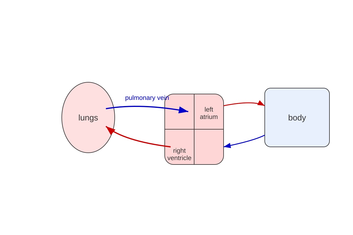
Q1. C – Pulmonary vein [1]
Teaching note: The pulmonary vein is the only vein that carries oxygenated blood (from lungs to left atrium). Pulmonary artery carries deoxygenated blood – a common trap.
Q2. A – 2 [1]
Teaching note: Pepsin works in the acidic stomach (HCl), optimum ~pH 2. Do not confuse with intestinal enzymes (pH ~7–8).
Q3. B – Bowman’s capsule [1]
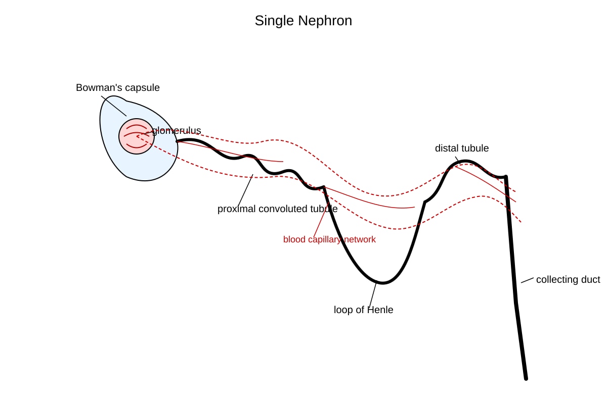
Teaching note: Filtration occurs at Bowman’s capsule / glomerulus. Ureter carries urine away; bladder stores; urethra expels.
Q4. B – contracts and flattens [1]
Teaching note: Inhalation → diaphragm contracts, flattens, thoracic volume increases, pressure drops, air in.
Q5. C – Plasma [1]
Teaching note: Plasma transports dissolved hormones, nutrients, wastes. Red cells carry O₂; platelets clot.
Section B
Q6. [2]
- (a) No nucleus → more space for haemoglobin [1]
- (b) Contains haemoglobin to bind O₂ / biconcave shape for large surface area [1]
Marking: any two correct adaptive features.
Q7. [2]
Enzyme: amylase [1] Product: maltose [1]
Teaching: Salivary amylase hydrolyses starch → maltose in mouth.
Q8. [2]
Water is reabsorbed by osmosis from the large intestine into the blood [1] to prevent dehydration / maintain water balance [1].
Common mistake: saying “absorbs food” – most food absorbed earlier in ileum.
Q9. [2]
Change: breathing rate increases (e.g. from 12 to 24) [1]
Reason: more CO₂ / need more O₂ for aerobic respiration during exercise [1]
Q10. [2]
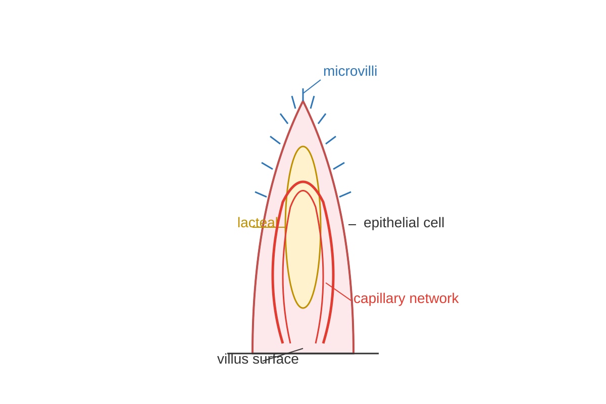
Lacteal: central vessel in villus (labelled) [1]
Capillary: loop in villus (labelled) [1]
Image must show finger-like villus, central lacteal, capillary loop.
Section C
Q11. [4]
- Enzymes are biological catalysts that speed up digestion without being used up [1]
- Amylase: starch → maltose (mouth/duodenum) [1]
- Pepsin: proteins → peptides (stomach, pH 2) [1]
- Lipase: fats → fatty acids + glycerol (duodenum) [1]
Marking: 1 for catalyst role, 3 for any two named enzyme–substrate–product sets (1.5 each if partial).
Q12. [3]
- Excess protein deaminated in liver → urea formed [1]
- Urea enters blood, raises solute concentration [1]
- Kidney reabsorbs more water by osmosis to concentrate urine / ADH released [1]
Trap: confusing volume with concentration.
Q13. [2]
Vessel: pulmonary artery [1] Blood type: deoxygenated [1]
Diagram shows right ventricle → lungs.
Q14. [2]
Working: % increase = ((24 − 12) / 12) × 100 = 100% [1]
Answer: 100% [1]
Q15. [4]
Left ventricle → aorta [1] → body artery to leg [1] → capillaries of toe (exchange) [1] → veins → vena cava → right atrium [1]
Marking: 1 mark per correct segment of double circulation pathway.
Q16. [3]
- Provides O₂ and nutrients from mother to fetus [1]
- Removes CO₂ and wastes from fetus [1]
- Allows gas/nutrient exchange without mixing blood [1]
Q17. [3]
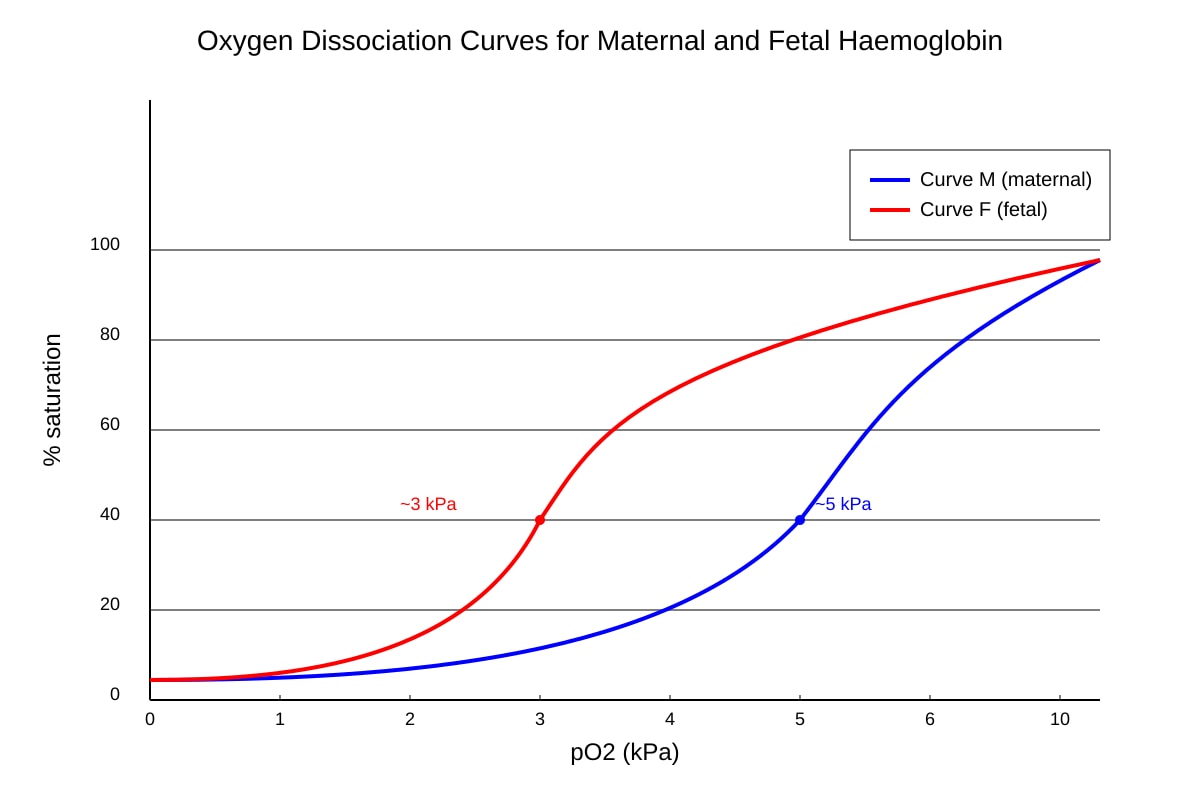
Difference: fetal haemoglobin curve is left-shifted (higher affinity at low pO₂) [1]
Advantage: fetus can load O₂ from maternal blood at low placental pO₂ [2]
Q18. [6]
- Zygote divides by mitosis → embryo [1]
- Embryo moves down fallopian tube [1]
- Blastocyst implants in uterine wall [1]
- Placenta develops for nutrient/O₂ supply [1]
- Umbilical cord links fetus to placenta [1]
- Amniotic fluid cushions fetus [1]
Q19. [4]
- Less O₂ transported → tissues receive less O₂, reduced aerobic respiration [2]
- Fatigue / anaemia symptoms due to low ATP production [2]
(or: CO₂ removal impaired)
Q20. [4]
(a) Glucose → proximal tubule [2]
(b) Water → collecting duct / loop of Henle [2]
(Other acceptable: amino acids – proximal tubule; salts – loop of Henle)
Image must show nephron structures labelled.