AI Generated Quiz
A Level Biology H3 Human Physiology Quiz
Free A Level Biology H3 Human Physiology quiz, AI version, with questions, answers, and A Level-style practice for Singapore students.
These static practice materials are generated from the site's syllabus and paper-generation workflow, with source and model context shown so students and parents can evaluate the material before use.
Questions

Free quiz and exam paper access
Enter your details to view this paper
Your access is remembered on this device.
Answers
Answer Key: A-Level Biology H3 Quiz - Human Physiology
Total Marks: 75
Section A: Homeostasis and Negative Feedback (Questions 1–5)
1. Define homeostasis and explain why it is essential for the proper functioning of enzymes in human cells. [3]
Answer:
- Definition: Homeostasis is the maintenance of a relatively constant internal environment within narrow limits, despite changes in the external environment. (1 mark)
- Essential for enzymes: Enzymes are proteins that have a specific three-dimensional shape, including an active site. Their activity is highly sensitive to changes in temperature and pH. (1 mark)
- Explanation: If temperature or pH deviates significantly from the optimum, the bonds (e.g., hydrogen bonds, ionic bonds) maintaining the enzyme's tertiary structure can be disrupted. This can lead to a change in the shape of the active site (denaturation), preventing the substrate from binding and reducing or abolishing enzyme activity. Homeostasis ensures conditions remain optimal for enzyme function, allowing metabolic reactions to proceed at appropriate rates. (1 mark)
Marking Notes: Award 1 mark for a clear definition. Award 1 mark for linking enzyme function to shape/active site. Award 1 mark for explaining how deviation from optimum conditions (temperature/pH) affects enzyme structure/function.
2. The control of blood glucose concentration involves both the nervous and endocrine systems.
(a) State the specific hormone secreted by β-cells of the pancreatic islets and its primary target organs. [2]
Answer:
- Hormone: Insulin (1 mark)
- Primary target organs: Liver, muscle (and adipose tissue). (1 mark for any two)
Marking Notes: Accept "muscle and liver" or "muscle, liver, and adipose tissue".
(b) Describe the cellular mechanism by which this hormone promotes the uptake of glucose into its target cells. [3]
Answer:
- Insulin binds to a specific receptor (a tyrosine kinase receptor) on the cell surface membrane of target cells. (1 mark)
- This binding triggers a signalling cascade within the cell, causing intracellular vesicles containing glucose transporter proteins (GLUT4) to move to and fuse with the cell surface membrane. (1 mark)
- The incorporation of GLUT4 transporters into the membrane increases the number of glucose transport channels, thereby increasing the rate of glucose uptake into the cell by facilitated diffusion. (1 mark)
Marking Notes: Award marks for the key steps: receptor binding, vesicle translocation, and increased transporter number leading to increased uptake.
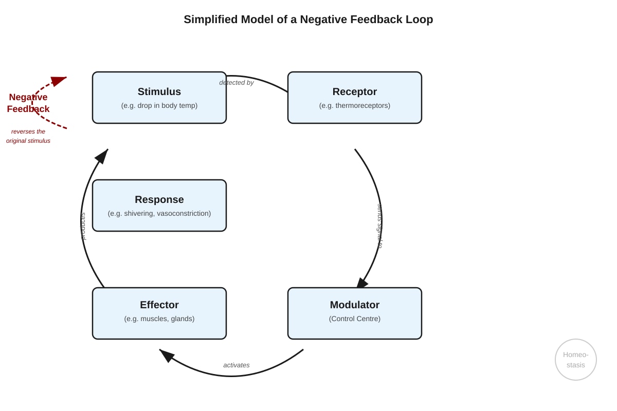
3. Using the diagram as a guide, explain how the body maintains a relatively constant core body temperature when the external environmental temperature drops. [5]
Answer:
- Stimulus: A drop in external temperature causes a decrease in core body temperature. (1 mark)
- Receptors: Thermoreceptors in the skin (peripheral) and in the hypothalamus (central) detect this decrease in temperature. (1 mark)
- Control Centre: The hypothalamus acts as the control centre (thermoregulatory centre). It receives input from the thermoreceptors and processes this information. (1 mark)
- Effectors: The hypothalamus sends nerve impulses (via the autonomic nervous system) and hormones to effectors, which include: (1 mark for naming effectors)
- Skeletal muscles: Shivering generates heat through rapid, involuntary muscle contractions.
- Arterioles in the skin: Vasoconstriction reduces blood flow to the skin, minimising heat loss.
- Adrenal medulla: Releases adrenaline, which increases metabolic rate.
- Sweat glands: Reduce or stop sweat production.
- Response and Negative Feedback: These responses increase heat production and decrease heat loss, causing core body temperature to rise back towards the set point. This change is detected by the receptors, and the response is switched off, demonstrating negative feedback. (1 mark)
Marking Notes: Award marks for identifying the stimulus, receptors, control centre, effectors, and the negative feedback loop. The answer must be in the context of a drop in temperature.
4. Osmoregulation is a key homeostatic process.
(a) State the location of the osmoreceptors that detect changes in blood water potential. [1]
Answer:
- The hypothalamus (specifically, the osmoreceptors are located in the hypothalamus). (1 mark)
(b) Describe the sequence of events that occurs when blood water potential decreases, leading to the production of more concentrated urine. [4]
Answer:
- A decrease in blood water potential (i.e., blood becomes more concentrated) is detected by osmoreceptors in the hypothalamus. (1 mark)
- The hypothalamus sends nerve impulses to the posterior pituitary gland, stimulating it to release more antidiuretic hormone (ADH) into the bloodstream. (1 mark)
- ADH travels in the blood to the kidneys, where it binds to receptors on the cells of the collecting ducts (and distal convoluted tubules). (1 mark)
- This binding triggers a signalling cascade that causes aquaporin water channels to be inserted into the apical membranes of these cells. This increases the permeability of the collecting duct to water, allowing more water to be reabsorbed by osmosis into the surrounding hypertonic medullary interstitial fluid. As a result, a smaller volume of more concentrated urine is produced. (1 mark)
Marking Notes: Award marks for the sequence: detection by hypothalamus, ADH release from posterior pituitary, action on collecting ducts, and increased water reabsorption leading to concentrated urine.
5. Compare and contrast the roles of the nervous system and the endocrine system in maintaining homeostasis. [4]
Answer:
- Similarity: Both systems are communication systems that work to maintain homeostasis by detecting changes and coordinating responses. Both often work together (e.g., the hypothalamus links the two systems). (1 mark)
- Difference 1 (Speed and Duration): The nervous system transmits signals rapidly (via electrical impulses) and its effects are short-lived. The endocrine system transmits signals more slowly (via hormones in the blood) but its effects are longer-lasting. (1 mark)
- Difference 2 (Nature of Signal): The nervous system uses electrical impulses and neurotransmitters at synapses. The endocrine system uses chemical messengers (hormones) transported in the bloodstream. (1 mark)
- Difference 3 (Target Specificity): The nervous system targets specific, precise locations (e.g., a specific muscle or gland). The endocrine system can have widespread effects, targeting many different cell types throughout the body that possess the specific receptor. (1 mark)
Marking Notes: Award 1 mark for a valid similarity and 1 mark for each of two valid, clearly explained differences. Accept other valid differences (e.g., nature of response, recovery time).
Section B: Neuronal Communication and the Action Potential (Questions 6–10)
6. The resting membrane potential of a neurone is approximately -70 mV.
(a) State the two main ions responsible for establishing this resting potential and the relative concentrations of each inside versus outside the cell. [2]
Answer:
- Potassium ions (K⁺): Higher concentration inside the cell than outside. (1 mark)
- Sodium ions (Na⁺): Higher concentration outside the cell than inside. (1 mark)
Marking Notes: Award 1 mark for each ion with its correct relative concentration.
(b) Explain the role of the Na⁺/K⁺-ATPase pump in maintaining the resting potential. [2]
Answer:
- The Na⁺/K⁺-ATPase pump actively transports 3 Na⁺ ions out of the cell and 2 K⁺ ions into the cell, using energy from ATP hydrolysis. (1 mark)
- This maintains the steep concentration gradients for Na⁺ and K⁺ across the membrane. These gradients are essential for the resting potential (as K⁺ diffuses out, leaving the inside negative) and for the generation of action potentials. (1 mark)
Marking Notes: Award 1 mark for describing the pump's action (3 Na⁺ out, 2 K⁺ in, ATP). Award 1 mark for linking this to maintaining the concentration gradients necessary for the resting potential.
7. Describe the sequence of ion movements across the membrane of an axon during the depolarisation and repolarisation phases of an action potential. [4]
Answer:
- Depolarisation: When a stimulus reaches threshold, voltage-gated Na⁺ channels open. (1 mark)
- Na⁺ ions diffuse rapidly into the axon down their electrochemical gradient, making the inside of the cell less negative and then positive (reaching ~+40 mV). (1 mark)
- Repolarisation: The voltage-gated Na⁺ channels become inactivated (close), and voltage-gated K⁺ channels open. (1 mark)
- K⁺ ions diffuse rapidly out of the axon down their concentration gradient, returning the membrane potential towards its negative resting value. (1 mark)
Marking Notes: Award marks for the key events: Na⁺ channel opening, Na⁺ influx, Na⁺ channel inactivation/K⁺ channel opening, and K⁺ efflux.
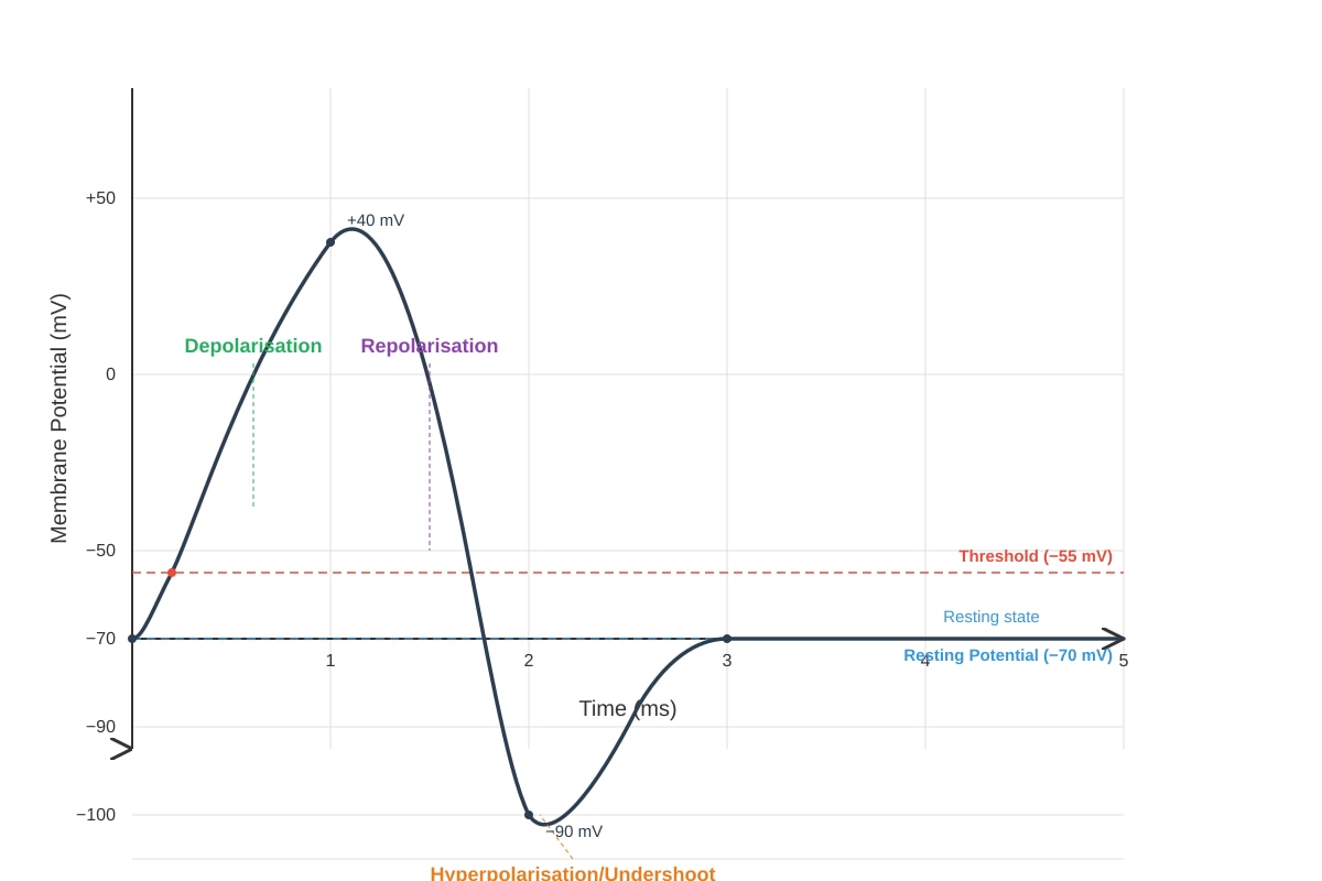
8. The diagram below shows the changes in membrane potential during an action potential.
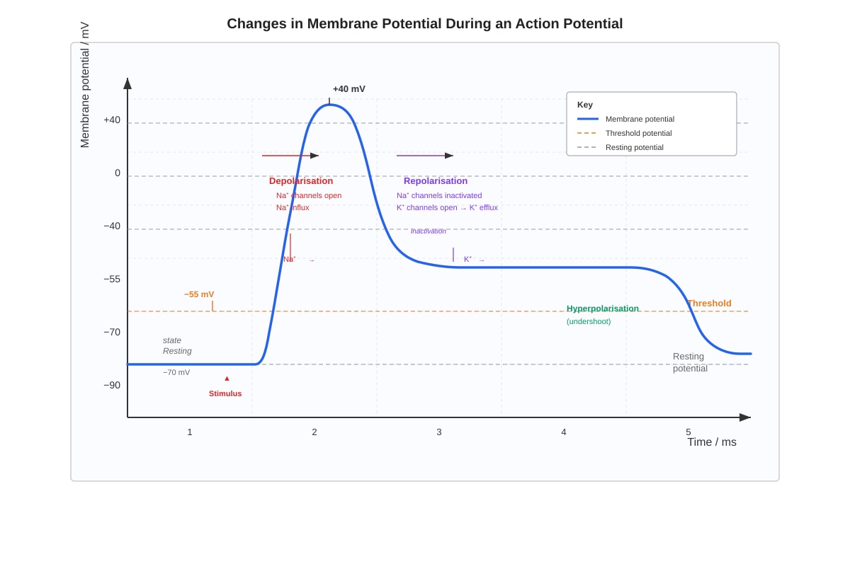
Generated diagram for this question.
(a) State the value of the threshold potential for a typical mammalian neurone. [1]
Answer:
- Approximately -55 mV. (1 mark)
(b) Explain the significance of the threshold potential in generating an action potential. [2]
Answer:
- The threshold potential is the critical level of depolarisation that must be reached for an action potential to be generated. (1 mark)
- If the membrane is depolarised to this level, it triggers the opening of voltage-gated Na⁺ channels, leading to a rapid, self-amplifying influx of Na⁺ (the all-or-nothing principle). If the threshold is not reached, no action potential is generated. (1 mark)
Marking Notes: Award 1 mark for the definition and 1 mark for linking it to the opening of voltage-gated Na⁺ channels and the all-or-nothing principle.
(c) With reference to the graph, explain the cause of the hyperpolarisation (undershoot) phase. [2]
Answer:
- During repolarisation, the voltage-gated K⁺ channels are slow to close. (1 mark)
- This means that for a brief period, the membrane is more permeable to K⁺ than at rest, causing an excessive efflux of K⁺. This makes the membrane potential temporarily more negative than the resting potential (e.g., -90 mV), before the K⁺ channels close and the Na⁺/K⁺ pump and leak channels restore the resting potential. (1 mark)
Marking Notes: Award 1 mark for identifying the slow closing of K⁺ channels and 1 mark for explaining the excessive K⁺ efflux leading to a more negative potential.
9. Explain how the myelin sheath increases the speed of conduction of an action potential along an axon. [3]
Answer:
- The myelin sheath is formed by Schwann cells (in the PNS) or oligodendrocytes (in the CNS) wrapping around the axon, creating a lipid-rich, insulating layer. (1 mark)
- The sheath is discontinuous, with gaps called Nodes of Ranvier. Voltage-gated Na⁺ and K⁺ channels are concentrated at these nodes and are absent in the myelinated regions. (1 mark)
- This forces the local circuits of ionic current to flow between the nodes, causing the action potential to "jump" from node to node. This is called saltatory conduction, which is much faster than continuous conduction along an unmyelinated axon. (1 mark)
Marking Notes: Award marks for the insulating property, the concentration of channels at Nodes of Ranvier, and the concept of saltatory conduction.
10. A local anaesthetic, such as lidocaine, blocks voltage-gated Na⁺ channels in sensory neurones.
(a) Predict the effect of lidocaine on the ability of a sensory neurone to generate an action potential. [1]
Answer:
- It will prevent the generation of an action potential (or it will stop the neurone from firing). (1 mark)
(b) Explain your prediction in terms of the mechanism of action potential generation. [2]
Answer:
- Generation of an action potential depends on the influx of Na⁺ through voltage-gated Na⁺ channels during depolarisation. (1 mark)
- By blocking these channels, lidocaine prevents the Na⁺ influx. Therefore, the membrane cannot depolarise to threshold, and no action potential can be generated or propagated along the neurone. (1 mark)
Marking Notes: Award 1 mark for identifying the role of Na⁺ influx and 1 mark for linking the block to the failure to reach threshold.
Section C: Synaptic Transmission (Questions 11–15)
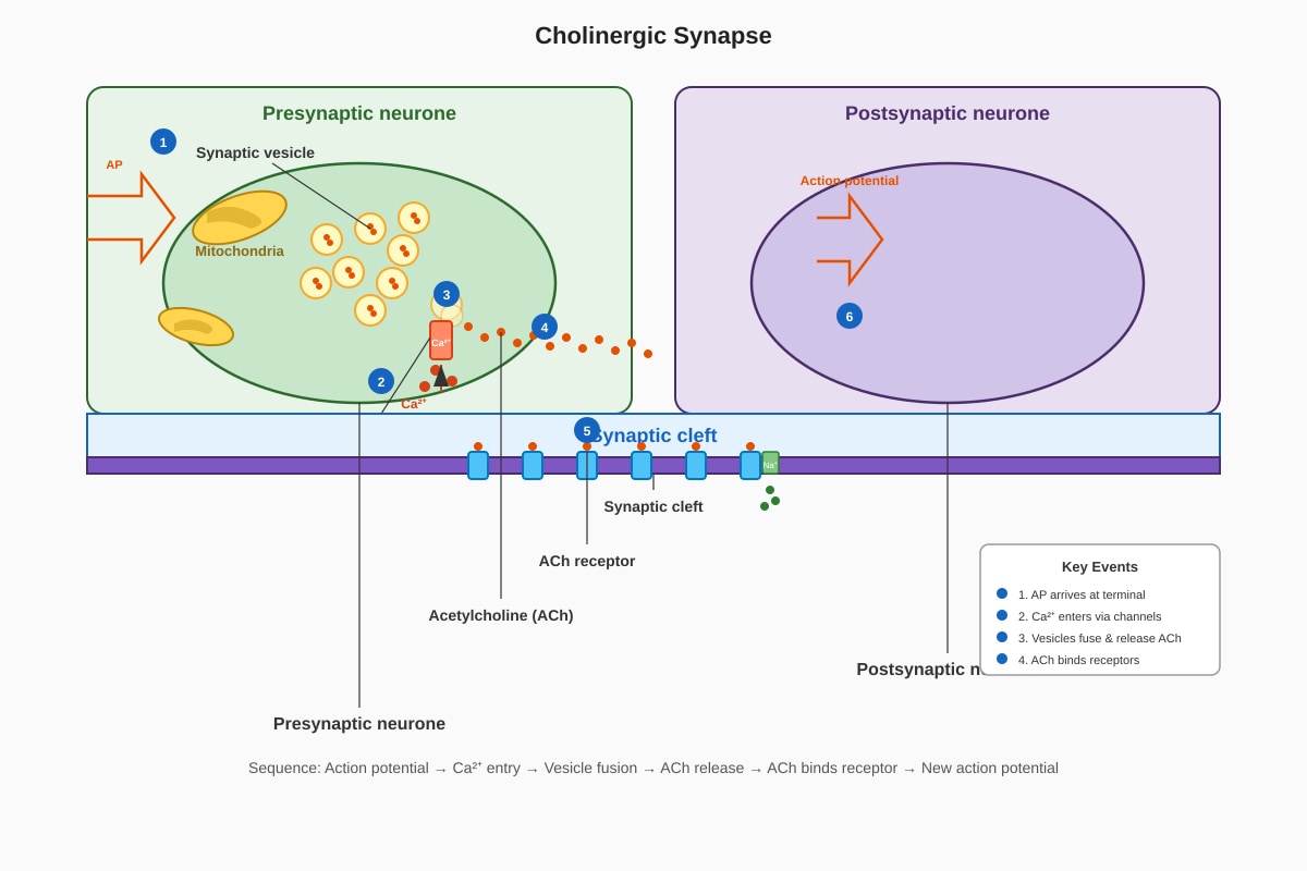
11. The diagram below shows a cholinergic synapse.
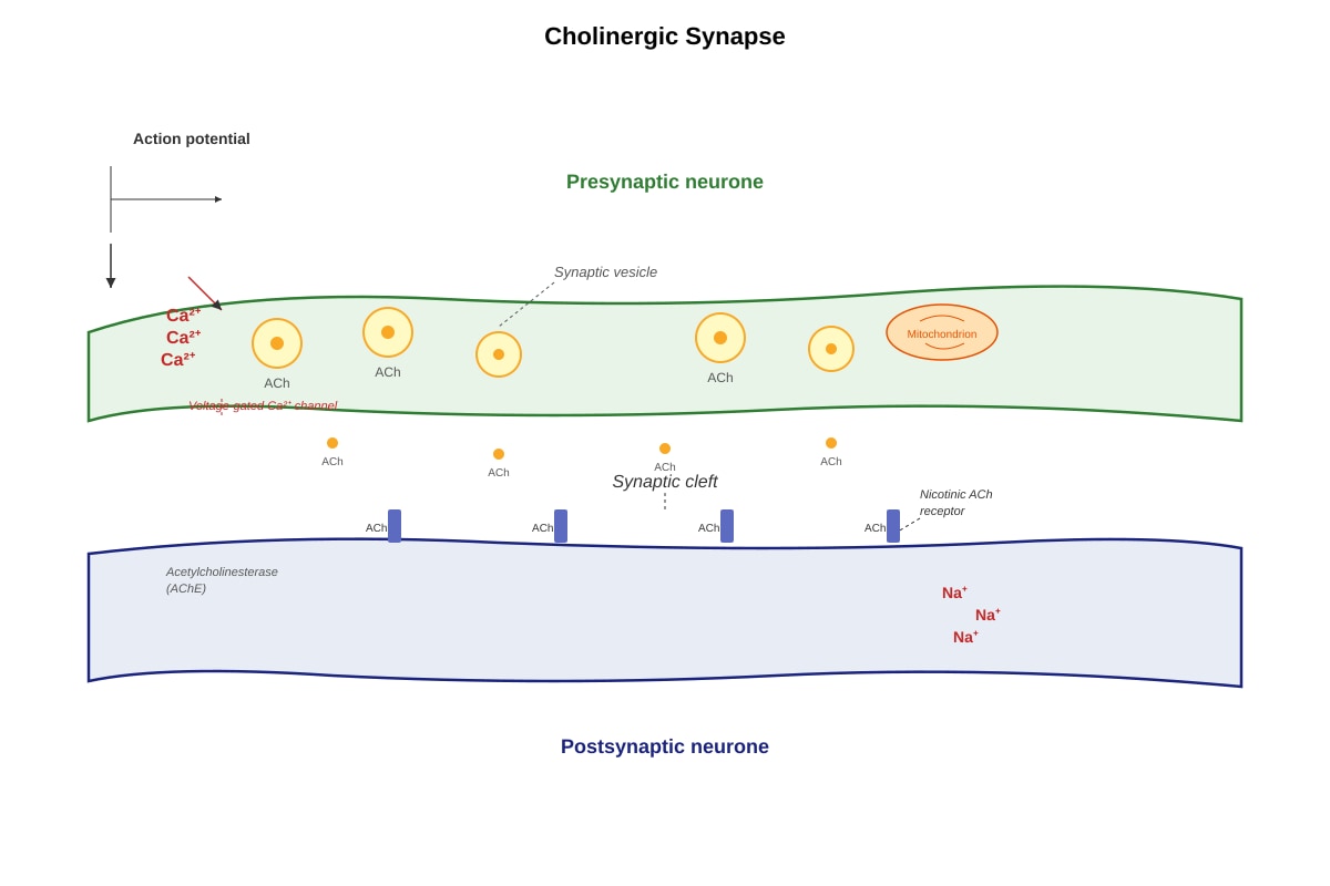
Generated diagram for this question.
(a) State the role of Ca²⁺ ions in synaptic transmission. [1]
Answer:
- Ca²⁺ ions enter the presynaptic terminal through voltage-gated Ca²⁺ channels when the action potential arrives, and this triggers the fusion of synaptic vesicles with the presynaptic membrane, causing the release of neurotransmitter (acetylcholine) into the synaptic cleft. (1 mark)
(b) Describe the sequence of events that occur at a cholinergic synapse, from the arrival of an action potential at the presynaptic terminal to the generation of a new action potential in the postsynaptic neurone. [5]
Answer:
- An action potential arrives at the presynaptic terminal, causing depolarisation of the membrane. (1 mark)
- This depolarisation opens voltage-gated Ca²⁺ channels, and Ca²⁺ ions diffuse into the presynaptic terminal. (1 mark)
- The influx of Ca²⁺ causes synaptic vesicles containing acetylcholine (ACh) to fuse with the presynaptic membrane, releasing ACh into the synaptic cleft by exocytosis. (1 mark)
- ACh diffuses across the cleft and binds to specific ACh receptors (nicotinic receptors) on the postsynaptic membrane. (1 mark)
- This binding causes ligand-gated Na⁺ channels to open. Na⁺ ions diffuse into the postsynaptic neurone, causing depolarisation (an excitatory postsynaptic potential, EPSP). If this depolarisation reaches the threshold, it triggers an action potential in the postsynaptic neurone. (1 mark)
Marking Notes: Award 1 mark for each of the 5 key steps in the correct sequence.
12. Explain how the enzyme acetylcholinesterase (AChE) in the synaptic cleft ensures that signalling at a cholinergic synapse is brief and unidirectional. [3]
Answer:
- Brief: AChE rapidly hydrolyses ACh in the synaptic cleft into choline and acetate. (1 mark) This removes the neurotransmitter from the cleft, preventing it from continuously binding to receptors on the postsynaptic membrane. This ensures that the postsynaptic response is brief and that the membrane can repolarise, ready for the next signal. (1 mark)
- Unidirectional: ACh is only released from the presynaptic terminal, and its receptors are only present on the postsynaptic membrane. The rapid destruction of ACh by AChE prevents it from diffusing back and stimulating the presynaptic terminal. This ensures that the signal can only pass in one direction: from presynaptic to postsynaptic. (1 mark)
Marking Notes: Award 1 mark for the role of AChE in breaking down ACh, 1 mark for linking this to a brief response, and 1 mark for linking the localisation of release/receptors and AChE action to unidirectionality.
13. Some nerve gases and insecticides are irreversible inhibitors of acetylcholinesterase.
(a) Predict the effect of an irreversible AChE inhibitor on synaptic transmission at a cholinergic synapse. [2]
Answer:
- ACh will not be broken down in the synaptic cleft. (1 mark)
- ACh will remain bound to its receptors on the postsynaptic membrane, causing continuous depolarisation and repeated firing of action potentials in the postsynaptic neurone. This leads to uncontrolled, excessive stimulation. (1 mark)
(b) Suggest why this effect can be lethal to an organism. [2]
Answer:
- If this occurs at neuromuscular junctions, it causes continuous, uncontrolled muscle contraction (spastic paralysis). (1 mark)
- If this affects muscles involved in breathing (e.g., the diaphragm), it can lead to respiratory failure and death. (1 mark)
Marking Notes: Award marks for linking to uncontrolled muscle contraction and respiratory failure. Accept other valid consequences (e.g., disruption of autonomic functions).
14. A student commented: "Synaptic transmission is simply the electrical signal jumping across the gap between two neurones." Evaluate this statement, explaining why it is inaccurate. [3]
Answer:
- The statement is inaccurate because the signal does not "jump" as an electrical current. (1 mark)
- The synaptic cleft is a physical gap that electrical impulses cannot cross. Instead, the electrical signal (action potential) is converted into a chemical signal. (1 mark)
- This involves the release of a neurotransmitter (e.g., acetylcholine) from the presynaptic terminal, its diffusion across the cleft, and its binding to receptors on the postsynaptic membrane, which then triggers a new electrical signal in the postsynaptic neurone. (1 mark)
Marking Notes: Award 1 mark for identifying the statement as inaccurate, 1 mark for explaining the need for a chemical signal, and 1 mark for describing the process of chemical transmission.
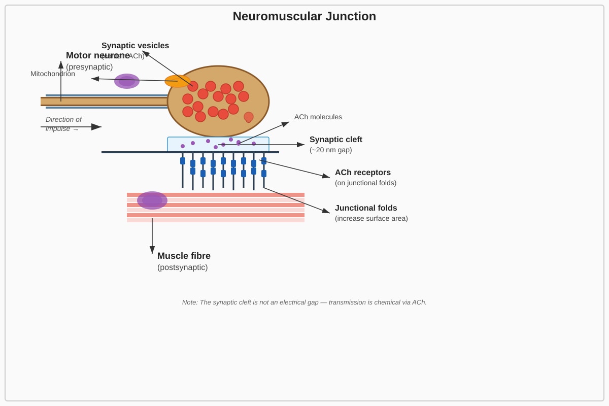
15. Explain how the structure of the neuromuscular junction is adapted to ensure rapid and reliable transmission of a signal from a motor neurone to a muscle fibre. [3]
Answer:
- Close apposition / narrow synaptic cleft: The presynaptic terminal and the muscle fibre membrane are very close together, reducing the diffusion distance for the neurotransmitter (ACh), making transmission rapid. (1 mark)
- Junctional folds: The postsynaptic membrane is folded, greatly increasing its surface area. This allows for a high density of ACh receptors to be packed into a small area, increasing the chance of ACh binding and ensuring a reliable response. (1 mark)
- Many synaptic vesicles: The presynaptic terminal contains a large number of synaptic vesicles filled with ACh, ensuring that a large amount of neurotransmitter is released in response to a single action potential, guaranteeing that the postsynaptic threshold is reached. (1 mark)
Marking Notes: Award 1 mark for each valid structural adaptation with its explanation.
Section D: Integration and Applied Physiology (Questions 16–20)
16. The graph below shows the changes in heart rate and stroke volume during moderate exercise.
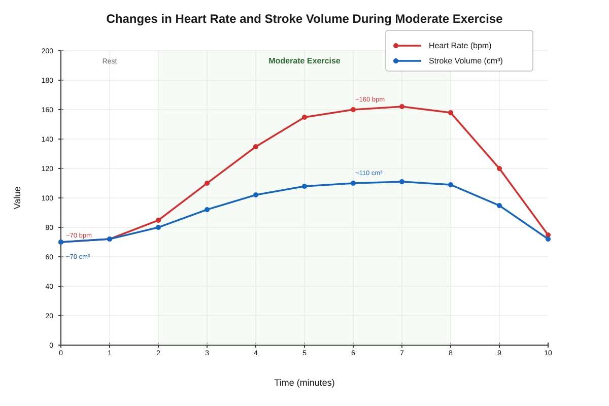
Generated graph for this question.
(a) Calculate the cardiac output at rest and during steady-state exercise using the data from the graph. Show your working. [2]
Answer:
- Cardiac Output (CO) = Heart Rate (HR) × Stroke Volume (SV)
- At rest: HR ≈ 70 bpm, SV ≈ 70 cm³. CO = 70 × 70 = 4900 cm³/min (or 4.9 dm³/min). (1 mark)
- During exercise: HR ≈ 160 bpm, SV ≈ 110 cm³. CO = 160 × 110 = 17600 cm³/min (or 17.6 dm³/min). (1 mark)
Marking Notes: Award 1 mark for each correct calculation, including the formula and working. Accept values within a reasonable range read from the graph.
(b) Explain how the changes in heart rate and stroke volume during exercise are brought about. [4]
Answer:
- Heart Rate: During exercise, muscle activity stimulates mechanoreceptors and chemoreceptors (detecting increased CO₂/decreased pH). These send impulses to the cardiovascular centre in the medulla oblongata. (1 mark) This centre increases sympathetic nervous system activity to the sinoatrial (SA) node, which increases the rate of depolarisation, leading to an increased heart rate. (1 mark)
- Stroke Volume: Increased sympathetic stimulation also increases the force of contraction of the ventricular muscle (positive inotropic effect). (1 mark) This, combined with increased venous return (due to the skeletal muscle pump and increased respiratory movements), leads to a greater end-diastolic volume. According to Starling's law, a greater stretch of the ventricular muscle leads to a more forceful contraction, increasing stroke volume. (1 mark)
Marking Notes: Award marks for the neural control of heart rate (sympathetic stimulation of SA node) and the mechanisms increasing stroke volume (increased contractility and increased venous return/Starling's law).
17. The hormone adrenaline is released during the "fight-or-flight" response.
(a) State the gland that releases adrenaline. [1]
Answer:
- The adrenal medulla (the inner part of the adrenal gland). (1 mark)
(b) Describe two physiological effects of adrenaline that prepare the body for physical activity. [2]
Answer:
- Effect 1: Increases heart rate and force of contraction, increasing cardiac output and blood flow to muscles. (1 mark)
- Effect 2: Causes vasodilation of arterioles in skeletal muscles and vasoconstriction of arterioles in the skin and digestive system, redirecting blood flow to where it is most needed. (1 mark)
- Effect 3: Stimulates glycogenolysis in the liver, raising blood glucose concentration to provide more substrate for respiration.
- Effect 4: Causes bronchodilation, increasing airflow to the lungs.
Marking Notes: Award 1 mark for each of any two valid effects. The effect must be clearly linked to preparing the body for physical activity.
18. A patient has a condition called diabetes insipidus, which is caused by a lack of antidiuretic hormone (ADH).
(a) Predict the effect of this condition on the volume and concentration of urine produced. [1]
Answer:
- The patient will produce a large volume of very dilute urine. (1 mark)
(b) Explain the physiological basis for your prediction. [3]
Answer:
- ADH normally acts on the collecting ducts of the nephron to increase their permeability to water by causing the insertion of aquaporin channels. (1 mark)
- Without ADH, the collecting ducts are relatively impermeable to water. (1 mark)
- Therefore, water is not reabsorbed from the filtrate back into the blood, and a large volume of dilute urine is excreted. (1 mark)
Marking Notes: Award 1 mark for the role of ADH on the collecting duct, 1 mark for the lack of permeability, and 1 mark for the consequence of reduced water reabsorption.
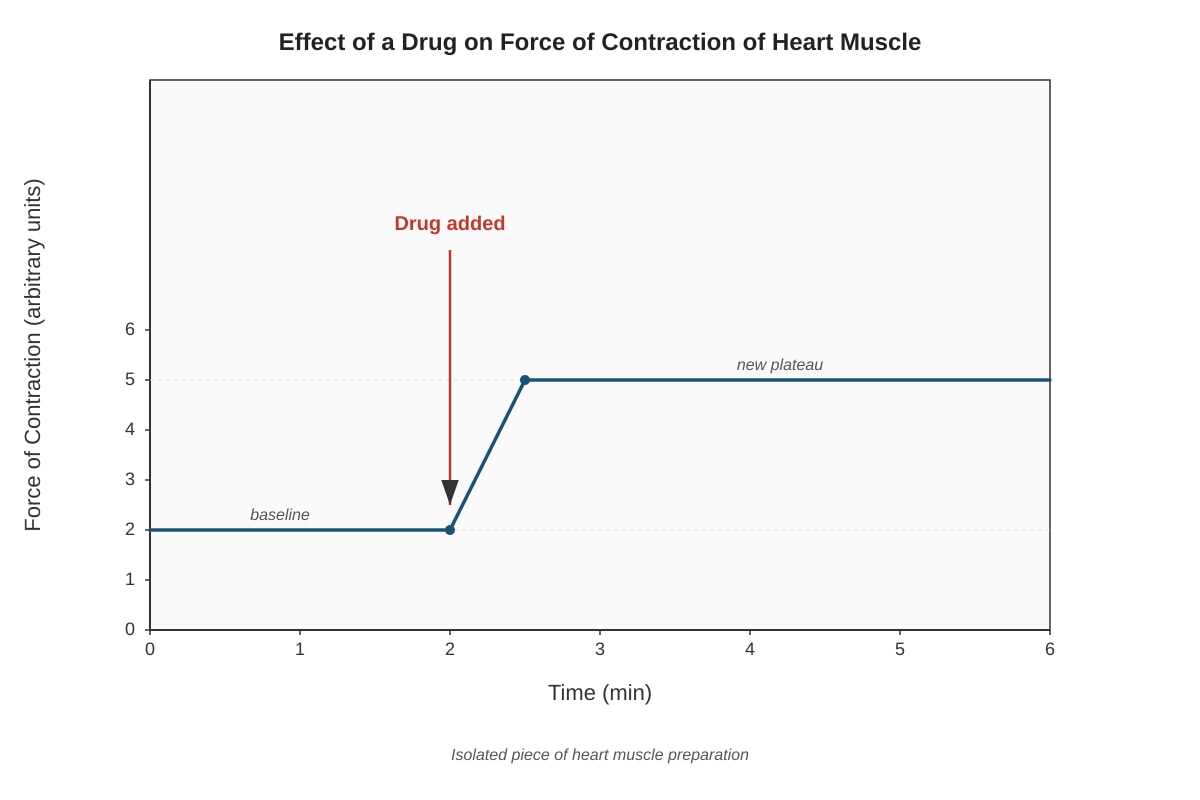
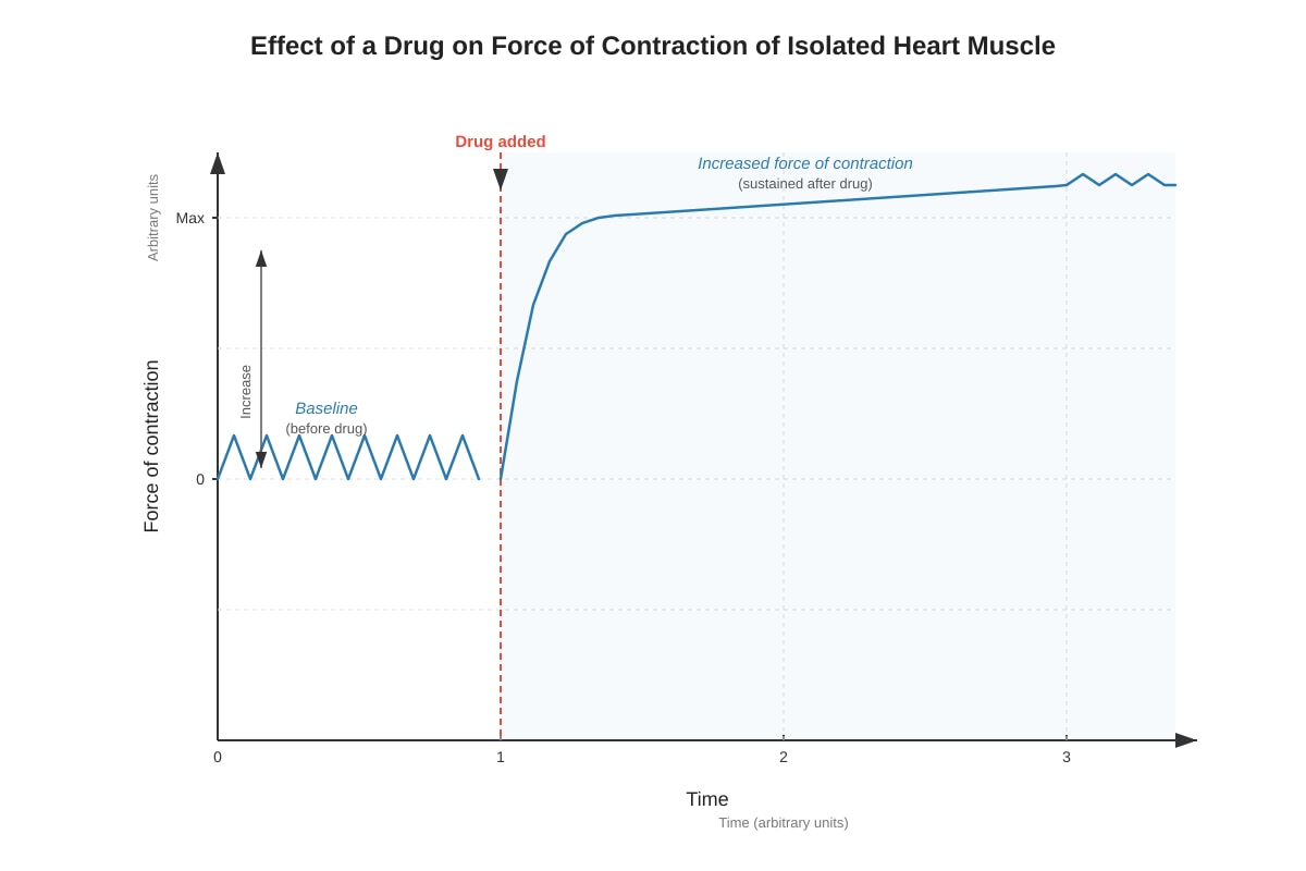
19. The graph below shows the effect of a drug on the force of contraction of an isolated piece of heart muscle.
Generated graph for this question.
(a) Describe the effect of the drug on the force of contraction. [1]
Answer:
- The drug causes a significant and sustained increase in the force of contraction. (1 mark)
(b) Suggest one mechanism by which this drug could produce this effect, relating your answer to the physiology of muscle contraction. [2]
Answer:
- Mechanism 1: The drug could increase the intracellular concentration of Ca²⁺ ions in the muscle cells. (1 mark) Ca²⁺ is essential for muscle contraction, as it binds to troponin, causing a conformational change that allows actin-myosin cross-bridges to form. More Ca²⁺ means more cross-bridges can form, leading to a stronger contraction. (1 mark)
- Mechanism 2: The drug could act as an agonist on β-adrenergic receptors (like adrenaline), activating a signalling pathway that increases Ca²⁺ release from the sarcoplasmic reticulum.
- Mechanism 3: The drug could inhibit the Na⁺/K⁺-ATPase pump (like digitalis), leading to an increase in intracellular Na⁺. This, in turn, reduces the activity of the Na⁺/Ca²⁺ exchanger, causing Ca²⁺ to accumulate inside the cell, increasing contractility.
Marking Notes: Award 1 mark for suggesting a plausible mechanism (e.g., increasing intracellular Ca²⁺) and 1 mark for linking it to the physiology of contraction (e.g., Ca²⁺ binding to troponin, cross-bridge formation).
20. Discuss the importance of the integration of the nervous and endocrine systems in coordinating the body's response to a sudden drop in blood glucose concentration. [6]
Answer: A high-quality answer should include the following points, structured logically:
1. Detection of the stimulus:
- A drop in blood glucose is detected by glucoreceptors, primarily in the hypothalamus (and also by the α-cells of the pancreatic islets directly). (1 mark)
2. The role of the endocrine system (direct response):
- The pancreas (α-cells) releases the hormone glucagon into the bloodstream. (1 mark)
- Glucagon travels to its primary target, the liver, and binds to receptors on hepatocytes.
- This activates a signalling cascade (via cAMP and protein kinase A) that promotes glycogenolysis (breakdown of glycogen to glucose) and gluconeogenesis (synthesis of glucose from non-carbohydrate sources). This raises blood glucose concentration. (1 mark)
3. The role of the nervous system (indirect response):
- The hypothalamus, acting as a control centre, also initiates a neural response. It increases sympathetic nervous system activity to the adrenal medulla. (1 mark)
- This stimulates the adrenal medulla to release adrenaline into the blood. (1 mark)
- Adrenaline reinforces and amplifies the effects of glucagon by also stimulating glycogenolysis in the liver, providing a rapid and powerful boost to blood glucose levels. (1 mark)
4. Integration and Homeostatic Outcome:
- The integration of the two systems ensures a rapid (nervous system → adrenaline) and sustained (endocrine → glucagon) response. The nervous system provides a fast, short-term response, while the endocrine system provides a slower, longer-lasting response. Together, they work synergistically to restore blood glucose to its set point, demonstrating effective negative feedback. (1 mark)
Marking Notes:
- Level 3 (5-6 marks): A detailed, well-structured answer that clearly explains the roles of both the endocrine (glucagon) and nervous (sympathetic → adrenaline) systems, explains how they integrate, and links this to the homeostatic outcome. The answer is logically organised and uses correct terminology.
- Level 2 (3-4 marks): A clear answer that describes the role of glucagon and the role of adrenaline, but the integration between the two systems may be less explicit or less well explained.
- Level 1 (1-2 marks): A basic answer that identifies one or two relevant hormones (e.g., glucagon) and their general effect (e.g., raises blood glucose) without a clear explanation of the mechanisms or integration.
END OF ANSWER KEY