AI Generated Quiz
A Level Biology H3 Human Physiology Quiz
Free A Level Biology H3 Human Physiology quiz, AI version, with questions, answers, and A Level-style practice for Singapore students.
These static practice materials are generated from the site's syllabus and paper-generation workflow, with source and model context shown so students and parents can evaluate the material before use.
Questions
Free quiz and exam paper access
Enter your details to view this paper
Your access is remembered on this device.
Answers
A-Level Biology H3 Quiz - Human Physiology: Answer Key
Total Marks: 75
Section A: Multiple-Choice Questions (Questions 1–5, 15 marks)
1. C) It insulates the axon, allowing action potentials to 'jump' between Nodes of Ranvier via saltatory conduction. [3]
- Explanation: The myelin sheath is a fatty insulating layer produced by Schwann cells (in the PNS) or oligodendrocytes (in the CNS). It prevents ion flow across the membrane in the myelinated regions. Action potentials can only occur at the Nodes of Ranvier, the unmyelinated gaps. This forces the depolarisation to 'jump' from node to node, a process called saltatory conduction, which is much faster than continuous conduction along an unmyelinated axon.
- Common Mistake: Students may think myelin actively pumps ions (A) or provides ATP (D). Myelin is passive; the Na⁺/K⁺ ATPase pumps are located at the nodes. Option B is incorrect because myelin decreases capacitance, which actually aids faster conduction.
2. D) Ca²⁺ [3]
- Explanation: When an action potential arrives at the presynaptic terminal, it depolarises the membrane, opening voltage-gated Ca²⁺ channels. Ca²⁺ ions flow into the terminal down their electrochemical gradient. This rise in intracellular Ca²⁺ concentration is the direct trigger that causes synaptic vesicles to fuse with the presynaptic membrane and release their neurotransmitter (acetylcholine) into the synaptic cleft via exocytosis.
- Common Mistake: Students often confuse the role of Na⁺ (which is key for the action potential itself) with Ca²⁺ (which is key for neurotransmitter release).
3. B) Antagonist [3]
- Explanation: An antagonist is a substance that binds to a receptor but does not activate it, blocking the receptor from being activated by the natural agonist (in this case, acetylcholine). Since the drug binds to the receptor but does not open the ion channel, it is an antagonist. An agonist would bind and activate the receptor, opening the channel.
- Common Mistake: Students may confuse antagonist with agonist. An agonist mimics the natural ligand, while an antagonist blocks it.
4. D) Positive feedback loop [3]
- Explanation: The core components of a homeostatic control system are a receptor (detects a change), a control centre (e.g., the brain, which determines the set point), and an effector (which carries out the response to restore the set point). This is typically achieved through negative feedback, which reverses the change. Positive feedback amplifies a change and is not a standard component of most homeostatic systems (though it does occur in specialised processes like childbirth and blood clotting).
- Common Mistake: Students may think positive feedback is a standard homeostatic mechanism. While it exists, it is the exception, not the rule, for maintaining stability.
5. C) The nervous system provides rapid, short-term responses to specific stimuli, while the endocrine system provides slower, longer-lasting, and more widespread regulation. [3]
- Explanation: The nervous system uses electrical impulses and neurotransmitters for fast, targeted communication (e.g., a reflex). The endocrine system uses hormones released into the bloodstream for slower, more prolonged, and widespread effects (e.g., growth and metabolism). Both are necessary because different physiological challenges require different response speeds and durations.
- Common Mistake: Option A reverses the speed characteristics. The nervous system is faster. Option D is incorrect because the nervous system uses electrical impulses and neurotransmitters, while the endocrine system uses hormones.
Section B: Structured Questions (Questions 6–15, 40 marks)
6. Describe the sequence of events that occur at a cholinergic synapse from the arrival of an action potential at the presynaptic terminal to the generation of a postsynaptic potential. [5]
Answer:
- An action potential arrives at the presynaptic terminal, depolarising the membrane. [1]
- This depolarisation opens voltage-gated Ca²⁺ channels, allowing Ca²⁺ ions to flow into the presynaptic terminal. [1]
- The influx of Ca²⁺ causes synaptic vesicles containing acetylcholine (ACh) to fuse with the presynaptic membrane and release ACh into the synaptic cleft via exocytosis. [1]
- ACh diffuses across the synaptic cleft and binds to specific ACh receptors on the postsynaptic membrane. [1]
- The binding of ACh opens ligand-gated Na⁺ channels, allowing Na⁺ to flow into the postsynaptic neurone. This depolarises the postsynaptic membrane, generating an excitatory postsynaptic potential (EPSP). [1]
Marking Notes: Award 1 mark for each of the five key steps. Accept "ACh" for acetylcholine. The final step must mention the generation of an EPSP or a postsynaptic potential.
7. Explain the role of the Na⁺/K⁺ ATPase pump in maintaining the resting membrane potential of a neurone. [3]
Answer: The Na⁺/K⁺ ATPase pump is an active transport protein that uses ATP to move 3 Na⁺ ions out of the cell and 2 K⁺ ions into the cell. [1] This creates and maintains concentration gradients for both ions: a high Na⁺ concentration outside the cell and a high K⁺ concentration inside the cell. [1] These gradients are essential for the resting membrane potential, as the leak of K⁺ ions out of the cell (down its concentration gradient) is the primary contributor to the negative charge inside the cell at rest (approximately -70 mV). [1]
Marking Notes: Award 1 mark for stating the pump moves 3 Na⁺ out and 2 K⁺ in. Award 1 mark for stating it establishes/maintains the concentration gradients. Award 1 mark for linking these gradients to the resting potential (e.g., via K⁺ leak channels).
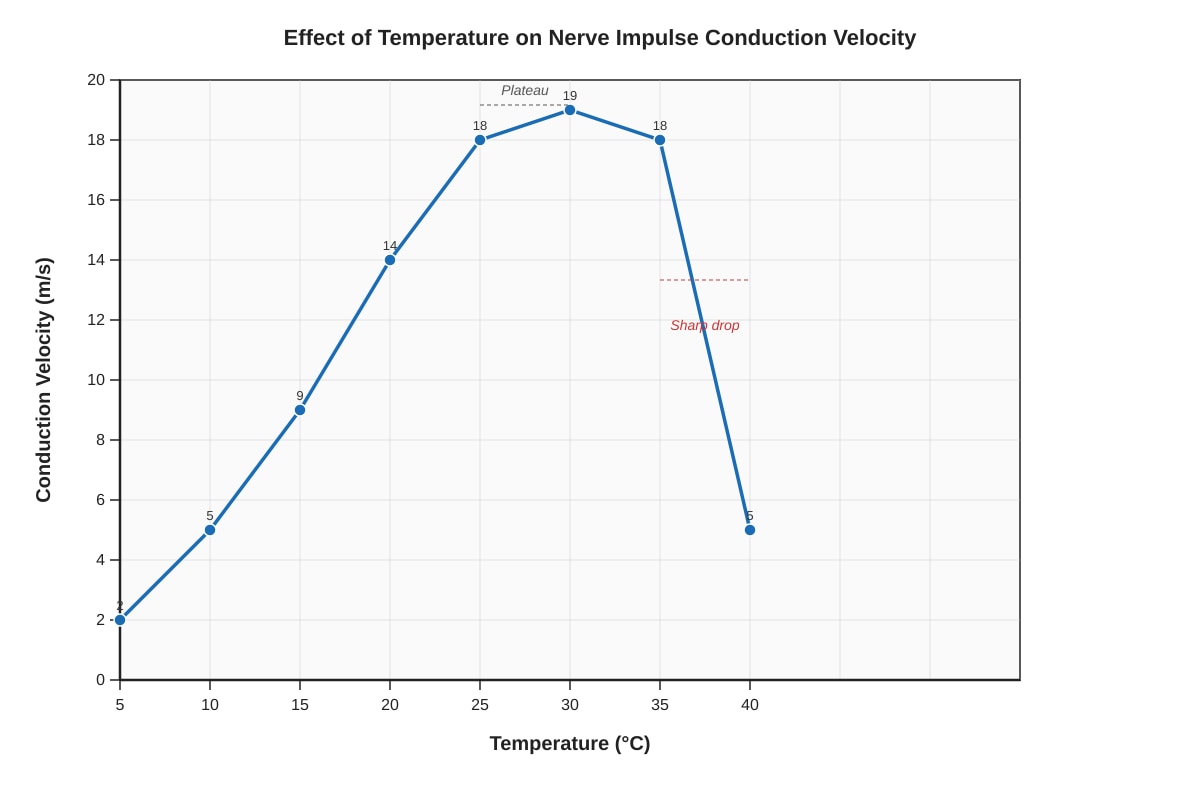
8. (a) Describe the trend shown in the graph. [2]
Answer: As temperature increases from 5°C to 30°C, the conduction velocity increases rapidly. [1] The velocity then plateaus between 30°C and 35°C, before dropping sharply at 40°C. [1]
Marking Notes: Award 1 mark for describing the initial increase. Award 1 mark for describing the plateau and the sharp drop.
(b) Explain the physiological reason for the sharp drop in conduction velocity at 40°C. [3]
Answer: At 40°C, the high temperature is likely denaturing the proteins involved in nerve impulse conduction. [1] This includes the voltage-gated Na⁺ and K⁺ channels, which are essential for generating and propagating action potentials. [1] Denaturation alters the 3D structure of these proteins, preventing them from functioning correctly, which drastically slows or stops impulse conduction. [1]
Marking Notes: Award 1 mark for the concept of protein denaturation. Award 1 mark for identifying specific proteins (e.g., ion channels, Na⁺/K⁺ ATPase). Award 1 mark for linking denaturation to loss of function and reduced conduction velocity.
9. Distinguish between the roles of the nervous system and the endocrine system in maintaining homeostasis. [4]
Answer:
- Nervous System: Uses electrical impulses and neurotransmitters for rapid, short-lived, and highly specific responses to stimuli. It is ideal for immediate adjustments, such as reflex actions or fine-tuning muscle movements. [2]
- Endocrine System: Uses hormones (chemical messengers) transported in the blood for slower, longer-lasting, and more widespread effects. It is ideal for regulating long-term processes like growth, metabolism, and reproduction. [2]
Marking Notes: Award 2 marks for each system, with 1 mark for the mode of communication and 1 mark for the nature of the response (speed/duration/scope).
10. A patient is diagnosed with a condition where her immune system produces antibodies that block the acetylcholine receptors at neuromuscular junctions. Predict the effect this would have on muscle function and explain the underlying mechanism. [4]
Answer: Prediction: The patient would experience muscle weakness, fatigue, and potentially paralysis. [1] Mechanism: Acetylcholine (ACh) is the neurotransmitter released at the neuromuscular junction. [1] It normally binds to ACh receptors on the muscle cell membrane, opening ion channels and triggering muscle contraction. [1] The antibodies block these receptors, preventing ACh from binding. This means that even when the nerve sends a signal, the muscle cannot be stimulated to contract effectively, leading to weakness. [1]
Marking Notes: Award 1 mark for the correct prediction. Award 1 mark for stating the role of ACh. Award 1 mark for explaining the normal binding process. Award 1 mark for explaining how blocking the receptor prevents contraction.
11. Describe the structure of a cholinergic synapse, naming the key components involved in signal transmission. [3]
Answer: A cholinergic synapse consists of a presynaptic terminal (containing synaptic vesicles filled with acetylcholine), a synaptic cleft (the narrow gap between the two neurones), and a postsynaptic membrane (containing specific acetylcholine receptors). [3]
Marking Notes: Award 1 mark for each of the three components correctly named and described. Accept "ACh" for acetylcholine.
12. Explain how the process of saltatory conduction increases the speed of action potential propagation along a myelinated neurone compared to an unmyelinated neurone. [4]
Answer: In an unmyelinated neurone, the action potential is propagated by continuous conduction, where depolarisation spreads to adjacent sections of the membrane, opening voltage-gated Na⁺ channels along the entire length of the axon. [1] This is a slow process. In a myelinated neurone, the myelin sheath acts as an electrical insulator, preventing ion flow across the membrane in the myelinated regions. [1] Voltage-gated Na⁺ channels are concentrated only at the Nodes of Ranvier (the unmyelinated gaps). [1] Therefore, the action potential 'jumps' from one node to the next (saltatory conduction), bypassing the myelinated sections. This is much faster because only the nodes need to be depolarised, not the entire axon membrane. [1]
Marking Notes: Award 1 mark for describing continuous conduction. Award 1 mark for the insulating role of myelin. Award 1 mark for the concentration of channels at nodes. Award 1 mark for explaining the 'jumping' and resulting speed increase.
13. A researcher is studying the effect of a toxin that prevents the breakdown of acetylcholine in the synaptic cleft. Predict the effect of this toxin on synaptic transmission and explain your reasoning. [4]
Answer: Prediction: The toxin would cause prolonged and excessive stimulation of the postsynaptic neurone or muscle cell, leading to continuous firing of action potentials, spasms, or paralysis. [2] Reasoning: Normally, acetylcholine is broken down by the enzyme acetylcholinesterase in the synaptic cleft to terminate the signal. [1] If this breakdown is prevented, acetylcholine will remain bound to its receptors, causing the postsynaptic membrane to remain depolarised and generate repeated action potentials. [1]
Marking Notes: Award 2 marks for the prediction (must mention prolonged/excessive stimulation). Award 1 mark for stating the normal role of acetylcholinesterase. Award 1 mark for explaining the consequence of its inhibition.
14. Explain why organisms need different communication systems, using the nervous and endocrine systems as examples. [4]
Answer: Different physiological challenges require different types of responses. [1] The nervous system provides rapid, short-term, and highly targeted responses (e.g., pulling your hand away from a hot surface). [1] The endocrine system provides slower, longer-lasting, and more widespread responses (e.g., regulating blood glucose levels over hours). [1] Having both systems allows an organism to respond effectively to a wide range of stimuli, from immediate threats to long-term metabolic needs. [1]
Marking Notes: Award 1 mark for the general principle. Award 1 mark for describing the nervous system's role. Award 1 mark for describing the endocrine system's role. Award 1 mark for explaining why both are needed.
15. Describe the role of negative feedback in the control of blood glucose concentration. [4]
Answer: When blood glucose rises (e.g., after a meal), the pancreas (receptor/control centre) detects the increase and releases insulin from beta cells. [1] Insulin acts on the liver and muscle cells (effectors) to increase glucose uptake and storage as glycogen, lowering blood glucose back to the set point. [1] When blood glucose falls (e.g., during exercise), the pancreas detects the decrease and releases glucagon from alpha cells. [1] Glucagon acts on the liver to break down glycogen into glucose, raising blood glucose back to the set point. [1] This is negative feedback because the response (lowering or raising glucose) reverses the initial stimulus.
Marking Notes: Award 1 mark for the response to high glucose (insulin release). Award 1 mark for the effect of insulin. Award 1 mark for the response to low glucose (glucagon release). Award 1 mark for the effect of glucagon. The final sentence about negative feedback is not essential for full marks but can be credited if the other points are missed.
Section C: Free-Response Question (Questions 16–20, 20 marks)
Answer any two of the following five questions. Each question is worth 10 marks. Quality of scientific argumentation and written communication will be assessed.
16. Discuss the interdependence of the nervous and endocrine systems in coordinating the body's response to a sudden drop in environmental temperature. [10]
Answer/ Marking Scheme:
This is a high-level question requiring integration of knowledge. Award marks for the following points:
- Detection (Nervous System): Thermoreceptors in the skin detect the drop in temperature. [1] This information is transmitted via sensory neurones to the hypothalamus (the body's thermoregulatory centre). [1]
- Integration (Hypothalamus): The hypothalamus acts as the control centre, comparing the input to the set point (approximately 37°C). [1]
- Rapid Response (Nervous System): The hypothalamus initiates immediate, short-term responses via the sympathetic nervous system:
- Vasoconstriction: Signals to smooth muscle in arterioles of the skin to constrict, reducing blood flow to the skin and minimising heat loss. [1]
- Shivering: Signals to skeletal muscles to contract involuntarily, generating heat through increased metabolic activity. [1]
- Piloerection: Signals to tiny muscles at the base of hairs to contract (raising hairs), which in humans is a minor effect but in other animals traps an insulating layer of air. [1]
- Sustained Response (Endocrine System): The hypothalamus also triggers a slower, longer-lasting response via the endocrine system:
- It releases thyrotropin-releasing hormone (TRH), which stimulates the anterior pituitary to release thyroid-stimulating hormone (TSH). [1]
- TSH stimulates the thyroid gland to release thyroxine (T4) and triiodothyronine (T3). [1]
- These hormones increase the basal metabolic rate of cells throughout the body, generating more heat over a prolonged period. [1]
- Interdependence: The nervous system provides the rapid detection and initial corrective action, while the endocrine system provides the sustained, systemic adjustment. The hypothalamus is the key link, receiving neural input and orchestrating both neural and hormonal outputs. [1]
Marking Notes:
- Award up to 9 marks for content.
- Award 1 mark for the quality of written communication (clear structure, logical flow, use of correct terminology).
- Accept other valid points, such as the role of adrenaline (released from the adrenal medulla via neural stimulation) in increasing metabolic rate.
17. Compare and contrast the mechanisms of action potential propagation along a myelinated neurone and signal transmission across a cholinergic synapse. [10]
Answer/ Marking Scheme:
| Feature | Action Potential Propagation (Myelinated Neurone) | Synaptic Transmission (Cholinergic Synapse) |
|---|---|---|
| Nature of Signal | Electrical (depolarisation) | Chemical (neurotransmitter) |
| Direction | Unidirectional (from cell body to axon terminal) | Unidirectional (from presynaptic to postsynaptic) |
| Speed | Very fast (saltatory conduction) | Slower (synaptic delay due to diffusion) |
| Amplification | No amplification; signal is regenerated at each node. | Can be amplified (one presynaptic AP can cause release of many ACh molecules). |
| Integration | No integration; all-or-nothing. | Can be integrated (summation of EPSPs and IPSPs). |
| Modulation | Not easily modulated. | Easily modulated (e.g., by drugs, toxins, or other neurotransmitters). |
| Key Structures | Myelin sheath, Nodes of Ranvier, voltage-gated Na⁺/K⁺ channels. | Presynaptic terminal, synaptic vesicles, synaptic cleft, postsynaptic receptors. |
| Ion Movement | Na⁺ influx (depolarisation), K⁺ efflux (repolarisation). | Ca²⁺ influx (triggers release), Na⁺ influx (generates EPSP). |
| Energy Use | Uses ATP for Na⁺/K⁺ pump to restore gradients. | Uses ATP for vesicle recycling and pump activity. |
| Role of Gap | No gap; signal travels along the membrane. | Signal crosses a physical gap (synaptic cleft). |
Marking Notes:
- Award up to 4 marks for a clear comparison (e.g., a table or structured paragraphs).
- Award up to 4 marks for contrasting the mechanisms (e.g., electrical vs. chemical, speed, modulation).
- Award 1 mark for mentioning the unidirectional nature of both.
- Award 1 mark for the quality of written communication.
- The answer must cover both topics for full marks.
18. Evaluate the importance of the human microbiota for our health, with reference to its role in immune system development and protection against pathogens. [10]
Answer/ Marking Scheme:
- Definition: The human microbiota refers to the trillions of microorganisms (bacteria, fungi, viruses) that live on and in our bodies, particularly in the gut. [1]
- Immune System Development:
- The microbiota is essential for the normal development of the immune system. Germ-free animals have underdeveloped immune systems (e.g., fewer Peyer's patches, lower antibody levels). [1]
- It helps to 'educate' the immune system, teaching it to distinguish between harmless commensals and dangerous pathogens. [1]
- It promotes the development of regulatory T cells (Tregs), which help maintain immune tolerance and prevent autoimmune diseases. [1]
- Protection Against Pathogens (Colonisation Resistance):
- Competitive Exclusion: Commensal bacteria occupy niches and consume resources, making it harder for pathogens to establish. [1]
- Production of Antimicrobial Substances: Some gut bacteria produce bacteriocins or other compounds that directly kill or inhibit pathogens. [1]
- Strengthening the Gut Barrier: The microbiota helps maintain the integrity of the intestinal epithelial barrier, preventing pathogens from crossing into the bloodstream. [1]
- Modulating Immune Responses: The microbiota can prime the immune system to respond more effectively to pathogens (e.g., by stimulating the production of antimicrobial peptides or enhancing phagocyte activity). [1]
- Evaluation: The microbiota is not just a passive passenger but an active and essential component of our health. Disruption of the microbiota (dysbiosis) through antibiotics, diet, or other factors is linked to increased susceptibility to infections, allergies, and autoimmune diseases. [1]
Marking Notes:
- Award up to 4 marks for points on immune system development.
- Award up to 4 marks for points on protection against pathogens.
- Award 1 mark for a concluding evaluation.
- Award 1 mark for the quality of written communication.
19. Discuss how an understanding of the principles of homeostasis and communication systems has contributed to the development of treatments for type 1 diabetes mellitus. [10]
Answer/ Marking Scheme:
- Understanding the Defect: Type 1 diabetes is an autoimmune condition where the immune system destroys the beta cells of the pancreas. This means the body can no longer produce insulin, a key hormone for glucose homeostasis. [1]
- Homeostatic Principle (Negative Feedback): The understanding that blood glucose is regulated by negative feedback (insulin lowers it, glucagon raises it) is fundamental. [1] The treatment aims to artificially restore this feedback loop.
- Treatment 1: Insulin Therapy:
- The most direct treatment is to replace the missing hormone. [1]
- Early treatments used animal-derived insulin. [1]
- The development of recombinant human insulin (using genetically engineered E. coli) was a major breakthrough, providing a pure, non-allergenic source. [1]
- Modern insulin analogues (e.g., rapid-acting lispro, long-acting glargine) are designed to better mimic the body's natural insulin secretion profile. [1]
- Treatment 2: Monitoring:
- Homeostasis requires monitoring. Blood glucose monitors allow patients to check their levels and adjust insulin doses accordingly. [1]
- Continuous glucose monitors (CGMs) provide real-time data, further improving control. [1]
- Treatment 3: Artificial Pancreas:
- This is a closed-loop system that integrates a CGM, an insulin pump, and a control algorithm. [1]
- It automatically adjusts insulin delivery based on glucose readings, mimicking the function of a healthy pancreas and the negative feedback loop. [1]
- Future Directions: Research into beta-cell transplantation and immunotherapy aims to restore the body's own homeostatic control. [1]
Marking Notes:
- Award up to 10 marks for a well-structured discussion. The answer must link the treatments back to the principles of homeostasis and communication.
- Award 1 mark for the quality of written communication.
20. Explain how the concept of immunological self-tolerance ensures that B and T lymphocytes do not normally attack correctly functioning host cells. [10]
Answer/ Marking Scheme:
- Definition: Immunological self-tolerance is the ability of the immune system to recognise and not mount a destructive response against the body's own (self) antigens. [1]
- Central Tolerance (in Primary Lymphoid Organs):
- T Cell Selection (Thymus): Developing T cells undergo positive and negative selection. [1] Positive selection ensures they can recognise self-MHC molecules. [1] Negative selection eliminates (via apoptosis) T cells that bind too strongly to self-antigens presented in the thymus. [1] This removes most self-reactive T cells before they mature.
- B Cell Selection (Bone Marrow): Developing B cells that bind strongly to self-antigens in the bone marrow are either eliminated (clonal deletion) or undergo receptor editing (changing their BCR to be non-self-reactive). [1]
- Peripheral Tolerance (in Secondary Lymphoid Organs/Tissues):
- Despite central tolerance, some self-reactive lymphocytes may escape to the periphery. [1] Several mechanisms exist to control them:
- Anergy: Self-reactive T cells that encounter self-antigen without the necessary co-stimulatory signals (e.g., from an infection) become functionally unresponsive (anergic). [1]
- Regulatory T Cells (Tregs): A specialised population of T cells (CD4+ FoxP3+) actively suppresses the activity of other self-reactive lymphocytes. [1]
- Immune Privilege: Some sites (e.g., the eye, brain) have mechanisms to limit immune responses. [1]
- Clonal Ignorance: Some self-antigens are sequestered (hidden) from the immune system, so self-reactive lymphocytes never encounter them. [1]
- Failure of Tolerance: When these mechanisms fail, autoimmunity can result (e.g., type 1 diabetes, rheumatoid arthritis). [1]
Marking Notes:
- Award up to 4 marks for central tolerance mechanisms.
- Award up to 5 marks for peripheral tolerance mechanisms.
- Award 1 mark for the quality of written communication.
- The answer must clearly explain how the mechanisms work, not just name them.
End of Answer Key