AI Generated Quiz
A Level Biology H3 Human Physiology Quiz
Free A Level Biology H3 Human Physiology quiz, AI version, with questions, answers, and A Level-style practice for Singapore students.
These static practice materials are generated from the site's syllabus and paper-generation workflow, with source and model context shown so students and parents can evaluate the material before use.
Questions
Free quiz and exam paper access
Enter your details to view this paper
Your access is remembered on this device.
Answers
A-Level Biology H3 Quiz - Human Physiology — Answer Key
Total Marks: 75
Section A: Multiple-Choice Questions (Questions 1–5)
1. B — The sinoatrial (SA) node is the natural pacemaker of the heart. It spontaneously generates action potentials that spread across the atria, initiating each heartbeat. Option A describes the AV node's role (delaying the impulse). Option C is incorrect because the SA node does not conduct directly to Purkinje fibres. Option D is incorrect because the SA node does not regulate contraction force. [1 mark]
2. C — The second heart sound ("dub") is caused by the closing of the semilunar valves (aortic and pulmonary valves) at the beginning of ventricular diastole. The first heart sound ("lub") is caused by the closing of the atrioventricular valves. [1 mark]
3. C — Saltatory conduction is the rapid propagation of an action potential that "jumps" from one node of Ranvier to the next in a myelinated neurone. Myelin sheaths insulate the axon, preventing ion flow except at the nodes, which speeds up transmission and conserves energy. [1 mark]
4. B — When an action potential reaches the presynaptic terminal, voltage-gated calcium channels open, allowing Ca²⁺ ions to enter the presynaptic knob. This influx of Ca²⁺ triggers the exocytosis of synaptic vesicles containing acetylcholine. Therefore, Ca²⁺ influx is the first event among the options. [1 mark]
5. C — The primary function of the loop of Henle is to create a concentration gradient in the medulla of the kidney. This gradient is essential for the subsequent reabsorption of water from the collecting duct under the influence of ADH. Filtration occurs in the Bowman's capsule (A), glucose reabsorption occurs in the proximal convoluted tubule (B), and H⁺ secretion occurs in the distal tubule and collecting duct (D). [1 mark]
Section B: Structured Questions (Questions 6–15)
6. (a) The AV node delays the electrical impulse from the atria before it passes to the ventricles. [1 mark]
(b) The delay at the AV node ensures that the atria have completed their contraction (atrial systole) and have fully emptied their blood into the ventricles before the ventricles begin to contract (ventricular systole). This allows for efficient filling of the ventricles, maximising stroke volume. Without this delay, the atria and ventricles would contract almost simultaneously, reducing the volume of blood ejected from the heart. [2 marks — 1 mark for explaining the timing of atrial vs. ventricular contraction, 1 mark for linking to efficient ventricular filling]
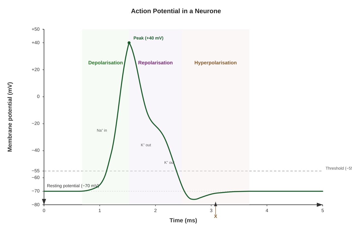
7. (a) Hyperpolarisation (or after-hyperpolarisation). [1 mark]
(b) During repolarisation, voltage-gated sodium channels become inactivated, stopping the influx of Na⁺. At the same time, voltage-gated potassium channels open, allowing K⁺ to diffuse rapidly out of the neurone down its electrochemical gradient. The loss of positive charge from the cell restores the negative membrane potential, repolarising the membrane. [3 marks — 1 mark for Na⁺ channel inactivation, 1 mark for K⁺ channel opening, 1 mark for K⁺ efflux repolarising the membrane]
8. (a) Homeostasis is the maintenance of a stable internal environment within narrow physiological limits, despite changes in the external environment. [1 mark]
(b) Example: Regulation of blood glucose concentration. When blood glucose rises after a meal, the pancreas (receptor and control centre) detects the increase. Beta cells of the islets of Langerhans secrete insulin into the blood. Insulin stimulates the uptake of glucose by liver and muscle cells (effectors), promoting glycogenesis (conversion of glucose to glycogen). This lowers blood glucose back towards the set point. When blood glucose falls, alpha cells secrete glucagon, which stimulates glycogenolysis (breakdown of glycogen to glucose), raising blood glucose. This is a negative feedback loop because the response (lowering or raising glucose) opposes the initial stimulus. [4 marks — 1 mark for naming a correct example, 1 mark for identifying the receptor/control centre, 1 mark for describing the effector response, 1 mark for explaining how it opposes the stimulus]
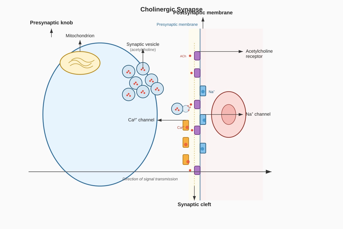
9. (a) Diagram should show: Presynaptic knob with synaptic vesicles containing acetylcholine (ACh), mitochondria, and Ca²⁺ channels. Synaptic cleft (the gap). Postsynaptic membrane with ACh receptors and Na⁺ channels. [2 marks — 1 mark for correct structures, 1 mark for correct labels]
(b) When an action potential arrives at the presynaptic knob, it depolarises the membrane, opening voltage-gated Ca²⁺ channels. Ca²⁺ ions diffuse into the presynaptic knob down their electrochemical gradient. The increase in intracellular Ca²⁺ concentration triggers the movement of synaptic vesicles to the presynaptic membrane. The vesicles fuse with the membrane and release their contents (acetylcholine) into the synaptic cleft via exocytosis. [3 marks — 1 mark for Ca²⁺ influx, 1 mark for vesicle movement and fusion, 1 mark for exocytosis of ACh]
10. (a) Glucose is small enough to be filtered from the blood into the Bowman's capsule, so it appears in the glomerular filtrate. However, all glucose is actively reabsorbed by the proximal convoluted tubule via specific transport proteins (SGLT co-transporters). In a healthy person, the transport maximum for glucose is not exceeded, so no glucose remains in the urine. [2 marks — 1 mark for filtration, 1 mark for active reabsorption in PCT]
(b) The concentration of sodium ions in urine is slightly higher than in plasma because water is reabsorbed from the collecting duct under the influence of ADH. This reabsorption of water concentrates the urine, increasing the concentration of solutes, including sodium ions, that remain in the tubular fluid. [2 marks — 1 mark for water reabsorption, 1 mark for concentrating effect]
11. (a) ADH increases the permeability of the collecting duct to water, allowing more water to be reabsorbed into the blood, thus concentrating the urine and conserving body water. [1 mark]
(b) ADH binds to receptors on the basolateral membrane of collecting duct cells. This triggers a signalling cascade involving cAMP, which causes aquaporin-2 (AQP2) water channel proteins to be inserted into the apical membrane of the cells. Water then moves from the tubular fluid, through the aquaporins, into the cell, and then out through aquaporin-3 and aquaporin-4 channels on the basolateral side into the blood. When ADH levels fall, the aquaporins are removed by endocytosis, reducing water permeability. [3 marks — 1 mark for ADH binding and signalling, 1 mark for aquaporin insertion, 1 mark for water movement]
12. (a) The sympathetic division increases heart rate and force of contraction (via noradrenaline acting on β₁ receptors), preparing the body for "fight or flight". The parasympathetic division (via the vagus nerve and acetylcholine acting on muscarinic receptors) decreases heart rate, promoting "rest and digest" activities. [2 marks — 1 mark for sympathetic, 1 mark for parasympathetic]
(b) When a person stands up, gravity causes blood to pool in the lower limbs, reducing venous return and blood pressure. Baroreceptors in the carotid sinus and aortic arch detect the decrease in blood pressure. They send fewer impulses to the cardiovascular centre in the medulla oblongata. The medulla responds by increasing sympathetic output to the heart (increasing heart rate and contractility) and constricting blood vessels (vasoconstriction). These responses increase cardiac output and total peripheral resistance, restoring blood pressure to normal. [3 marks — 1 mark for detecting the drop in pressure, 1 mark for the medullary response, 1 mark for the effector responses]
13. (a) A motor unit consists of a single motor neurone and all the skeletal muscle fibres it innervates. The number of muscle fibres per motor unit varies; for example, in muscles requiring fine control (e.g., extraocular muscles), a motor unit may innervate only a few fibres, whereas in large postural muscles, a motor unit may innervate hundreds of fibres. [2 marks — 1 mark for definition, 1 mark for variation in size]
(b) Spatial summation occurs when multiple presynaptic neurones release neurotransmitter onto a single postsynaptic neurone at the same time. The combined effect of the individual EPSPs (excitatory postsynaptic potentials) can depolarise the postsynaptic membrane to threshold, generating an action potential. This allows for integration of signals from different sources. [2 marks — 1 mark for multiple presynaptic inputs, 1 mark for reaching threshold]
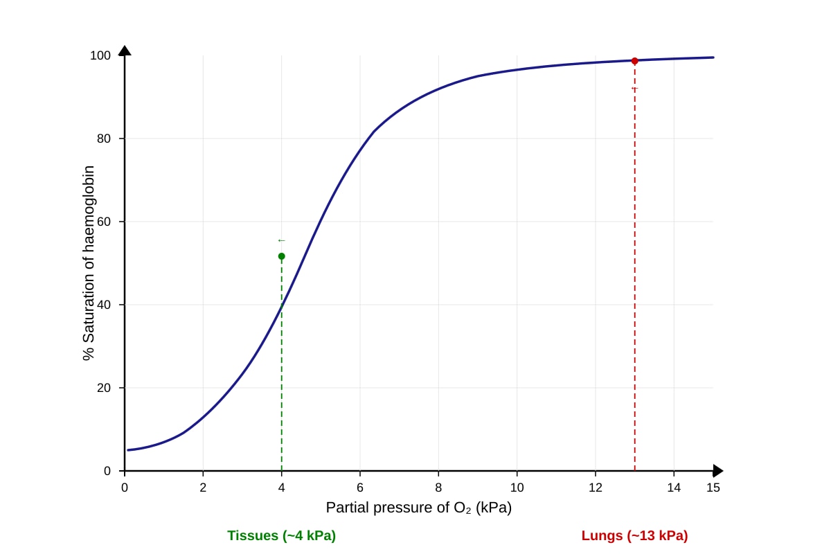
14. (a) An oxygen dissociation curve is a graph showing the relationship between the partial pressure of oxygen (pO₂) and the percentage saturation of haemoglobin with oxygen. [1 mark]
(b) The curve should be sigmoidal (S-shaped), with the y-axis labelled "% Saturation of haemoglobin" (0–100%) and the x-axis labelled "Partial pressure of O₂ (kPa)" (0–15 kPa). The curve should be steepest between approximately 2–6 kPa and plateau near 100% saturation at high pO₂. The approximate pO₂ in the lungs (~13 kPa) should be marked on the plateau, and the pO₂ in the tissues (~4 kPa) should be marked on the steep part of the curve. [2 marks — 1 mark for correct sigmoidal shape, 1 mark for labelled axes and pO₂ points]
(c) An increase in temperature or a decrease in pH (increase in H⁺ concentration) shifts the oxygen dissociation curve to the right (Bohr shift). This means that at any given pO₂, haemoglobin has a lower affinity for oxygen and releases oxygen more readily. During exercise, active muscles produce more CO₂ and lactic acid, lowering pH and increasing temperature. This rightward shift facilitates the unloading of oxygen to the metabolically active tissues, meeting their increased demand for oxygen. [3 marks — 1 mark for rightward shift, 1 mark for decreased affinity, 1 mark for significance during exercise]
15. (a) During inspiration, the diaphragm contracts and flattens, and the external intercostal muscles contract, pulling the rib cage upwards and outwards. These actions increase the volume of the thoracic cavity, which decreases the intrapleural pressure below atmospheric pressure, causing air to flow into the lungs. [2 marks — 1 mark for diaphragm action, 1 mark for intercostal action and pressure change]
(b) Oxygen diffuses from the alveoli (where pO₂ is high, ~13 kPa) into the pulmonary capillaries (where pO₂ is low, ~5 kPa) down its partial pressure gradient. Carbon dioxide diffuses in the opposite direction, from the capillaries (where pCO₂ is high, ~6 kPa) into the alveoli (where pCO₂ is low, ~0.04 kPa) down its partial pressure gradient. This passive diffusion is driven by the concentration differences established by ventilation and blood flow. [3 marks — 1 mark for O₂ gradient, 1 mark for CO₂ gradient, 1 mark for passive diffusion]
Section C: Free-Response Questions (Questions 16–20)
16. The cardiac cycle begins with atrial systole. The atria contract, forcing blood through the open atrioventricular (AV) valves into the relaxed ventricles. Atrial pressure then falls, and the atria relax. Ventricular systole begins: the ventricles contract, raising ventricular pressure above atrial pressure, which forces the AV valves to close (producing the first heart sound). As ventricular pressure continues to rise and exceeds the pressure in the aorta and pulmonary artery, the semilunar valves open, and blood is ejected into the arteries. Ventricular pressure then falls below arterial pressure, causing the semilunar valves to close (producing the second heart sound). Ventricular diastole follows: the ventricles relax, pressure falls below atrial pressure, the AV valves open, and blood flows passively from the atria into the ventricles. The cycle then repeats. [5 marks — 1 mark for atrial systole, 1 mark for AV valve closure, 1 mark for semilunar valve opening and ejection, 1 mark for semilunar valve closure, 1 mark for ventricular diastole and filling. Award marks for clear description of pressure changes controlling valve movements.]
17. (a) Both insulin and glucagon are peptide hormones secreted by the pancreas that regulate blood glucose, but they have opposite effects. Insulin is secreted by beta cells in response to high blood glucose; it promotes glucose uptake by cells, glycogenesis, and lipogenesis, lowering blood glucose. Glucagon is secreted by alpha cells in response to low blood glucose; it promotes glycogenolysis and gluconeogenesis, raising blood glucose. Insulin acts via tyrosine kinase receptors, while glucagon acts via G-protein-coupled receptors and cAMP. [3 marks — 1 mark for contrasting stimuli, 1 mark for contrasting effects, 1 mark for receptor mechanism difference]
(b) In type 2 diabetes, target cells (e.g., muscle, liver, adipose) become resistant to insulin. This means that even though insulin is secreted, the cells do not respond effectively by taking up glucose. The negative feedback loop fails because the rise in blood glucose does not trigger an adequate reduction in blood glucose. Over time, the pancreas may also become unable to produce enough insulin to overcome the resistance, leading to chronically high blood glucose levels. [2 marks — 1 mark for insulin resistance, 1 mark for failure of negative feedback]
18. (a) A neuromuscular junction is a specialised synapse between a motor neurone and a skeletal muscle fibre. The presynaptic terminal contains synaptic vesicles filled with acetylcholine (ACh). The synaptic cleft is a narrow gap. The postsynaptic membrane (motor end plate) contains ACh receptors and is folded to increase surface area. [2 marks — 1 mark for presynaptic structures, 1 mark for postsynaptic structures]
(b) An action potential arrives at the presynaptic terminal, causing Ca²⁺ influx. This triggers exocytosis of ACh into the synaptic cleft. ACh diffuses across the cleft and binds to nicotinic ACh receptors on the motor end plate. These receptors are ligand-gated Na⁺ channels; when ACh binds, they open, allowing Na⁺ influx. This depolarises the muscle fibre membrane, generating an end-plate potential. If the depolarisation reaches threshold, voltage-gated Na⁺ channels open, propagating an action potential along the muscle fibre. The action potential travels along the sarcolemma and down the T-tubules, triggering Ca²⁺ release from the sarcoplasmic reticulum, which initiates the sliding filament mechanism of contraction. [3 marks — 1 mark for ACh release and binding, 1 mark for end-plate potential and action potential generation, 1 mark for excitation-contraction coupling]
19. (a) The hypothalamus acts as the body's thermostat. It contains thermoreceptors that detect the temperature of the blood and receives input from peripheral thermoreceptors. It compares the actual temperature to the set point and coordinates responses to maintain core temperature. [1 mark]
(b) When core temperature rises above the set point, the hypothalamus initiates cooling responses:
- Vasodilation of skin arterioles: Blood vessels near the skin surface dilate, increasing blood flow to the skin. This allows more heat to be lost to the environment by radiation and convection.
- Sweating: Sweat glands secrete sweat onto the skin surface. As sweat evaporates, it absorbs latent heat from the skin, cooling the body.
- Reduction in metabolic heat production: The hypothalamus reduces shivering and other heat-generating activities.
- Behavioural changes: The person may seek shade, remove clothing, or reduce activity. [4 marks — 1 mark for each of two well-described responses with explanation, up to a maximum of 4 marks. Award marks for vasodilation, sweating, reduced heat production, and behavioural changes.]
20. (a) Organisms need different communication systems because different physiological processes require different response characteristics. The nervous system provides rapid, short-lived, and precisely targeted responses, which are essential for immediate reactions (e.g., reflex actions, movement). The endocrine system provides slower, longer-lasting, and more widespread responses, which are necessary for long-term regulation (e.g., growth, metabolism, reproduction). Having both systems allows the body to coordinate a wide range of activities efficiently. [2 marks — 1 mark for contrasting speed/duration, 1 mark for contrasting specificity/scope]
(b) Speed: The nervous system uses electrical impulses and neurotransmitter release, allowing responses in milliseconds (e.g., withdrawal reflex). The endocrine system relies on hormone transport via the bloodstream, which takes seconds to hours (e.g., growth hormone's effects on growth). Duration: Nervous system responses are brief because neurotransmitters are rapidly broken down or reabsorbed (e.g., muscle contraction stops quickly after nerve stimulation ceases). Endocrine responses are prolonged because hormones are cleared slowly and can trigger cascades of cellular effects (e.g., thyroid hormones regulate metabolism for days). Specificity: The nervous system targets specific cells via precise synaptic connections (e.g., a motor neurone innervates a specific muscle fibre). The endocrine system is less specific; hormones travel throughout the blood but only affect cells with the appropriate receptors (e.g., insulin affects many cell types, but only those with insulin receptors). [3 marks — 1 mark for each well-explained comparison (speed, duration, specificity) with appropriate examples. Award marks for clear integration of the two systems.]
End of Answer Key