AI Generated Quiz
A Level Biology H3 Cells Biomolecules Quiz
Free A Level Biology H3 Cells Biomolecules quiz, AI version, with questions, answers, and A Level-style practice for Singapore students.
These static practice materials are generated from the site's syllabus and paper-generation workflow, with source and model context shown so students and parents can evaluate the material before use.
Questions
Free quiz and exam paper access
Enter your details to view this paper
Your access is remembered on this device.
Answers
A-Level Biology H3 Quiz - Cells Biomolecules: Answer Key
Total Marks: 50
Section A: Multiple-Choice Questions (10 marks)
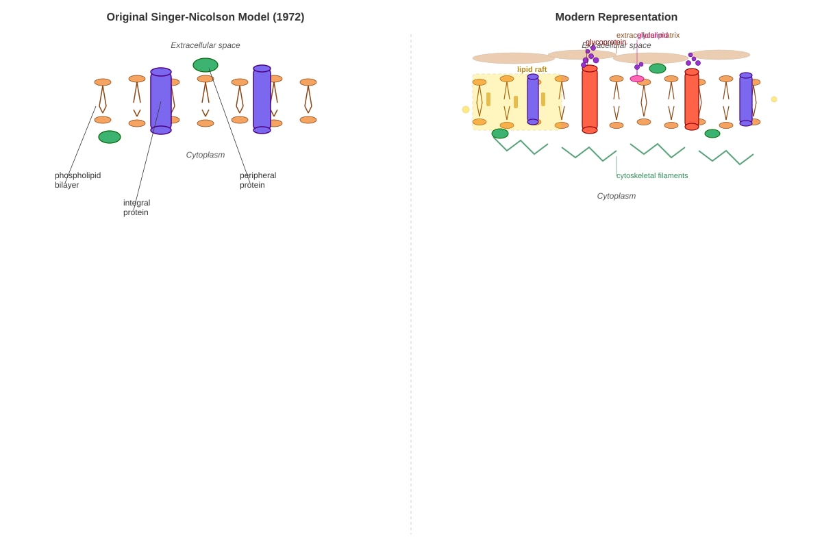
1. C. It did not explain the existence of membrane microdomains or lipid rafts.
- Explanation: The original Singer-Nicolson model proposed a homogeneous, fluid bilayer where lipids and proteins could diffuse freely. It did not account for the existence of more ordered, cholesterol- and sphingolipid-rich microdomains called lipid rafts, which are now known to play crucial roles in signaling and membrane organization. Option A is incorrect because the model did include integral proteins. Option B is incorrect because the model proposed that lipids were mobile. Option D is incorrect because the model did allow for protein mobility.
- Marks: 1 mark for correct option.
2. B. Are composed solely of protein and lack any nucleic acid.
- Explanation: The cell theory states that all living things are composed of cells and that genetic information is carried by nucleic acids (DNA/RNA). Prions are infectious proteins that replicate by converting normal cellular proteins into the misfolded prion form. They contain no nucleic acid, challenging the idea that all life must have a genome. Option A is false. Option C is false; they do not have ribosomes. Option D is false; they are not cellular.
- Marks: 1 mark for correct option.
3. B. These organelles have double membranes and their own ribosomes that resemble those of prokaryotes.
- Explanation: The endosymbiotic theory proposes that mitochondria and chloroplasts were once free-living prokaryotes that were engulfed by a host cell. Evidence includes: they have their own circular DNA (like prokaryotes), their own 70S ribosomes (like prokaryotes), and a double membrane (the inner from the prokaryote, the outer from the host's vesicle). Option A is incorrect; they have circular DNA. Option C is incorrect; they reproduce by binary fission. Option D is incorrect; they can synthesize some of their own proteins.
- Marks: 1 mark for correct option.
4. C. Phosphorylation
- Explanation: The removal of a phosphate group is called dephosphorylation. The question states the protein was treated with an enzyme that removes phosphate groups, which is a phosphatase. The modification that was originally present and then removed was phosphorylation (the addition of a phosphate group). The loss of activity upon dephosphorylation shows that the phosphorylated form was the active state. Cleavage is cutting the protein. Glycosylation is adding sugars. Denaturation is loss of 3D structure.
- Marks: 1 mark for correct option.
5. A. The binding of oxygen to one subunit causing a conformational change that increases the affinity of other subunits for oxygen.
- Explanation: Cooperativity is a hallmark of allosteric proteins like haemoglobin. When oxygen binds to the iron in the haem group of one subunit, it pulls the iron into the plane of the porphyrin ring, causing a conformational change in that subunit. This change is transmitted to adjacent subunits, altering their shape and increasing their affinity for oxygen. This makes the oxygen-binding curve sigmoidal. Option B describes non-cooperative binding. Option C is a necessary step but doesn't explain cooperativity. Option D is incorrect; BPG decreases oxygen affinity.
- Marks: 1 mark for correct option.
6. A. The presence of septate hyphae with distinct cellular compartments.
- Explanation: The classical cell theory states that organisms are made of cells, each with a single nucleus. Coenocytic hyphae (option B) challenge this because they are multinucleate and lack septa (cross-walls), meaning the "cell" is a single continuous cytoplasmic mass with many nuclei. Option A describes septate hyphae, which do have cross-walls and are more consistent with the cell theory, as each compartment can be considered a cell. Options C and D are general fungal characteristics that don't directly challenge the cell theory in the same way.
- Marks: 1 mark for correct option.
7. B. The variable regions of the light and heavy chains.
- Explanation: An antibody (immunoglobulin) has a Y-shaped structure. The tips of the Y are formed by the variable (V) regions of both the light and heavy chains. These V regions have hypervariable loops that form the antigen-binding site. The specific amino acid sequence in these loops determines the shape and chemical properties of the binding site, giving the antibody its specificity for a particular antigen. The constant region (A) determines the antibody's class and effector function. Disulfide bonds (C) hold the chains together. Carbohydrate groups (D) are involved in stability and effector functions, not antigen binding.
- Marks: 1 mark for correct option.
8. D. Random diffusion of enzymes throughout the cytoplasm without spatial organization.
- Explanation: Eukaryotic cells use several strategies for efficient enzyme regulation: compartmentalization (A) concentrates enzymes and substrates in organelles; allosteric regulation (B) allows for rapid feedback control; and covalent modification (C) like phosphorylation provides a fast, reversible on/off switch. Option D is the least likely to contribute to efficient regulation. While diffusion does occur, relying on random diffusion without any spatial organization (e.g., scaffolding proteins, metabolic channeling) would be slow, inefficient, and prone to unwanted cross-talk between pathways.
- Marks: 1 mark for correct option.
9. C. It can affect protein folding, stability, and cell-cell recognition.
- Explanation: Glycosylation is the covalent attachment of carbohydrate (sugar) chains to proteins. It occurs in the endoplasmic reticulum and Golgi apparatus (not the cytoplasm, so A is wrong). It does not involve lipids (B is wrong). It is generally not a reversible on/off switch like phosphorylation (D is wrong). The attached glycans can act as "tags" for protein folding quality control, protect proteins from proteolysis (stability), and serve as recognition sites for other cells or molecules (e.g., blood group antigens).
- Marks: 1 mark for correct option.
10. B. The presence of clefts, pockets, and grooves with specific chemical properties (e.g., hydrophobic, charged).
- Explanation: Protein surfaces are not uniform. They have complex topography with invaginations (clefts, pockets, grooves) and protrusions. The amino acid residues lining these features create a unique chemical environment (hydrophobic, polar, charged, etc.) that allows the protein to bind specifically to a complementary molecule. A single rigid site (C) would only bind one thing. A uniform structure (A) would lack specificity. The absence of charges (D) would prevent binding to many polar or charged molecules.
- Marks: 1 mark for correct option.
Section B: Structured Questions (20 marks)
11. (a) Describe how the fluid mosaic model has evolved from the original proposal by Singer and Nicolson to our current understanding. (3 marks)
- Answer:
- The original Singer-Nicolson model (1972) proposed a simple, homogeneous phospholipid bilayer with proteins embedded or attached, free to diffuse laterally. It was a good first approximation but lacked detail. (1 mark)
- Our current understanding is much more complex. We now know the membrane is not homogeneous. It contains lipid rafts—cholesterol- and sphingolipid-enriched microdomains that are more ordered and function as platforms for signaling and protein sorting. (1 mark)
- The model now includes a dynamic glycocalyx (carbohydrate chains of glycoproteins and glycolipids) on the extracellular surface, involved in cell recognition and adhesion. It also incorporates connections to the cytoskeleton (via proteins like spectrin), which can restrict the lateral mobility of some membrane proteins, contradicting the original idea of completely free diffusion. (1 mark)
- Marks: 1 mark for describing the original model; 1 mark for lipid rafts; 1 mark for glycocalyx and/or cytoskeletal connections. Accept other valid modern additions like protein crowding, transient interactions, etc.
12. (b) Explain how the concept of lipid rafts challenges the original idea of a homogeneous fluid mosaic. (2 marks)
- Answer:
- The original fluid mosaic model proposed that the lipid bilayer was a homogeneous, uniform fluid where all lipids and proteins could diffuse freely and randomly. (1 mark)
- Lipid rafts challenge this by demonstrating that the membrane is heterogeneous. Rafts are localized, ordered microdomains enriched in cholesterol and sphingolipids that are less fluid than the surrounding bilayer. They concentrate specific proteins and can act as signaling platforms, showing that the membrane is not a uniform "sea" of lipids but a structured, compartmentalized environment. (1 mark)
- Marks: 1 mark for stating the original idea of homogeneity; 1 mark for explaining how rafts introduce heterogeneity and structure.
13. The cell theory states that all living organisms are composed of cells, and that all cells arise from pre-existing cells. Discuss how the following biological entities challenge this theory, and to what extent they conform to it: (i) Prions, (ii) Coenocytic hyphae of filamentous fungi, (iii) Mitochondria (in the context of endosymbiosis). (6 marks)
- Answer:
- (i) Prions: Prions are infectious proteinaceous particles that lack any nucleic acid (DNA or RNA). They challenge the cell theory because they are not cells and do not contain genetic material, yet they can replicate (by converting normal proteins) and cause disease. They conform to the cell theory only in that they require a host cell to replicate; they cannot do so independently. (2 marks)
- (ii) Coenocytic hyphae: These are multinucleate, continuous cytoplasmic masses without septa (cross-walls). They challenge the cell theory because the classical definition of a cell includes a single nucleus bounded by a membrane. In coenocytic hyphae, there is no clear division into individual cells; the entire hypha is a single "supercell" with many nuclei. They conform to the cell theory in that they are still composed of cytoplasm and are bounded by a plasma membrane, and they arise from pre-existing hyphae. (2 marks)
- (iii) Mitochondria: According to the endosymbiotic theory, mitochondria were once free-living prokaryotes that were engulfed by a host cell. They challenge the cell theory because they have their own circular DNA and 70S ribosomes (like prokaryotes) and replicate by binary fission, independently of the host cell's division. This suggests they were once independent organisms. They conform to the cell theory in that they are now obligate endosymbionts; they cannot live independently outside the host cell and are passed on from parent to daughter cells during cell division. (2 marks)
- Marks: 2 marks for each entity (1 for challenge, 1 for conformity). Accept well-reasoned alternative points.
14. (a) Define the term "protein subunit" and explain how the quaternary structure of haemoglobin () is an example of a protein composed of subunits. (2 marks)
- Answer:
- A protein subunit is a single polypeptide chain that assembles with other polypeptide chains (which may be identical or different) to form a functional protein complex. Subunits are held together by non-covalent interactions (e.g., hydrogen bonds, hydrophobic interactions, ionic bonds). (1 mark)
- Haemoglobin has a quaternary structure of , meaning it is composed of four subunits: two -globin chains and two -globin chains. Each chain is a separate polypeptide that folds independently, and they associate non-covalently to form the functional tetrameric haemoglobin molecule. (1 mark)
- Marks: 1 mark for definition; 1 mark for correct application to haemoglobin.
15. (b) The binding of oxygen to haemoglobin is cooperative. Describe the molecular mechanism that underlies this cooperativity, including the role of conformational changes. (3 marks)
- Answer:
- Haemoglobin exists in two main conformations: a T (tense) state with low oxygen affinity and an R (relaxed) state with high oxygen affinity. In the deoxygenated state, haemoglobin is predominantly in the T state. (1 mark)
- When the first oxygen molecule binds to the iron atom in the haem group of one subunit, it pulls the iron into the plane of the porphyrin ring. This causes a small conformational change in that subunit, which is transmitted to the interface. (1 mark)
- This change stabilizes the R state, making it easier for subsequent oxygen molecules to bind to the remaining subunits. The binding of oxygen thus increases the affinity of the other subunits for oxygen, resulting in a sigmoidal oxygen-binding curve. This is positive cooperativity. (1 mark)
- Marks: 1 mark for T/R states; 1 mark for conformational change upon oxygen binding; 1 mark for increased affinity of subsequent subunits.
16. (a) Name two types of post-translational protein modification and briefly describe how each can alter a protein's function. (2 marks)
- Answer:
- Phosphorylation: The addition of a phosphate group (by a kinase) to specific amino acids (e.g., serine, threonine, tyrosine). This can activate or inactivate an enzyme by inducing a conformational change, or create binding sites for other proteins. (1 mark)
- Glycosylation: The addition of carbohydrate chains (glycans) to proteins (e.g., asparagine for N-linked glycosylation). This can affect protein folding, stability, targeting to membranes, and cell-cell recognition (e.g., blood group antigens). (1 mark)
- Marks: 1 mark for each modification with a correct functional consequence. Accept other valid modifications (e.g., acetylation, ubiquitination, methylation, disulfide bond formation).
17. (b) A specific protein kinase adds a phosphate group to a target enzyme, activating it. A protein phosphatase then removes this phosphate group, inactivating the enzyme. Explain why this reversible phosphorylation is a more efficient regulatory mechanism for a cell than synthesizing and degrading the enzyme each time its activity needs to change. (2 marks)
- Answer:
- Reversible phosphorylation is much faster than synthesizing and degrading an enzyme. Synthesis requires transcription, translation, and folding, which can take minutes to hours. Phosphorylation and dephosphorylation occur in seconds to minutes, allowing rapid responses to changing conditions. (1 mark)
- It is also more energy-efficient. Synthesizing a new protein requires a large amount of ATP (for transcription, translation, and folding). Degrading a protein also requires energy (e.g., ubiquitin-proteasome pathway). Reversible phosphorylation only requires the energy of one ATP to add the phosphate, and no energy to remove it, making it a "cheap" on/off switch. (1 mark)
- Marks: 1 mark for speed; 1 mark for energy efficiency.
Section C: Free-Response Questions (20 marks)
18. A eukaryotic cell must coordinate the activities of thousands of enzymes within its tiny volume. Describe the various mechanisms, including compartmentalization, allosteric regulation, and covalent modification, that allow a cell to achieve this precise and efficient regulation. In your answer, explain how these mechanisms work together to prevent metabolic chaos and allow the cell to respond rapidly to changing conditions. (10 marks)
- Answer:
- Introduction: Eukaryotic cells face the challenge of regulating thousands of simultaneous enzymatic reactions in a crowded cytoplasm. They use multiple, integrated mechanisms to ensure order, efficiency, and rapid responsiveness.
- Compartmentalization: Enzymes and their substrates are segregated into membrane-bound organelles (e.g., mitochondria for respiration, lysosomes for digestion, peroxisomes for oxidation). This prevents conflicting reactions (e.g., protein synthesis in the cytosol vs. degradation in lysosomes), concentrates substrates and enzymes for efficiency, and creates specific microenvironments (e.g., acidic pH in lysosomes). (2 marks)
- Allosteric Regulation: Many enzymes are allosterically regulated by metabolites (e.g., feedback inhibition). The end product of a pathway binds to a regulatory site on an early enzyme, causing a conformational change that reduces its activity. This provides rapid, reversible, and precise control of flux through metabolic pathways, preventing overproduction. (2 marks)
- Covalent Modification: Reversible modifications like phosphorylation (by kinases/phosphatases) provide a fast, reversible on/off switch. This allows enzymes to be activated or inactivated in seconds, without needing to be synthesized or degraded. It is ideal for signal transduction cascades (e.g., MAP kinase pathway) where a signal is amplified. (2 marks)
- Integration of Mechanisms: These mechanisms work together. For example, a hormone signal can trigger a phosphorylation cascade (covalent modification) that activates an allosteric enzyme, which then controls a metabolic pathway in a specific compartment. This allows the cell to integrate external signals with internal metabolic state. (2 marks)
- Preventing Metabolic Chaos: Without these mechanisms, enzymes would act indiscriminately, leading to wasteful cycles and toxic intermediates. Compartmentalization physically separates conflicting pathways. Allosteric regulation ensures that pathways are only active when needed. Covalent modification allows rapid shutdown of pathways when conditions change. Together, they create a highly ordered, responsive, and efficient metabolic network. (2 marks)
- Marks: Award marks based on: clear description of each mechanism (2 marks each for compartmentalization, allosteric regulation, covalent modification); explanation of integration (2 marks); explanation of preventing chaos (2 marks). Quality of scientific argumentation and written communication is assessed holistically.
19. The ability of proteins to bind to a vast array of molecules—from small ions to large macromolecules—is fundamental to life. With reference to specific examples (e.g., haemoglobin, immunoglobulins, and enzymes), discuss how the structural features of proteins, including their surfaces, clefts, and binding sites, enable this remarkable binding diversity and specificity. (10 marks)
- Answer:
- Introduction: Protein binding is highly specific and diverse due to the unique three-dimensional structure of each protein, which creates complementary binding sites for target molecules.
- Haemoglobin (Oxygen Binding): Haemoglobin binds oxygen cooperatively. The binding site is a cleft (the haem pocket) that holds the haem group with an iron atom. The iron coordinates oxygen. The quaternary structure allows conformational changes (T to R state) that alter the affinity of other subunits, enabling cooperative binding. The surface of the protein has charged residues that interact with allosteric regulators like BPG. (3 marks)
- Immunoglobulins (Antigen Binding): Antibodies have a Y-shaped structure. The antigen-binding site is formed by the variable regions of the light and heavy chains. These regions have hypervariable loops that create a pocket or groove complementary in shape and chemistry to the antigen (epitope). The diversity of binding is generated by somatic recombination, creating millions of different variable region sequences. (3 marks)
- Enzymes (Substrate Binding): Enzymes have active sites that are often deep clefts or pockets that exclude water and provide a specific chemical environment. The amino acid residues lining the active site form specific interactions (hydrogen bonds, ionic bonds, hydrophobic interactions) with the substrate. This induced fit model shows that the enzyme can change shape upon substrate binding to optimize interactions, ensuring high specificity. (2 marks)
- Conclusion: The binding diversity and specificity of proteins arise from their complex surfaces, which feature clefts, pockets, grooves, and protrusions lined with specific amino acids. This allows proteins to recognize and bind a vast array of molecules with high precision, from small ions to large macromolecules, underpinning all cellular processes. (2 marks)
- Marks: Award marks based on: clear explanation for each example (3 marks for haemoglobin, 3 marks for immunoglobulins, 2 marks for enzymes); synthesis and conclusion (2 marks). Quality of scientific argumentation and written communication is assessed holistically.
20. (a) Describe the role of the glycocalyx in cell recognition and adhesion. (2 marks)
- Answer:
- The glycocalyx is a carbohydrate-rich layer on the extracellular surface of the plasma membrane, composed of glycoproteins and glycolipids. (1 mark)
- It plays a key role in cell-cell recognition (e.g., blood group antigens, immune cell recognition) and cell adhesion (e.g., mediating interactions between cells or between cells and the extracellular matrix via lectins or adhesion molecules). (1 mark)
- Marks: 1 mark for definition; 1 mark for roles.
20. (b) Explain how the cytoskeleton contributes to the dynamic nature of the cell membrane. (2 marks)
- Answer:
- The cytoskeleton (e.g., actin filaments, spectrin) is attached to the cytoplasmic side of the plasma membrane via linker proteins. (1 mark)
- It provides mechanical support and shape, but also restricts the lateral mobility of some membrane proteins (e.g., by anchoring them to specific domains). It can also actively move membrane proteins (e.g., via motor proteins) and drive membrane deformation (e.g., during endocytosis or cell movement). (1 mark)
- Marks: 1 mark for attachment; 1 mark for dynamic roles (restriction, movement, deformation).