AI Generated Quiz
A Level Biology H3 Cells Biomolecules Quiz
Free A Level Biology H3 Cells Biomolecules quiz, AI version, with questions, answers, and A Level-style practice for Singapore students.
These static practice materials are generated from the site's syllabus and paper-generation workflow, with source and model context shown so students and parents can evaluate the material before use.
Questions
Free quiz and exam paper access
Enter your details to view this paper
Your access is remembered on this device.
Answers
A-Level Biology H3 Quiz - Cells Biomolecules — Answer Key
Total Marks: 60
Section A: Multiple-Choice Questions (Questions 1–5)
1. B [1 mark]
- Explanation: The fluid mosaic model has been refined over time. Early models were static, but later evidence from techniques like FRAP (fluorescence recovery after photobleaching) showed that membrane components are mobile. The discovery of membrane rafts—cholesterol- and sphingolipid-enriched microdomains—and the role of the cytoskeleton in anchoring proteins have added complexity to the original model.
- Common mistake: Students may think the model is fixed (A) or only applies to eukaryotes (D). The model applies to all cell membranes, and it is dynamic, not static.
2. B [1 mark]
- Explanation: A prion is an infectious protein particle that lacks nucleic acid. It is a misfolded form of a normal cellular protein (PrP^C) that can induce other normal proteins to misfold, leading to disease (e.g., Creutzfeldt-Jakob disease, BSE).
- Common mistake: Students may confuse prions with viruses (which have nucleic acid) or viroids (which are RNA). Prions are unique in being protein-only infectious agents.
3. B [1 mark]
- Explanation: The cell theory states that all living things are composed of one or more cells, and that the cell is the basic unit of life. Filamentous fungi have hyphae that are multinucleate (coenocytic), meaning there are no septa (cross-walls) separating individual cells. This challenges the idea that the cell is the fundamental unit, as the hypha is a continuous tube with many nuclei but no distinct cellular compartments.
- Common mistake: Students may choose C (chitin cell walls) or D (heterotrophic), but these do not directly challenge the cell theory. The multinucleate condition is the key challenge.
4. B [1 mark]
- Explanation: The endosymbiotic theory, proposed by Lynn Margulis, states that mitochondria and chloroplasts originated from free-living prokaryotes that were engulfed by a host eukaryotic cell. Over time, they became permanent organelles.
- Common mistake: Students may think they originated from viruses (A) or host DNA fragments (C). The key evidence includes their own circular DNA, double membranes, and ribosomes similar to prokaryotes.
5. C [1 mark]
- Explanation: Phosphorylation is the addition of a phosphate group to a protein, typically by a kinase enzyme. This often alters the protein's conformation and activity, acting as a molecular switch (e.g., in signalling pathways).
- Common mistake: Students may confuse phosphorylation with cleavage (A) or glycosylation (B). Phosphorylation adds a phosphate group, not a carbohydrate.
Section B: Structured Questions (Questions 6–15)
6. Explain how the fluid mosaic model has developed over time to incorporate the concept of membrane rafts. [3]
Answer:
- The original Singer-Nicolson model (1972) proposed a fluid lipid bilayer with randomly distributed proteins. [1]
- Later research showed that the membrane is not homogeneous. Membrane rafts are cholesterol- and sphingolipid-enriched microdomains that are more ordered and less fluid than the surrounding bilayer. [1]
- These rafts selectively concentrate certain proteins (e.g., signalling proteins) and are involved in processes like signal transduction and membrane trafficking, refining the model to include lateral heterogeneity. [1]
Marking notes: Award 1 mark for each key point. Accept any reasonable description of how rafts add complexity to the original model.
7. Describe the morphology and replication cycle of a prion. How does its acellular nature challenge the traditional definition of a living organism? [4]
Answer:
- Morphology: Prions are misfolded isoforms (PrP^Sc) of a normal cellular protein (PrP^C). They are proteinaceous particles with no nucleic acid. [1]
- Replication cycle: PrP^Sc acts as a template, inducing normal PrP^C to misfold into the PrP^Sc conformation. This creates a chain reaction, leading to the accumulation of misfolded protein aggregates. [1]
- Challenge to definition of life: Prions are acellular—they have no cells, no metabolism, no ability to reproduce independently. They rely entirely on host cells to provide the normal PrP^C substrate. [1]
- This challenges the cell theory, which states that all living things are made of cells and that cells arise from pre-existing cells. Prions are not cells and do not reproduce in a cellular manner, yet they can propagate and cause disease. [1]
Marking notes: Award 1 mark for morphology, 1 mark for replication, 1 mark for acellularity, and 1 mark for the challenge to cell theory/life definition.
8. Compare and contrast the morphology of yeasts and filamentous fungi. In your answer, explain how the multinucleate condition of filamentous fungal hyphae relates to the cell theory. [4]
Answer:
- Yeasts: Unicellular, oval or spherical cells. Reproduce by budding or fission. [1]
- Filamentous fungi: Multicellular, composed of long, branching filaments called hyphae. Hyphae may be septate (with cross-walls) or coenocytic (aseptate, multinucleate). [1]
- Comparison: Both are fungi, have cell walls of chitin, and are heterotrophic. Yeasts are unicellular, while filamentous fungi are multicellular. [1]
- Multinucleate condition and cell theory: In coenocytic hyphae, there are no septa, so the hypha is a continuous tube with many nuclei sharing a common cytoplasm. This challenges the cell theory, which states that the cell is the basic structural and functional unit of life. Here, the hypha functions as a single unit but is not divided into discrete cells. [1]
Marking notes: Award 1 mark for yeast morphology, 1 mark for filamentous fungi morphology, 1 mark for comparison, and 1 mark for the relationship to cell theory.
9. The endosymbiotic theory is supported by several lines of evidence. State three pieces of evidence that support this theory. [3]
Answer: Any three of the following:
- Mitochondria and chloroplasts have their own circular DNA, similar to bacterial DNA. [1]
- They have double membranes: the inner membrane is derived from the endosymbiont, and the outer membrane from the host cell's vesicle. [1]
- They have their own ribosomes (70S), which are similar to prokaryotic ribosomes, not eukaryotic (80S) ribosomes. [1]
- They replicate independently of the host cell via binary fission, similar to bacteria. [1]
- Phylogenetic analysis shows that mitochondrial and chloroplast genes are closely related to certain bacterial groups (e.g., α-proteobacteria for mitochondria, cyanobacteria for chloroplasts). [1]
Marking notes: Award 1 mark for each correct piece of evidence, up to a maximum of 3.
10. Justify why cell differentiation is necessary in multicellular organisms. Use a specific example to illustrate your answer. [3]
Answer:
- Cell differentiation is the process by which cells become specialised to perform specific functions. [1]
- It is necessary because multicellular organisms have complex structures and functions that cannot be performed by a single cell type. Specialisation allows for division of labour, increasing efficiency. [1]
- Example: In humans, red blood cells (erythrocytes) differentiate to lose their nucleus and organelles, becoming biconcave discs packed with haemoglobin. This maximises space for oxygen transport. Without differentiation, all cells would be identical and unable to perform this specialised function efficiently. [1]
Marking notes: Award 1 mark for definition, 1 mark for justification, and 1 mark for a specific, well-explained example. Accept other examples (e.g., neurones, muscle cells, xylem vessels).
11. Haemoglobin is a protein composed of multiple subunits. Describe how protein binding sites and subunit interactions allow haemoglobin to carry out its function of oxygen transport. [4]
Answer:
- Haemoglobin is a tetramer composed of two α-globin and two β-globin subunits. Each subunit contains a haem group with an iron ion (Fe²⁺) that can bind one oxygen molecule. [1]
- The binding of oxygen to one subunit induces a conformational change that increases the affinity of the remaining subunits for oxygen (cooperative binding). This is due to interactions between subunits. [1]
- The binding sites (haem pockets) are positioned such that oxygen binding causes the iron ion to move into the plane of the porphyrin ring, pulling the attached histidine residue and triggering a shift in subunit orientation. [1]
- This cooperative binding allows haemoglobin to efficiently load oxygen in the lungs (high pO₂) and unload it in the tissues (low pO₂), resulting in a sigmoidal oxygen dissociation curve. [1]
Marking notes: Award 1 mark for subunit composition, 1 mark for cooperative binding, 1 mark for conformational change mechanism, and 1 mark for physiological significance.
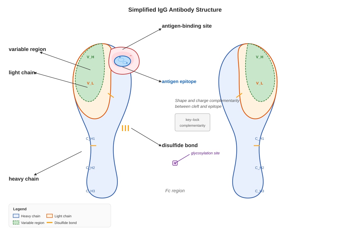
12. Immunoglobulin G (IgG) is a glycoprotein. Explain how protein modification, specifically glycosylation, contributes to the function of IgG. [3]
Answer:
- Glycosylation is the addition of carbohydrate (sugar) chains to the protein. In IgG, glycosylation occurs at the Fc (constant) region. [1]
- The carbohydrate moieties help stabilise the IgG structure and maintain the correct conformation of the Fc region. [1]
- Glycosylation is essential for IgG to bind to Fc receptors on immune cells (e.g., macrophages, NK cells) and to activate the complement system. Without proper glycosylation, IgG cannot effectively trigger effector functions. [1]
Marking notes: Award 1 mark for identifying glycosylation site, 1 mark for structural role, and 1 mark for functional role in immunity.
13. Using the diagram and your knowledge of protein structure, explain how the properties and shapes of protein surfaces and clefts allow antibodies to recognise and bind to a highly diverse range of molecules. [4]
Answer:
- Antibodies (IgG) have variable regions that form a binding site (cleft) complementary in shape and charge to a specific antigen epitope. [1]
- The hypervariable loops (CDRs) within the variable regions create a diverse array of surface shapes and chemical properties (hydrophobic, hydrophilic, charged). [1]
- The binding site is a cleft or groove that can accommodate antigens of various sizes and shapes. The specificity comes from the precise three-dimensional fit and non-covalent interactions (hydrogen bonds, ionic bonds, van der Waals forces, hydrophobic interactions). [1]
- The enormous diversity of antibody binding sites (generated by V(D)J recombination and somatic hypermutation) allows the immune system to recognise virtually any antigen. [1]
Marking notes: Award 1 mark for complementarity, 1 mark for CDRs/diversity generation, 1 mark for binding interactions, and 1 mark for overall diversity. The diagram should show the antigen-binding cleft and the complementary antigen.
14. Prokaryotic RNA polymerase is a large multi-subunit enzyme. Describe the roles of its protein subunits in recognising and binding to promoter regions of DNA. [3]
Answer:
- The core enzyme (α₂ββ'ω) has catalytic activity but cannot specifically bind to promoters. [1]
- The sigma (σ) subunit is required for promoter recognition. It binds to the core enzyme to form the holoenzyme. [1]
- The σ subunit recognises specific sequences in the promoter region (e.g., the -10 and -35 boxes in E. coli), positioning the core enzyme to initiate transcription. [1]
Marking notes: Award 1 mark for core enzyme function, 1 mark for sigma subunit role, and 1 mark for promoter recognition details.
15. A living eukaryotic cell must regulate thousands of enzymes efficiently. Describe two mechanisms by which a cell achieves this regulation, and explain why such regulation is essential for cellular homeostasis. [4]
Answer: Mechanism 1: Compartmentalisation. Enzymes are localised in specific organelles (e.g., glycolytic enzymes in the cytoplasm, Krebs cycle enzymes in the mitochondria). This prevents conflicting reactions from occurring in the same space and allows for optimal conditions (pH, substrate concentration) for each set of reactions. [1] Mechanism 2: Allosteric regulation. Many enzymes are regulated by allosteric effectors that bind at sites distinct from the active site, causing conformational changes that increase or decrease activity. This allows for rapid, reversible control in response to cellular needs (e.g., feedback inhibition). [1] Why essential: Without such regulation, metabolic pathways would run uncontrollably, leading to waste of energy and resources, accumulation of toxic intermediates, and failure to respond to changing conditions. Homeostasis requires precise control of enzyme activity to maintain stable internal conditions. [2]
Marking notes: Award 1 mark for each mechanism (2 total) and 2 marks for explanation of importance. Accept other valid mechanisms (e.g., covalent modification, proteolytic activation, gene expression control, enzyme sequestration).
Section C: Free-Response Questions (Questions 16–20)
16. Discuss how the concepts of acellularity (prions and viruses), multinucleation (filamentous fungi), and endosymbiosis each challenge the classical cell theory. To what extent does each of these phenomena conform to or violate the theory? [6]
Answer: Acellularity (prions and viruses):
- Challenge: Prions and viruses are acellular—they have no cells, no metabolism, and cannot reproduce independently. This violates the cell theory's claim that all living things are made of cells. [1]
- Conformity: Prions and viruses are not considered truly alive by many definitions. They are obligate parasites that require host cells to replicate. Thus, they do not disprove the cell theory for living organisms; they simply exist at the boundary of life. [1]
Multinucleation (filamentous fungi):
- Challenge: Coenocytic hyphae are multinucleate and lack septa, meaning there are no discrete cells. This challenges the idea that the cell is the basic structural unit. [1]
- Conformity: The hypha is still a membrane-bound compartment with cytoplasm and organelles. Some argue that the entire hypha can be considered a single, large, multinucleate cell. Thus, it partially conforms to the cell theory. [1]
Endosymbiosis:
- Challenge: Mitochondria and chloroplasts were once free-living prokaryotes. This suggests that cells can arise from the incorporation of other cells, not just by division of pre-existing cells. [1]
- Conformity: Endosymbiosis is a special case of cell evolution. Once incorporated, organelles replicate within the host cell and are passed on during cell division. The theory does not invalidate the cell theory; it explains the origin of eukaryotic cells. [1]
Overall assessment: Each phenomenon challenges a strict interpretation of the cell theory but does not entirely disprove it. The theory remains a foundational principle of biology, with these exceptions highlighting the diversity of life.
Marking notes: Award up to 2 marks for each phenomenon (1 for challenge, 1 for conformity/assessment). Accept well-reasoned alternative interpretations.
17. Protein modification (cleavage, phosphorylation, and glycosylation) confers new capabilities to proteins. Using specific examples, explain how each type of modification alters protein function and why these modifications are crucial for cellular regulation. [6]
Answer: Cleavage:
- Example: Proinsulin is cleaved to remove the C-peptide, producing active insulin. [1]
- Effect: Cleavage activates the protein by removing an inhibitory domain. This is irreversible and is often used for zymogen activation (e.g., digestive enzymes) or hormone maturation. [1]
Phosphorylation:
- Example: Glycogen phosphorylase is activated by phosphorylation in response to glucagon, promoting glycogen breakdown. [1]
- Effect: Addition of a phosphate group induces a conformational change that alters enzyme activity. This is reversible (kinases/phosphatases) and allows rapid, dynamic regulation of signalling pathways and metabolism. [1]
Glycosylation:
- Example: IgG is glycosylated at its Fc region. [1]
- Effect: The carbohydrate chains stabilise the protein structure and are required for binding to Fc receptors on immune cells. Glycosylation also affects protein folding, trafficking, and cell-cell recognition. [1]
Crucial for regulation: These modifications allow cells to rapidly alter protein function without synthesising new proteins. They provide a layer of post-translational control that is essential for processes like signal transduction, cell cycle control, and immune responses.
Marking notes: Award 1 mark for each example and 1 mark for each explanation of effect (total 6 marks). Accept other valid examples (e.g., caspase cleavage in apoptosis, phosphorylation of transcription factors, glycosylation of mucins).
18. Describe how the structure of a eukaryotic cell allows it to regulate the activity of thousands of enzymes simultaneously. In your answer, consider compartmentalisation, enzyme localisation, and the role of protein complexes. [6]
Answer: Compartmentalisation:
- Eukaryotic cells have membrane-bound organelles (nucleus, mitochondria, ER, Golgi, lysosomes, peroxisomes) that create distinct chemical environments. [1]
- Each organelle contains a specific set of enzymes and substrates, allowing simultaneous, non-interfering reactions. For example, fatty acid synthesis occurs in the cytoplasm, while β-oxidation occurs in the mitochondria. [1]
Enzyme localisation:
- Enzymes are often targeted to specific organelles or membranes via signal sequences. This ensures that enzymes are in the right place at the right time. [1]
- Localisation also concentrates enzymes and substrates, increasing reaction efficiency. For example, enzymes of the electron transport chain are embedded in the inner mitochondrial membrane. [1]
Role of protein complexes:
- Many enzymes function as part of multi-enzyme complexes (e.g., pyruvate dehydrogenase complex, fatty acid synthase). [1]
- These complexes channel intermediates from one active site to the next, preventing diffusion and increasing efficiency. They also allow for coordinated regulation—one regulatory signal can affect the entire complex. [1]
Overall: The structural organisation of the eukaryotic cell allows for spatial and temporal regulation of thousands of enzymes, enabling complex metabolic networks to function simultaneously without conflict.
Marking notes: Award 2 marks for compartmentalisation, 2 marks for enzyme localisation, and 2 marks for protein complexes. Accept well-developed answers that integrate these concepts.
19. With reference to haemoglobin and immunoglobulin G, explain how the assembly of protein subunits and post-translational modifications contribute to the functional diversity of proteins. [6]
Answer: Haemoglobin:
- Subunit assembly: Haemoglobin is a tetramer of two α and two β subunits. The quaternary structure allows cooperative binding of oxygen, which is essential for efficient oxygen transport. [1]
- Post-translational modification: Haemoglobin undergoes no major PTMs, but the haem group (a prosthetic group) is essential for oxygen binding. The assembly of globin chains with haem creates the functional protein. [1]
- Functional diversity: Different globin chains (α, β, γ, δ, ε, ζ) combine to form different haemoglobin variants (HbA, HbF, HbA₂) with different oxygen affinities, adapted to different developmental stages. [1]
Immunoglobulin G:
- Subunit assembly: IgG is composed of two heavy chains and two light chains, held together by disulfide bonds. The variable regions of these chains form the antigen-binding site. [1]
- Post-translational modification: Glycosylation of the Fc region is essential for effector functions (complement activation, Fc receptor binding). Disulfide bonds stabilise the structure. [1]
- Functional diversity: V(D)J recombination and somatic hypermutation generate millions of different antibody specificities from a limited number of gene segments. This allows the immune system to recognise a vast array of antigens. [1]
Conclusion: Both examples show that subunit assembly and PTMs are critical for creating functional diversity from a limited genome. Haemoglobin uses different subunit combinations for different oxygen needs, while IgG uses genetic recombination and PTMs for immune diversity.
Marking notes: Award up to 3 marks for each protein (haemoglobin and IgG). For each, award 1 mark for subunit assembly, 1 mark for PTM/prosthetic group, and 1 mark for how this contributes to functional diversity.
20. "The fluid mosaic model is a dynamic and evolving concept that has been refined by new evidence." Evaluate this statement, describing the key refinements to the model and the evidence that led to them. [6]
Answer: Original model (Singer-Nicolson, 1972):
- Proposed a phospholipid bilayer with proteins embedded in a fluid mosaic. Proteins were thought to be freely diffusing. [1]
Refinement 1: Membrane rafts
- Evidence: Biochemical isolation of detergent-resistant membranes and microscopy showing clustered lipids and proteins. [1]
- Refinement: The membrane contains cholesterol- and sphingolipid-enriched microdomains (rafts) that are more ordered and less fluid. These rafts concentrate signalling proteins and are involved in membrane trafficking. [1]
Refinement 2: Cytoskeletal anchoring
- Evidence: FRAP experiments showed that some membrane proteins are immobile or have restricted diffusion. [1]
- Refinement: The cytoskeleton (actin, spectrin) anchors and restricts the movement of certain membrane proteins, creating domains within the membrane. [1]
Refinement 3: Asymmetric distribution of lipids
- Evidence: Lipid asymmetry (e.g., phosphatidylserine on the inner leaflet) is maintained by flippases and floppases. [1]
- Refinement: The two leaflets of the bilayer have different lipid compositions, and this asymmetry is functionally important (e.g., in cell signalling and apoptosis). [1]
Evaluation:
- The statement is accurate. The fluid mosaic model has been refined from a simple, homogeneous fluid bilayer to a complex, heterogeneous, and dynamic structure. [1]
- New techniques (FRAP, single-particle tracking, super-resolution microscopy) continue to reveal new levels of complexity. [1]
- The model remains a useful framework, but it is now understood that the membrane is far more organised than originally proposed. [1]
Marking notes: Award up to 2 marks for describing the original model, up to 3 marks for refinements with evidence, and up to 2 marks for evaluation. Accept other valid refinements (e.g., glycocalyx, protein-protein interactions, curvature). Maximum 6 marks.
End of Answer Key