AI Generated Quiz
A Level H2 Biology Cells Biomolecules Quiz
Free A Level H2 Biology Cells Biomolecules quiz, LongCat AI version, with questions, answers, and A Level-style practice for Singapore students.
These static practice materials are generated from the site's syllabus and paper-generation workflow, with source and model context shown so students and parents can evaluate the material before use.
Questions
A-Level Biology H2 Quiz - Cells Biomolecules
Name: ___________________________
Class: ___________________________
Date: ___________________________
Score: ________ / 60
Duration: 60 minutes
Total Marks: 60
Instructions:
- Answer ALL questions in the spaces provided.
- Write in dark blue or black pen.
- You may use a pencil for any diagrams or graphs.
- The number of marks for each question or part question is shown in brackets [ ].
- The total marks for this paper is 60.
- Where a question requires a description or explanation, use precise biological terminology.
- For questions involving calculations, show all working and include units where appropriate.
Section A: Multiple Choice (Questions 1–5) [10 marks]
Each question is worth 2 marks. Choose the one best answer.
1. Which of the following correctly describes a structural difference between prokaryotic and eukaryotic cells?
A. Prokaryotic cells have linear DNA associated with histones, while eukaryotic cells have circular DNA without histones.
B. Prokaryotic cells have 80S ribosomes, while eukaryotic cells have 70S ribosomes.
C. Prokaryotic cells lack a membrane-bound nucleus, while eukaryotic cells possess a true nucleus enclosed by a double membrane.
D. Prokaryotic cells have membrane-bound organelles such as mitochondria, while eukaryotic cells do not.
Answer: _________________________________ [2]
2. A student observes a cell under an electron microscope and identifies an organelle with a double membrane, cristae, and a matrix containing circular DNA. Which process occurs in this organelle?
A. Transcription of mRNA from DNA
B. The Krebs cycle and oxidative phosphorylation
C. Assembly of ribosomal subunits
D. Packaging and modification of secretory proteins
Answer: _________________________________ [2]
3. Which of the following statements about water is incorrect?
A. Water is a polar molecule due to the unequal sharing of electrons between oxygen and hydrogen atoms.
B. Water's high specific heat capacity helps organisms resist rapid temperature changes.
C. Cohesion between water molecules is due to covalent bonds forming between adjacent water molecules.
D. Water acts as a universal solvent for ionic and polar substances.
Answer: _________________________________ [2]
4. A polypeptide is hydrolysed and found to contain the amino acids cysteine, alanine, and leucine. Which type of bond is responsible for linking these amino acids in the polypeptide chain?
A. Hydrogen bonds
B. Ionic bonds
C. Peptide bonds
D. Disulphide bonds
Answer: _________________________________ [2]
5. An enzyme-catalysed reaction was carried out at 35 °C and pH 7. The reaction rate was measured at increasing substrate concentrations in the absence and presence of an inhibitor. The results showed that Vmax remained unchanged but the apparent Km increased. What type of inhibition is occurring?
A. Non-competitive inhibition
B. Uncompetitive inhibition
C. Competitive inhibition
D. Allosteric activation
Answer: _________________________________ [2]
Section B: Structured Questions (Questions 6–15) [30 marks]
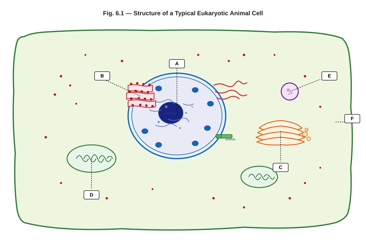
6. Fig. 6.1 shows the structure of a typical eukaryotic cell as seen under an electron microscope.
Generated diagram for Q6.
(a) Identify the organelles labelled A and C in Fig. 6.1. [2]
A: _________________________________
C: _________________________________
(b) State one function of the organelle labelled B. [1]
(c) Explain why the organelle labelled D is described as the "powerhouse of the cell". [2]
(d) Describe the role of the organelle labelled E in the cell. [2]
7. Table 7.1 shows the approximate composition of four biological molecules found in living organisms.
| Biological molecule | Carbon / % | Hydrogen / % | Oxygen / % | Nitrogen / % | Sulphur / % |
|---|---|---|---|---|---|
| Molecule W | 50 | 7 | 23 | 16 | 0 |
| Molecule X | 35 | 5 | 30 | 0 | 0 |
| Molecule Y | 50 | 7 | 23 | 15 | 1 |
| Molecule Z | 40 | 6 | 45 | 0 | 0 |
(a) Identify which molecule (W, X, Y, or Z) is most likely to be a protein. Explain your reasoning. [2]
(b) Molecule X is most likely a lipid. Explain how the data in Table 7.1 supports this conclusion. [2]
(c) State two functions of lipids in living organisms. [2]
8. Fig. 8.1 shows the effect of temperature on the rate of an enzyme-catalysed reaction.
Generated graph for Q8.
(a) With reference to Fig. 8.1, describe the effect of temperature on the rate of the enzyme-catalysed reaction between 0 °C and 37 °C. [2]
(b) Explain the decrease in the rate of reaction above 37 °C. [3]
(c) A student repeated the experiment using the same enzyme from a thermophilic bacterium. Predict and explain how the graph would differ. [2]
9. Describe the structure of the fluid mosaic model of the plasma membrane. In your answer, refer to the roles of phospholipids, cholesterol, and proteins. [5]
10. Explain how the structure of DNA is related to its function as the genetic material. In your answer, refer to complementary base pairing, the double helix, and the sugar-phosphate backbone. [4]
11. A student carried out an experiment to test for the presence of non-reducing sugars in a food sample. The student first tested the sample with Benedict's reagent and observed no colour change. The student then hydrolysed the sample with dilute hydrochloric acid, neutralised it with sodium hydrogencarbonate, and re-tested with Benedict's reagent. An orange-red precipitate was observed.
(a) Explain why the first test with Benedict's reagent showed no colour change. [1]
(b) Name the type of sugar that was present in the food sample. [1]
(c) Explain the purpose of adding dilute hydrochloric acid in the second part of the procedure. [2]
(d) Why was it necessary to neutralise the mixture with sodium hydrogencarbonate before re-testing with Benedict's reagent? [1]
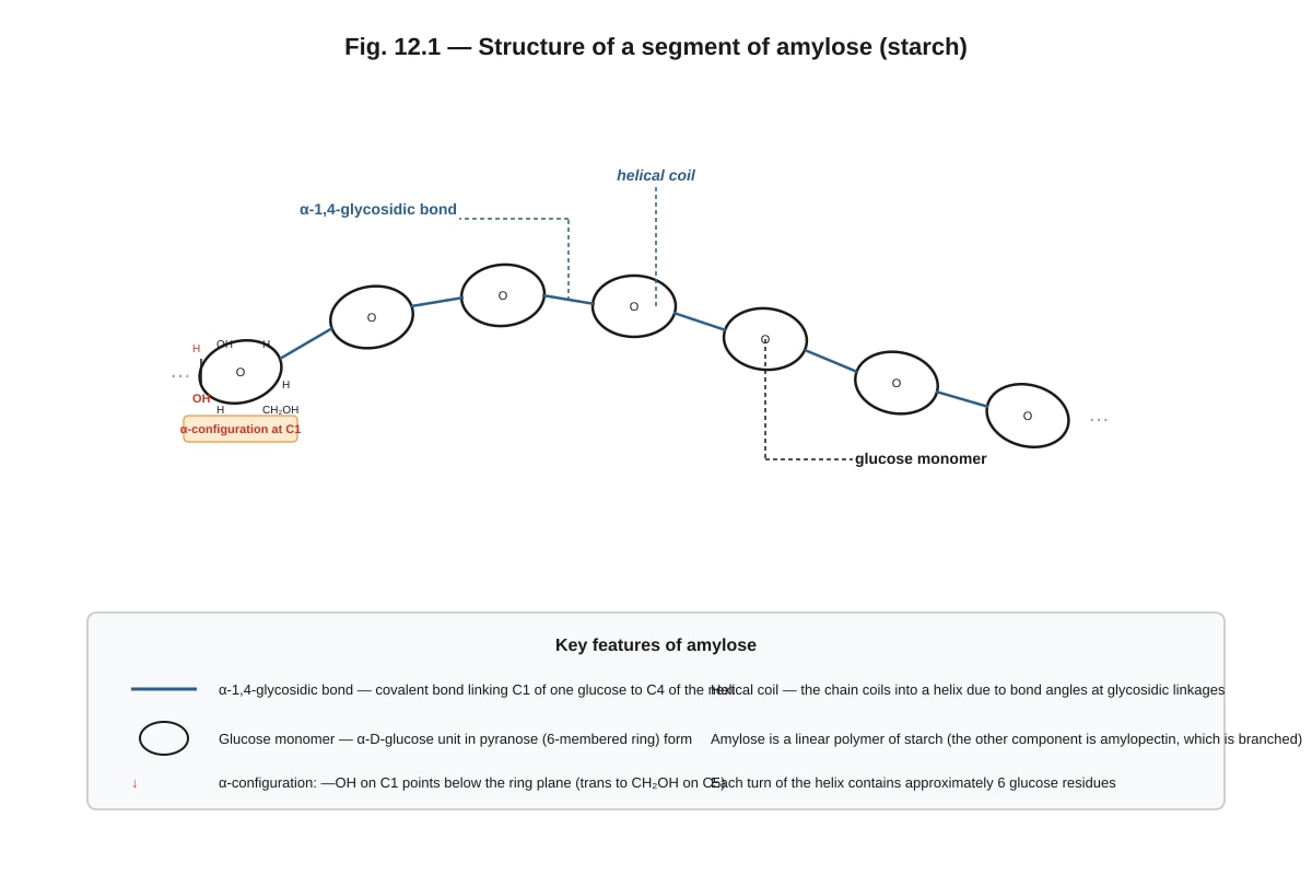
12. Fig. 12.1 shows the structure of a molecule of starch.
Generated diagram for Q12.
(a) With reference to Fig. 12.1, name the type of reaction that joins glucose monomers to form starch. [1]
(b) Name the type of bond labelled in Fig. 12.1. [1]
(c) Explain why starch is a suitable molecule for energy storage in plants. Refer to its structure in your answer. [3]
13. Compare and contrast the processes of facilitated diffusion and active transport across the plasma membrane. In your answer, refer to the role of carrier proteins, the direction of movement relative to the concentration gradient, and the requirement for energy. [4]
14. Describe the four levels of protein structure (primary, secondary, tertiary, and quaternary). For each level, state the type(s) of bond or interaction that stabilise the structure. [6]
Primary structure: _______________________________________________________________
Secondary structure: _______________________________________________________________
Tertiary structure: _______________________________________________________________
Quaternary structure: _______________________________________________________________
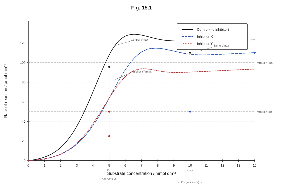
15. Fig. 15.1 shows the effect of substrate concentration on the rate of an enzyme-catalysed reaction in the absence and presence of two different inhibitors, X and Y.
Generated graph for Q15.
(a) With reference to Fig. 15.1, state the type of inhibition shown by inhibitor X. Explain your answer. [2]
(b) With reference to Fig. 15.1, state the type of inhibition shown by inhibitor Y. Explain your answer. [2]
(c) Suggest a molecular mechanism by which inhibitor X might act on the enzyme. [1]
Section C: Free Response (Questions 16–20) [20 marks]
16. Explain how the structure of a mitochondrion is adapted for its role in aerobic respiration. In your answer, refer to the outer membrane, inner membrane (cristae), matrix, and the presence of circular DNA and 70S ribosomes. [5]
17. A student investigated the effect of pH on the activity of the enzyme catalase using the apparatus shown in Fig. 17.1. Filter discs soaked in catalase solution were placed in hydrogen peroxide solutions at different pH values. The time taken for each disc to rise to the surface (due to oxygen gas production) was recorded.
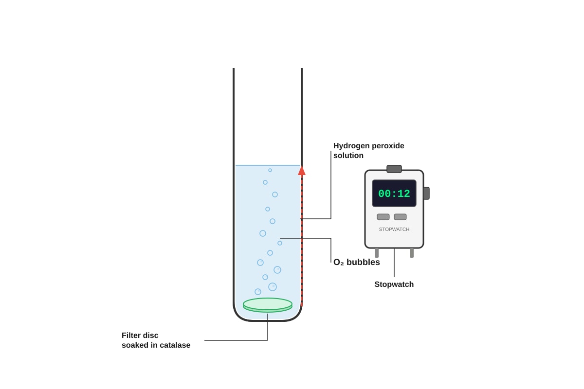
Generated experimental_setup for Q17.
The results are shown in Table 17.1.
| pH | Time for disc to rise / s | Rate of reaction / s⁻¹ |
|---|---|---|
| 3 | 85 | 0.012 |
| 5 | 32 | 0.031 |
| 7 | 12 | 0.083 |
| 9 | 28 | 0.036 |
| 11 | 78 | 0.013 |
(a) Explain why the rate of reaction is calculated as 1/time. [1]
(b) Describe the trend shown in Table 17.1. [2]
(c) Explain the effect of pH on catalase activity at pH values below and above the optimum. [3]
(d) Identify two variables that should be kept constant in this experiment. [2]
18. Compare and contrast the structure and function of rough endoplasmic reticulum (RER) and smooth endoplasmic reticulum (SER). In your answer, explain how the structural differences between RER and SER relate to their distinct roles in the cell. [5]
19. Explain the significance of the complementary base pairing rule in DNA. In your answer, refer to DNA replication, transcription, and the maintenance of genetic stability across cell divisions. [5]
20. A food sample was tested for the presence of biological molecules using the following tests:
| Test | Reagent used | Observation |
|---|---|---|
| Test 1 | Iodine solution | Blue-black colour observed |
| Test 2 | Benedict's reagent (heated) | Green precipitate observed |
| Test 3 | Biuret reagent | Violet/purple colour observed |
| Test 4 | Ethanol emulsion test | Cloudy white emulsion observed |
(a) State what each positive result indicates about the biological molecules present in the food sample. [4]
Test 1: _______________________________________________________________
Test 2: _______________________________________________________________
Test 3: _______________________________________________________________
Test 4: _______________________________________________________________
(b) For Test 2, explain why a green precipitate (rather than an orange-red precipitate) was observed. [1]
(c) Describe the procedure for the ethanol emulsion test (Test 4). [2]
END OF QUIZ
Answers
A-Level Biology H2 Quiz - Cells Biomolecules
Answer Key and Marking Scheme
Section A: Multiple Choice (Questions 1–5)
1. C [2]
Explanation: Prokaryotic cells lack a membrane-bound nucleus; their DNA is located in a region called the nucleoid. Eukaryotic cells possess a true nucleus enclosed by a double nuclear envelope with nuclear pores. Option A is incorrect because it is prokaryotes that have circular DNA (eukaryotes have linear DNA with histones). Option B is incorrect because prokaryotes have 70S ribosomes and eukaryotes have 80S ribones. Option D is incorrect because prokaryotes lack membrane-bound organelles.
Common mistake: Students often confuse the ribosome sizes (70S vs 80S) between prokaryotes and eukaryotes. Remember: Prokaryotes = Smaller (70S).
2. B [2]
Explanation: The organelle described is a mitochondrion, identified by its double membrane, cristae (infoldings of the inner membrane), and matrix containing circular DNA. The Krebs cycle occurs in the matrix, and oxidative phosphorylation (electron transport chain and chemiosmosis) occurs on the inner mitochondrial membrane (cristae). Transcription (A) occurs in the nucleus. Ribosomal subunit assembly (C) occurs in the nucleolus. Packaging and modification of secretory proteins (D) occurs in the Golgi apparatus.
Common mistake: Students may select A because mitochondria do contain DNA and can transcribe mRNA, but the primary and most significant function of the mitochondrion is aerobic respiration (Krebs cycle and oxidative phosphorylation).
3. C [2]
Explanation: Cohesion between water molecules is due to hydrogen bonds, not covalent bonds. Covalent bonds exist within a single water molecule (between O and H atoms). Hydrogen bonds form between adjacent water molecules due to the partial charges on the polar water molecule. Options A, B, and D are all correct statements about water.
Common mistake: Students frequently confuse intramolecular bonds (covalent bonds within a water molecule) with intermolecular forces (hydrogen bonds between water molecules). Cohesion is an intermolecular phenomenon.
4. C [2]
Explanation: Amino acids are linked together by peptide bonds (—CO—NH—) formed by a condensation reaction between the amino group (—NH₂) of one amino acid and the carboxyl group (—COOH) of another. Hydrogen bonds (A) stabilise secondary protein structure. Ionic bonds (B) can form between charged R-groups in tertiary structure. Disulphide bonds (D) form between the —SH groups of cysteine residues and stabilise tertiary structure, but they are not the primary bonds linking amino acids in the polypeptide chain.
Common mistake: Students may select D (disulphide bonds) because cysteine is listed among the amino acids. However, disulphide bonds are not the bonds that link amino acids in the primary chain — they are additional bonds that form between cysteine residues in the folded protein.
5. C [2]
Explanation: Competitive inhibition is characterised by an increased apparent Km (the enzyme requires a higher substrate concentration to reach ½ Vmax) but unchanged Vmax (at very high substrate concentrations, the substrate outcompetes the inhibitor, and the same maximum rate can be achieved). The inhibitor competes with the substrate for the active site because it has a similar molecular structure.
- Non-competitive inhibition (A): Vmax decreases, Km unchanged.
- Uncompetitive inhibition (B): Both Vmax and Km decrease.
- Allosteric activation (D): Would increase the rate, not decrease it.
Common mistake: Students often confuse competitive and non-competitive inhibition. A useful memory aid: In competitive inhibition, the inhibitor competes for the active site — so adding more substrate can overcome it (Vmax unchanged, Km increases).
Section B: Structured Questions (Questions 6–15)
6.
(a) [2]
- A: Nucleus [1]
- C: Golgi apparatus (Golgi body) [1]
Teaching note: The nucleus is the largest organelle, typically central, with a double membrane and nuclear pores. The Golgi apparatus appears as a stack of flattened membrane-bound sacs (cisternae), often near the nucleus or ER.
(b) [1]
- Protein synthesis — ribosomes attached to the rough endoplasmic reticulum synthesise proteins, particularly secretory proteins and membrane proteins. [1]
Acceptable alternatives: Transport of proteins; folding of newly synthesised proteins.
(c) [2]
- The mitochondrion is the site of aerobic respiration [1], where ATP is produced through the Krebs cycle (in the matrix) and oxidative phosphorylation / electron transport chain (on the cristae of the inner membrane) [1].
Marking: 1 mark for identifying aerobic respiration / ATP production; 1 mark for specifying the processes or locations (Krebs cycle and oxidative phosphorylation / electron transport chain).
(d) [2]
- Lysosomes contain hydrolytic (digestive) enzymes [1] that break down worn-out organelles, engulfed pathogens, and other cellular waste materials through intracellular digestion [1].
Marking: 1 mark for hydrolytic enzymes; 1 mark for the function of breaking down waste / worn-out organelles / pathogens.
7.
(a) [2]
- Molecule W is most likely a protein [1]. This is because it contains nitrogen (16%), which is a characteristic element found in amino acids (the building blocks of proteins). It does not contain sulphur, which is consistent with many proteins that may or may not contain sulphur-containing amino acids [1].
Alternative acceptable answer: Molecule Y could also be identified as a protein because it contains nitrogen (15%) and a small amount of sulphur (1%), which is consistent with proteins containing cysteine or methionine residues.
Marking: 1 mark for correct identification (W or Y); 1 mark for correct reasoning based on nitrogen content.
(b) [2]
- Molecule X has a high carbon and hydrogen content relative to oxygen (C:H:O ratio is approximately 35:5:30, or simplified, a high proportion of C and H compared to O) [1]. Lipids have a much lower proportion of oxygen compared to carbohydrates and are rich in carbon and hydrogen, which makes them have a high energy yield upon oxidation [1].
Marking: 1 mark for identifying the high C:H ratio relative to O; 1 mark for linking this to the characteristic composition of lipids.
(c) [2] Any two of the following:
- Energy storage (e.g., triglycerides store more energy per gram than carbohydrates) [1]
- Insulation (e.g., adipose tissue under the skin reduces heat loss) [1]
- Protection of internal organs (e.g., fat around kidneys) [1]
- Component of cell membranes (phospholipids form the bilayer) [1]
- Waterproofing (e.g., waxes on leaves and skin) [1]
- Hormone production (e.g., steroid hormones are derived from lipids) [1]
8.
(a) [2]
- As temperature increases from 0 °C to 37 °C, the rate of the enzyme-catalysed reaction increases [1]. This is because increasing temperature increases the kinetic energy of both enzyme and substrate molecules, leading to more frequent and more energetic successful collisions between enzyme and substrate, thus increasing the rate of formation of enzyme-substrate complexes [1].
Marking: 1 mark for stating the rate increases; 1 mark for explaining in terms of kinetic energy and collisions.
(b) [3]
- Above 37 °C, the rate of reaction decreases sharply because the enzyme denatures [1]. High temperatures cause the hydrogen bonds and other weak interactions (e.g., ionic bonds, hydrophobic interactions) that maintain the enzyme's three-dimensional tertiary structure to break [1]. This changes the shape of the active site so that the substrate can no longer bind effectively (the enzyme-substrate complex cannot form), and the enzyme is permanently inactivated [1].
Marking: 1 mark for denaturation; 1 mark for bonds/interactions breaking; 1 mark for change in active site shape / loss of function.
(c) [2]
- The graph would show an optimum temperature higher than 37 °C (e.g., around 60–80 °C) [1]. This is because enzymes from thermophilic bacteria have evolved to function at high temperatures; they have more stable tertiary structures (e.g., more disulphide bonds, stronger hydrophobic interactions, more compact folding) that resist denaturation at elevated temperatures [1].
Marking: 1 mark for higher optimum temperature; 1 mark for explanation in terms of structural stability / adaptation to high temperature.
9. [5]
The fluid mosaic model describes the plasma membrane as follows:
-
Phospholipid bilayer: The basic structure consists of a double layer of phospholipids. Each phospholipid has a hydrophilic (polar) phosphate head and two hydrophobic (non-polar) fatty acid tails. The heads face the aqueous environments (outside the cell and cytoplasm), while the tails face inward, away from water. This arrangement forms a stable barrier. [1]
-
Fluid nature: The phospholipids can move laterally (sideways) within their own monolayer, giving the membrane a fluid, flexible character. This fluidity is influenced by temperature and the presence of cholesterol. [1]
-
Cholesterol: Cholesterol molecules are interspersed among the phospholipid tails. At high temperatures, cholesterol reduces fluidity by restraining phospholipid movement. At low temperatures, it prevents the membrane from becoming too rigid by preventing close packing of phospholipids. [1]
-
Proteins: Proteins are embedded in or attached to the phospholipid bilayer in a mosaic pattern. Intrinsic (integral) proteins span the entire bilayer (transmembrane proteins) and function as channels, carriers, receptors, or enzymes. Extrinsic (peripheral) proteins are loosely attached to the surface of the membrane and may be involved in cell signalling or maintaining cell shape. [1]
-
Glycoproteins and glycolipids: Some membrane proteins and lipids have carbohydrate chains attached on the extracellular surface, forming glycoproteins and glycolipids. These are involved in cell recognition, cell signalling, and immune responses. [1]
Marking scheme: 1 mark each for the five points above. Answers must refer to phospholipids, cholesterol, and proteins to achieve full marks.
10. [4]
-
Complementary base pairing: Adenine (A) pairs with thymine (T) via two hydrogen bonds, and guanine (G) pairs with cytosine (C) via three hydrogen bonds. This specific pairing ensures that the two strands are complementary, allowing accurate replication and transcription. [1]
-
Double helix: The two antiparallel strands twist around each other to form a stable double helix. The sugar-phosphate backbones are on the outside, protecting the nitrogenous bases inside. This structure provides stability and protects the genetic information from chemical and physical damage. [1]
-
Sugar-phosphate backbone: The alternating sugar (deoxyribose) and phosphate groups form a strong, covalently bonded backbone that provides structural integrity to the DNA molecule. [1]
-
Function as genetic material: The sequence of bases along the DNA strand encodes genetic information. Complementary base pairing allows DNA to replicate itself accurately during cell division (semiconservative replication) and to be transcribed into mRNA for protein synthesis. The double-stranded nature also allows for repair — if one strand is damaged, the complementary strand can serve as a template for repair. [1]
Marking scheme: 1 mark each for the four points. Answers must refer to complementary base pairing, the double helix, and the sugar-phosphate backbone.
11.
(a) [1]
- The food sample contained a non-reducing sugar (e.g., sucrose), which does not react with Benedict's reagent because it does not have a free aldehyde or ketone group available to act as a reducing agent. [1]
(b) [1]
- Sucrose (a non-reducing disaccharide) [1]
(c) [2]
- Dilute hydrochloric acid hydrolyses the glycosidic bond in the non-reducing sugar (e.g., sucrose) [1], breaking it down into its constituent monosaccharides (glucose and fructose), which are reducing sugars and can react with Benedict's reagent [1].
Marking: 1 mark for hydrolysis of glycosidic bond; 1 mark for production of reducing sugars (glucose/fructose).
(d) [1]
- Benedict's reagent requires an alkaline conditions to react with reducing sugars. The hydrochloric acid would create acidic conditions, which would prevent the redox reaction from occurring. Neutralisation ensures the correct pH for the Benedict's test. [1]
12.
(a) [1]
- Condensation reaction (dehydration synthesis) [1]
(b) [1]
- α-1,4-glycosidic bond [1]
(c) [3]
- Starch is a polysaccharide made up of many glucose monomers joined by α-1,4-glycosidic bonds (and α-1,6-glycosidic bonds in amylopectin branches) [1]. It is insoluble in water, so it does not affect the water potential of the cell and can be stored without causing osmotic problems [1]. The helical structure of amylose makes starch compact, allowing large amounts of glucose to be stored in a small space. Starch can be hydrolysed by amylase enzymes to release glucose when energy is needed [1].
Marking: 1 mark for polysaccharide / glucose monomers; 1 mark for insolubility / no osmotic effect; 1 mark for compact / can be hydrolysed for energy.
13. [4]
| Feature | Facilitated Diffusion | Active Transport |
|---|---|---|
| Direction | Down the concentration gradient (from high to low concentration) | Against the concentration gradient (from low to high concentration) |
| Energy requirement | Does not require energy (passive process) | Requires energy in the form of ATP |
| Carrier/channel proteins | Uses carrier proteins or channel proteins to transport molecules that cannot diffuse directly through the lipid bilayer (e.g., large polar molecules, ions) | Uses carrier proteins (pumps) that undergo conformational changes to transport molecules |
| Specificity | Specific to particular molecules (depends on the shape of the carrier/channel protein) | Highly specific — each carrier protein transports specific molecules or ions |
Marking scheme: 1 mark for each of the following (maximum 4):
- Direction of movement (down gradient vs against gradient) [1]
- Energy requirement (no ATP vs requires ATP) [1]
- Role of carrier/channel proteins in both processes [1]
- Specificity of transport [1]
Common mistake: Students often state that facilitated diffusion does not involve proteins — it does. The key difference is the direction of movement and the energy requirement.
14. [6]
Primary structure: [1.5]
- The sequence of amino acids in a polypeptide chain, linked together by peptide bonds (covalent bonds) [1]. The primary structure is determined by the gene encoding the protein [0.5].
Secondary structure: [1.5]
- Local folding of the polypeptide chain into regular structures such as the α-helix or β-pleated sheet [1]. Stabilised by hydrogen bonds between the C=O group of one amino acid and the N—H group of another amino acid in the backbone [0.5].
Tertiary structure: [1.5]
- The overall three-dimensional folding of the polypeptide chain into its final shape [1]. Stabilised by interactions between R-groups, including: hydrogen bonds, ionic bonds (between charged R-groups), disulphide bonds (covalent bonds between cysteine residues), and hydrophobic interactions (between non-polar R-groups) [0.5 — any two interactions for the half mark].
Quaternary structure: [1.5]
- The arrangement of two or more polypeptide chains (subunits) into a single functional protein [1]. Stabilised by the same types of bonds and interactions as tertiary structure (hydrogen bonds, ionic bonds, disulphide bonds, hydrophobic interactions) between the subunits [0.5]. Example: haemoglobin has four polypeptide subunits.
Marking: 1.5 marks per level (1 mark for description, 0.5 for bonds/interactions). Total: 6 marks.
15.
(a) [2]
- Inhibitor X shows competitive inhibition [1]. This is because the Vmax is unchanged (same as the control) but a higher substrate concentration is needed to reach Vmax (the curve is shifted to the right), indicating an increased apparent Km [1].
Teaching note: In competitive inhibition, the inhibitor competes with the substrate for the active site. At high substrate concentrations, the substrate outcompetes the inhibitor, so the same Vmax can be achieved.
(b) [2]
- Inhibitor Y shows non-competitive inhibition [1]. This is because the Vmax is lower than the control (reduced to 50 μmol min⁻¹), but the substrate concentration needed to reach ½ Vmax is the same (Km is unchanged) [1].
Teaching note: In non-competitive inhibition, the inhibitor binds to a site other than the active site (allosteric site), changing the enzyme's shape. Increasing substrate concentration cannot overcome this inhibition, so Vmax is reduced.
(c) [1]
- Inhibitor X likely has a similar molecular shape to the substrate and competes for the active site of the enzyme, preventing the substrate from binding [1].
Acceptable alternative: Inhibitor X binds reversibly to the active site, blocking substrate access.
Section C: Free Response (Questions 16–20)
16. [5]
The structure of a mitochondrion is highly adapted for aerobic respiration:
-
Outer membrane: The outer membrane is permeable to small molecules and ions, allowing the entry of pyruvate, ADP, and NADH from the cytoplasm. It also contains porins (channel proteins) that facilitate the passage of molecules. [1]
-
Inner membrane (cristae): The inner membrane is highly folded into cristae, which greatly increase the surface area available for the electron transport chain and ATP synthase complexes. The inner membrane is impermeable to protons (H⁺), which is essential for maintaining the proton gradient (electrochemical gradient) needed for chemiosmosis and ATP synthesis. The electron transport chain proteins (NADH dehydrogenase, cytochrome complexes, cytochrome c oxidase) and ATP synthase are embedded in the inner membrane. [1]
-
Matrix: The matrix is the fluid-filled space inside the inner membrane. It contains the enzymes of the Krebs cycle (citric acid cycle), as well as enzymes for the oxidation of fatty acids. The matrix also contains circular DNA and 70S ribosomes, allowing the mitochondrion to synthesise some of its own proteins (those needed for respiration) independently of the nucleus. [1]
-
Circular DNA and 70S ribosomes: These allow the mitochondrion to produce its own proteins required for the electron transport chain and ATP synthesis, enabling rapid, localised protein production without relying on nuclear gene expression. This supports the endosymbiotic theory that mitochondria were once free-living prokaryotes. [1]
-
Intermembrane space: The narrow space between the outer and inner membranes accumulates protons (H⁺ ions) pumped out by the electron transport chain, creating a proton gradient that drives ATP synthesis via chemiosmosis. [1]
Marking scheme: 1 mark each for the five points above. Answers must refer to the outer membrane, inner membrane/cristae, matrix, and circular DNA/70S ribosomes.
17.
(a) [1]
- The rate of reaction is inversely proportional to the time taken. Calculating 1/time gives a measure of how fast the reaction proceeds — a shorter time means a faster rate, and 1/time will be larger [1].
(b) [2]
- As pH increases from 3 to 7, the time for the disc to rise decreases (from 85 s to 12 s), meaning the rate of reaction increases [1]. As pH increases from 7 to 11, the time increases (from 12 s to 78 s), meaning the rate of reaction decreases. The optimum pH for catalase is pH 7 [1].
Marking: 1 mark for describing the trend (rate increases then decreases); 1 mark for identifying the optimum pH as 7.
(c) [3]
- At pH values below 7 (acidic conditions), the excess H⁺ ions disrupt the hydrogen bonds and ionic bonds that maintain the enzyme's tertiary structure [1]. This causes the enzyme to denature — the active site changes shape so the substrate (hydrogen peroxide) can no longer bind effectively, reducing the rate of reaction [1].
- At pH values above 7 (alkaline conditions), the excess OH⁻ ions similarly disrupt the bonds maintaining the enzyme's three-dimensional structure, leading to denaturation and a decrease in enzyme activity [1].
Marking: 1 mark for effect of acidic pH (H⁺ disrupts bonds); 1 mark for denaturation / active site change; 1 mark for effect of alkaline pH (OH⁻ disrupts bonds / denaturation).
(d) [2] Any two of the following:
- Concentration of hydrogen peroxide solution [1]
- Volume of hydrogen peroxide solution [1]
- Size / surface area of the filter disc [1]
- Volume of catalase solution used to soak the disc [1]
- Temperature [1]
Marking: 1 mark each for any two valid controlled variables.
18. [5]
Similarities:
- Both RER and SER are part of the endomembrane system and are composed of flattened, membrane-bound sacs (cisternae) continuous with the outer nuclear membrane [1].
- Both are involved in the synthesis and transport of molecules within the cell [1].
Differences:
| Feature | Rough Endoplasmic Reticulum (RER) | Smooth Endoplasmic Reticulum (SER) |
|---|---|---|
| Structure | Studded with ribosomes on the cytoplasmic surface | No ribosomes attached; smooth appearance |
| Function | Synthesis and processing of proteins (especially secretory proteins, membrane proteins, and lysosomal proteins). Proteins are folded and modified (e.g., glycosylation) in the RER lumen. | Synthesis of lipids (including phospholipids and cholesterol), steroid hormone production, and detoxification of drugs and poisons (especially in liver cells). |
| Transport | Proteins are transported in vesicles from the RER to the Golgi apparatus for further processing and sorting | Lipids are transported in vesicles to the Golgi apparatus or to other parts of the cell |
Structural-functional relationship: The presence of ribosomes on the RER provides the machinery for protein synthesis directly at the site where proteins are needed. The absence of ribosomes on the SER allows more membrane surface area for lipid-synthesising enzymes. The extensive membrane network of both RER and SER provides a large surface area for their respective biochemical reactions [1].
Marking scheme: 1 mark for similarities; 2 marks for structural and functional differences; 1 mark for structural-functional relationship; 1 mark for clarity and use of biological terminology. Total: 5 marks.
19. [5]
-
Complementary base pairing rule: In DNA, adenine (A) always pairs with thymine (T) via two hydrogen bonds, and guanine (G) always pairs with cytosine (C) via three hydrogen bonds. This specificity is fundamental to DNA's role as the genetic material [1].
-
DNA replication: During semiconservative replication, the two DNA strands separate, and each strand serves as a template for the synthesis of a new complementary strand. Because A always pairs with T and G always pairs with C, the new strands are exact complements of the template strands. This ensures that genetic information is accurately copied and passed on to daughter cells [1].
-
Transcription: During transcription, one strand of DNA (the template/antisense strand) is used to synthesise a complementary mRNA molecule. The complementary base pairing rule ensures that the mRNA sequence is an accurate copy of the genetic code (with uracil replacing thymine). This mRNA is then translated into a specific sequence of amino acids in a protein [1].
-
Maintenance of genetic stability: The specificity of complementary base pairing ensures that mutations are minimised during replication. If an incorrect base is inserted, the mismatch can often be detected and repaired by DNA repair enzymes because the incorrect pairing does not follow the A-T and G-C rule. This maintains the fidelity of genetic information across many cell divisions [1].
-
Universal significance: The complementary base pairing rule is universal across almost all living organisms, supporting the idea of a common ancestor and enabling genetic engineering techniques (e.g., PCR, DNA sequencing, recombinant DNA technology) that rely on predictable base pairing [1].
Marking scheme: 1 mark each for the five points above. Answers must refer to DNA replication, transcription, and maintenance of genetic stability.
20.
(a) [4]
- Test 1 (Iodine → blue-black): The food sample contains starch [1]
- Test 2 (Benedict's → green precipitate): The food sample contains reducing sugar (e.g., glucose, maltose, fructose) at a low concentration [1]
- Test 3 (Biuret → violet/purple): The food sample contains protein [1]
- Test 4 (Ethanol emulsion → cloudy white): The food sample contains lipid (fat) [1]
(b) [1]
- A green precipitate indicates a low concentration of reducing sugar. Benedict's test produces a colour gradient: blue (no reducing sugar) → green → yellow → orange → brick-red precipitate (high concentration of reducing sugar). The green colour means some reducing sugar is present but in relatively small amounts [1].
(c) [2]
- The food sample is dissolved in ethanol (e.g., by shaking or grinding the sample with ethanol) [1]. The ethanol solution is then poured into water. If lipids are present, the ethanol dissolves the lipid, and when the ethanol mixes with water, the lipid is precipitated out as tiny droplets, forming a cloudy white emulsion [1].
Marking: 1 mark for dissolving in ethanol; 1 mark for adding to water and observing cloudy white emulsion.
Total: 60 marks
Free quiz and exam paper access
Enter your details to view this paper
Your access is remembered on this device.