From Real Exams Quiz
A Level H2 Biology Cells Biomolecules Quiz
Free A Level H2 Biology Cells Biomolecules quiz, LongCat Exam version, with questions, answers, and A Level-style practice for Singapore students.
These static practice materials are generated from the site's syllabus and paper-generation workflow, with source and model context shown so students and parents can evaluate the material before use.
Questions
A-Level Biology H2 Quiz - Cells Biomolecules
Name: ___________________________
Class: ___________________________
Date: ___________________________
Score: ________ / 50
Duration: 60 minutes
Total Marks: 50
Instructions:
- Answer ALL questions in the spaces provided.
- The number of marks for each question or part-question is shown in brackets [ ].
- You are advised to spend no more than 60 minutes on this quiz.
- Where a question requires explanation or description, use precise biological terminology.
- Where diagrams are referenced, study the figure carefully before answering.
Section A: Multiple Choice & Short Answer (Questions 1–10)
Questions 1–5: Multiple Choice. Circle the single best answer. Each question carries 1 mark.
1. Which of the following organelles is found in both prokaryotic and eukaryotic cells?
A. Nucleus
B. Mitochondrion
C. Ribosome
D. Endoplasmic reticulum
2. A student observed a cell under an electron microscope and noted an organelle with a double membrane, cristae, and a matrix containing circular DNA. This organelle is most likely involved in:
A. Protein synthesis
B. Aerobic respiration
C. Photosynthesis
D. Lipid synthesis
3. Which property of water is most directly responsible for its role as a solvent for ionic biological molecules?
A. High specific heat capacity
B. Cohesion and surface tension
C. Polarity of the water molecule
D. High latent heat of vaporisation
4. During the formation of a triglyceride, three fatty acid molecules combine with one glycerol molecule. This is an example of:
A. Hydrolysis
B. Condensation
C. Oxidation
D. Phosphorylation
5. Which of the following best describes the role of cholesterol in the cell membrane?
A. It provides energy for active transport across the membrane.
B. It increases membrane fluidity at all temperatures.
C. It regulates membrane fluidity by restricting phospholipid movement at high temperatures and preventing tight packing at low temperatures.
D. It acts as a receptor for all signalling molecules.
Questions 6–10: Short Answer. Each question carries 2 marks.
6. State two structural differences between prokaryotic and eukaryotic cells.
7. Explain why the term "fluid mosaic" is used to describe the structure of the cell surface membrane.
8. Describe the biological test for the presence of a reducing sugar. Include the reagent used, the procedure, and the positive result.
9. State two functions of proteins in living organisms and give one named example for each function.
10. Explain what is meant by the term "hydrophobic" and explain why the hydrophobic nature of the fatty acid tails is important for the function of the cell membrane.
Section B: Structured Response (Questions 11–17)
11. [4 marks]
Generated diagram for Q11.
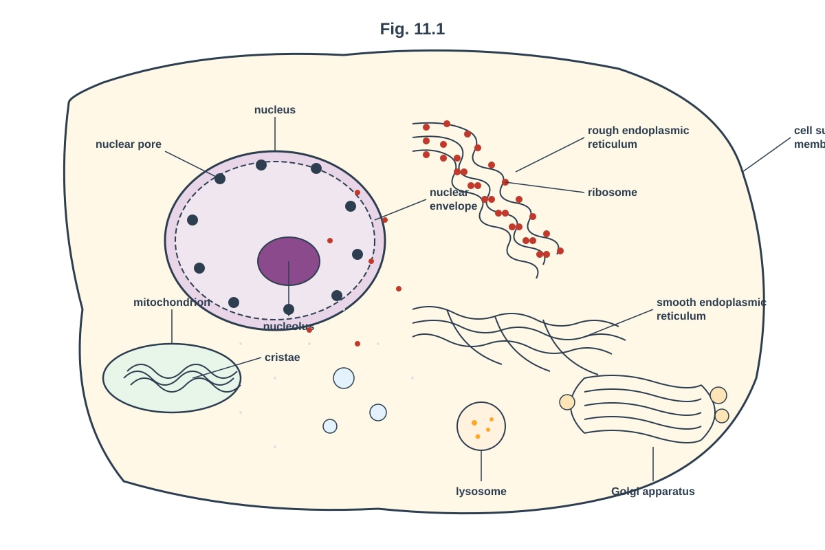
Fig. 11.1 shows an animal cell as seen under an electron microscope.
(a) Name the organelles labelled A (nucleus) and B (rough endoplasmic reticulum). [2]
A: _______________________________________________________________
B: _______________________________________________________________
(b) Describe the function of the organelle labelled C (Golgi apparatus). [2]
12. [5 marks]
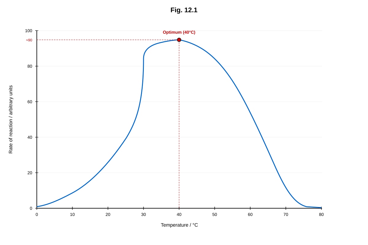
Generated graph for Q12.
Fig. 12.1 shows the effect of temperature on the rate of an enzyme-catalysed reaction.
(a) Describe the relationship between temperature and the rate of reaction as shown in Fig. 12.1. [2]
(b) Explain the shape of the curve between 0 °C and 40 °C. [2]
(c) Explain the sharp decrease in the rate of reaction above 40 °C. [1]
13. [4 marks]
Generated diagram for Q13.
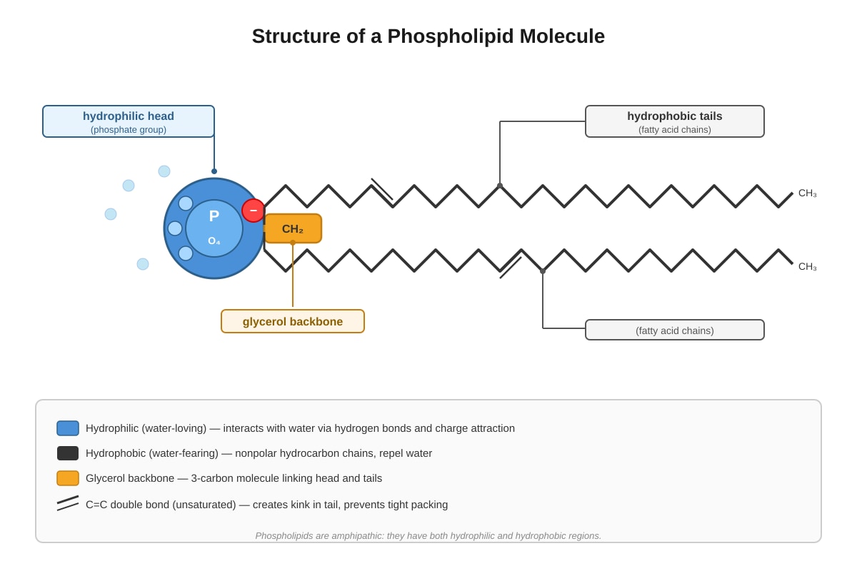
Fig. 13.1 shows the structure of a phospholipid molecule.
(a) With reference to Fig. 13.1, explain why phospholipids spontaneously form a bilayer when placed in water. [3]
(b) Explain how the phospholipid bilayer acts as a barrier to the movement of ions and large polar molecules. [1]
14. [5 marks]
A student carried out an experiment to investigate the effect of pH on the activity of the enzyme catalase, which breaks down hydrogen peroxide into water and oxygen. The volume of oxygen gas collected in 2 minutes at different pH values was recorded.
Table 14.1
| pH | Volume of O₂ collected in 2 min / cm³ |
|---|---|
| 3 | 4.2 |
| 5 | 12.8 |
| 7 | 28.6 |
| 9 | 18.4 |
| 11 | 3.1 |
(a) Describe the trend shown in Table 14.1. [2]
(b) Explain why the enzyme activity is highest at pH 7. [2]
(c) Suggest why the enzyme activity at pH 11 is similar to that at pH 3. [1]
15. [4 marks]
Describe the structure of DNA and explain how its structure enables it to carry genetic information and to replicate itself accurately.
16. [5 marks]
Generated diagram for Q16.
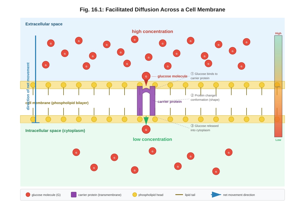
Fig. 16.1 shows the movement of glucose molecules across a cell membrane by facilitated diffusion.
(a) With reference to Fig. 16.1, describe the process of facilitated diffusion. [3]
(b) Explain how facilitated diffusion differs from active transport. [2]
17. [4 marks]
Explain the roles of the following organelles in the production and secretion of a protein such as insulin:
(a) Ribosome [1]
(b) Rough endoplasmic reticulum [1]
(c) Golgi apparatus [1]
(d) Mitochondrion [1]
Section C: Data & Source-Based Response (Questions 18–20)
Read the following passage and answer Questions 18–20.
The Discovery of the Cell Membrane Structure
In 1972, Singer and Nicolson proposed the fluid mosaic model of the cell membrane. This model described the membrane as a fluid phospholipid bilayer with proteins floating within it, much like icebergs in a sea. The proteins can be integral (embedded within or spanning the bilayer) or peripheral (loosely attached to the surface). Cholesterol molecules are found between the phospholipid tails, helping to regulate membrane fluidity.
Earlier models, such as the Davson-Danielli model (1935), proposed that the lipid bilayer was coated on both sides with a layer of protein. However, evidence from electron microscopy and freeze-fracture studies showed that proteins were actually embedded within the bilayer, supporting the fluid mosaic model.
The fluid mosaic model explains many properties of the cell membrane, including its selective permeability, the ability of cells to fuse, and the lateral movement of proteins within the membrane. Membrane proteins serve diverse functions: some act as channels or carriers for transport, others as receptors for cell signalling, and some as enzymes.
18. [3 marks]
With reference to the passage, describe the key difference between the Davson-Danielli model and the Singer-Nicolson fluid mosaic model of the cell membrane.
19. [3 marks]
Using information from the passage, explain why the fluid mosaic model is considered a better model of the cell membrane than the Davson-Danielli model.
20. [3 marks]
State three functions of membrane proteins mentioned in the passage and explain how each function is important for the cell.
Answers
A-Level Biology H2 Quiz - Cells Biomolecules
Answer Key
Question 1 [1 mark]
Answer: C (Ribosome)
Explanation: Ribosomes are the only organelle listed that are found in both prokaryotic and eukaryotic cells. Prokaryotic cells lack membrane-bound organelles such as the nucleus (A), mitochondrion (B), and endoplasmic reticulum (D). Ribosomes are responsible for protein synthesis in all cells. In prokaryotes, ribosomes are 70S (smaller), while in eukaryotes they are 80S, but both types possess them.
Common mistake: Students may select mitochondrion, thinking all cells need energy production. However, prokaryotes carry out respiration at the cell membrane, not in mitochondria.
Question 2 [1 mark]
Answer: B (Aerobic respiration)
Explanation: The description — double membrane, cristae, and circular DNA in the matrix — matches the mitochondrion. Mitochondria are the sites of aerobic respiration (specifically the Krebs cycle in the matrix and oxidative phosphorylation/electron transport chain on the cristae). The circular DNA is a remnant of their evolutionary origin as endosymbiotic prokaryotes.
Common mistake: Students may confuse this with the chloroplast (photosynthesis), which also has a double membrane and circular DNA, but chloroplasts have thylakoids, not cristae.
Question 3 [1 mark]
Answer: C (Polarity of the water molecule)
Explanation: Water is a polar molecule because oxygen is more electronegative than hydrogen, creating a partial negative charge (δ⁻) on the oxygen and partial positive charges (δ⁺) on the hydrogens. This polarity allows water molecules to surround and stabilise ions (e.g., Na⁺, Cl⁻) through electrostatic interactions, dissolving them effectively. The other properties listed are important for other biological roles (e.g., temperature buffering, cohesion in transpiration) but are not directly responsible for dissolving ionic substances.
Question 4 [1 mark]
Answer: B (Condensation)
Explanation: A condensation reaction is a reaction in which two molecules join together with the removal of a water molecule. In triglyceride formation, each of the three hydroxyl groups (–OH) of glycerol reacts with the carboxyl group (–COOH) of a fatty acid, forming an ester bond and releasing one molecule of water per bond (three water molecules in total). Hydrolysis (A) is the reverse process — breaking bonds by adding water.
Teaching note: Condensation builds polymers/large molecules; hydrolysis breaks them down. Students should remember: "Condensation = construct (remove water); Hydrolysis = destroy (add water)."
Question 5 [1 mark]
Answer: C
Explanation: Cholesterol is a lipid molecule found within the phospholipid bilayer of animal cell membranes. At high temperatures, cholesterol restricts the movement of phospholipid fatty acid tails, reducing membrane fluidity and preventing it from becoming too permeable. At low temperatures, cholesterol prevents the phospholipids from packing too closely together, maintaining fluidity and preventing the membrane from becoming too rigid. This dual role makes cholesterol a fluidity buffer.
Common mistake: Students often think cholesterol only increases or only decreases fluidity. The key is that it regulates fluidity in both directions.
Question 6 [2 marks]
Answer (1 mark each, any two of the following):
- Prokaryotic cells do not have a nucleus (DNA is free in the cytoplasm/nucleoid region), whereas eukaryotic cells have a true nucleus enclosed by a double nuclear envelope.
- Prokaryotic cells do not have membrane-bound organelles (e.g., mitochondria, ER, Golgi), whereas eukaryotic cells do.
- Prokaryotic cells have 70S ribosomes, whereas eukaryotic cells have 80S ribosomes.
- Prokaryotic cells have a cell wall made of murein (peptidoglycan), whereas eukaryotic plant cells have a cell wall made of cellulose.
- Prokaryotic cells are generally much smaller (1–5 μm) than eukaryotic cells (10–100 μm).
Marking note: Award 1 mark per valid structural difference. The difference must compare prokaryotic and eukaryotic — stating a feature of one without the other scores 0 for that point.
Question 7 [2 marks]
Answer:
- "Fluid" — The phospholipid bilayer is not rigid; the phospholipids and proteins can move laterally (sideways) within the membrane, giving it a fluid, dynamic nature. [1]
- "Mosaic" — The proteins are scattered throughout the bilayer at irregular intervals, creating a mosaic pattern when viewed from above. [1]
Teaching note: Students should be able to explain both parts of the term. "Fluid" refers to the dynamic, flexible nature of the membrane (phospholipids and proteins can move laterally). "Mosaic" refers to the scattered arrangement of different proteins within the bilayer, like tiles in a mosaic artwork.
Question 8 [2 marks]
Answer:
- Reagent: Benedict's solution (an alkaline solution of copper(II) sulfate). [½]
- Procedure: Add Benedict's solution to the sample and heat in a water bath at approximately 60–80 °C for 2–5 minutes. [½]
- Positive result: A colour change from blue → green → yellow → orange → brick-red precipitate indicates the presence of a reducing sugar. [1]
Marking note: Award marks for correct reagent, correct procedure (heating required), and correct positive result (colour change / brick-red precipitate). If the student only says "turns red" without mentioning the starting colour (blue) or the precipitate, award ½ for the result.
Common mistake: Confusing Benedict's test with the iodine test (for starch) or the biuret test (for protein). Also, students sometimes forget that heating is required.
Question 9 [2 marks]
Answer (1 mark per function + valid example):
| Function | Named Example |
|---|---|
| Structural | Collagen (in connective tissue) / Keratin (in hair and nails) |
| Enzymatic (catalysis) | Catalase / Amylase / DNA polymerase |
| Transport | Haemoglobin (transports oxygen) |
| Defence / Immune | Antibodies (immunoglobulins) |
| Signalling / Hormonal | Insulin (regulates blood glucose) |
| Motor / Contractile | Actin and Myosin (muscle contraction) |
| Storage | Ovalbumin (in egg white) / Ferritin (stores iron) |
| Receptor | Receptor proteins on cell surface (e.g., insulin receptor) |
Marking note: Award 1 mark for each correct function with a valid named example. Generic answers like "enzymes" without a named example score 0 for that point.
Question 10 [2 marks]
Answer:
- Hydrophobic means "water-fearing" — hydrophobic molecules are non-polar and do not interact favourably with water molecules; they are repelled by water. [1]
- The hydrophobic fatty acid tails face inwards, away from the aqueous environment, forming the interior of the bilayer. This creates a hydrophobic core that acts as a barrier to the passage of water-soluble substances (ions, polar molecules) across the membrane, enabling the membrane to control what enters and leaves the cell. [1]
Teaching note: Students should connect the chemical property (hydrophobicity) to the structural arrangement (tails facing inwards) and then to the functional consequence (selective permeability / barrier function).
Question 11 [4 marks]
(a) [2 marks — 1 mark each]
- A: Nucleus
- B: Rough endoplasmic reticulum (rough ER)
(b) [2 marks]
The Golgi apparatus:
- Receives proteins from the rough ER (in vesicles). [½]
- Modifies proteins (e.g., by adding carbohydrate groups — glycosylation). [½]
- Packages proteins into vesicles for transport to their final destinations (e.g., secretion via exocytosis, delivery to lysosomes, or insertion into the cell membrane). [½]
- Also produces lysosomes. [½]
Marking note: Award 1 mark for each correct organelle name in (a). In (b), award up to 2 marks for a clear description of Golgi function. Key points: modification/packaging/sorting of proteins, formation of vesicles, production of lysosomes.
Image note for Q11: The diagram should show a clearly labelled animal cell with nucleus (showing double membrane, nuclear pores, nucleolus), rough ER (with visible ribosomes), smooth ER, Golgi apparatus (stacked cisternae), mitochondrion (with cristae), lysosomes, and cell surface membrane. Labels A, B, and C should correspond to nucleus, rough ER, and Golgi apparatus respectively.
Question 12 [5 marks]
(a) [2 marks]
As temperature increases from 0 °C to 40 °C, the rate of reaction increases, reaching a maximum at 40 °C (the optimum temperature). Above 40 °C, the rate of reaction decreases sharply, approaching zero at approximately 70 °C. [2]
Marking note: Award 1 mark for describing the increase up to the optimum, and 1 mark for describing the decrease after the optimum. Students must reference the data (temperatures and trend), not just state "increases then decreases."
(b) [2 marks]
As temperature increases, the kinetic energy of both the enzyme and substrate molecules increases. [1] This leads to more frequent successful collisions between the enzyme's active site and the substrate molecules, forming more enzyme-substrate complexes per unit time, hence a higher rate of reaction. [1]
(c) [1 mark]
Above 40 °C, the enzyme denatures — the high temperature disrupts the hydrogen bonds and other weak interactions that maintain the enzyme's tertiary structure, causing the active site to lose its specific shape. The substrate can no longer bind to the active site, so the rate of reaction drops sharply.
Common mistake: Students often say "the enzyme is killed" — enzymes are not living and cannot be "killed." The correct term is "denatured."
Question 13 [4 marks]
(a) [3 marks]
- The phospholipid has a hydrophilic (water-loving) phosphate head and two hydrophobic (water-fearing) fatty acid tails. [1]
- When placed in water, the hydrophilic heads orient themselves towards the water (aqueous environment), while the hydrophobic tails are repelled by water and orient themselves away from it. [1]
- This spontaneous arrangement results in a bilayer, where the heads face the aqueous environment on both sides (extracellular and intracellular) and the tails face each other in the interior, shielded from water. [1]
(b) [1 mark]
The hydrophobic interior (fatty acid tail region) of the bilayer acts as a barrier because ions and large polar molecules cannot dissolve in or pass through this non-polar hydrophobic core. They require transport proteins (channel or carrier proteins) to cross the membrane.
Image note for Q13: The diagram should clearly show a phospholipid with a hydrophilic phosphate head (labelled) and two hydrophobic fatty acid tails (labelled). The head should be depicted as a polar/charged group, and the tails as non-polar hydrocarbon chains.
Question 14 [5 marks]
(a) [2 marks]
As pH increases from 3 to 7, the volume of oxygen collected (enzyme activity) increases, reaching a maximum at pH 7 (28.6 cm³). As pH increases further from 7 to 11, the volume of oxygen collected decreases. [2]
Marking note: Award 1 mark for describing the increase from pH 3 to 7, and 1 mark for describing the decrease from pH 7 to 11. Students must reference the data values or the trend clearly.
(b) [2 marks]
At pH 7, the enzyme's active site has the correct shape and charge distribution to bind the substrate (hydrogen peroxide) most effectively. [1] The tertiary structure of the enzyme is maintained at this optimum pH, allowing the maximum number of enzyme-substrate complexes to form, resulting in the highest rate of reaction. [1]
(c) [1 mark]
At both pH 3 (strongly acidic) and pH 11 (strongly alkaline), the enzyme is denatured — the extreme pH disrupts the ionic bonds and hydrogen bonds that maintain the enzyme's tertiary structure, altering the shape of the active site so that the substrate can no longer bind effectively.
Common mistake: Students may say the enzyme "stops working" without explaining the structural change (denaturation of tertiary structure / active site shape change).
Question 15 [4 marks]
Answer (marking scheme — award 1 mark for each valid point, max 4):
Structure of DNA:
- DNA is a double helix composed of two polynucleotide strands (chains) running antiparallel to each other.
- Each nucleotide consists of a deoxyribose sugar, a phosphate group, and a nitrogenous base (adenine, thymine, guanine, or cytosine).
- The two strands are held together by complementary base pairing: adenine pairs with thymine (via 2 hydrogen bonds), and guanine pairs with cytosine (via 3 hydrogen bonds).
- The sugar-phosphate backbone is on the outside, with the bases projecting inwards.
How structure enables function: 5. The sequence of bases along the strand encodes genetic information (the genetic code), as different sequences code for different amino acids/proteins. 6. Complementary base pairing ensures accurate replication: during replication, each strand serves as a template for the synthesis of a new complementary strand, producing two identical DNA molecules (semi-conservative replication). 7. The double-stranded structure provides stability and protects the genetic information from damage.
Marking note: Award up to 2 marks for describing DNA structure and up to 2 marks for explaining how the structure enables information storage and accurate replication. Answers should include specific details (complementary base pairing, hydrogen bonds, antiparallel strands, semi-conservative replication).
Question 16 [5 marks]
(a) [3 marks]
- Facilitated diffusion is the movement of molecules (e.g., glucose) across the cell membrane from a region of higher concentration to a region of lower concentration, down the concentration gradient. [1]
- It requires transmembrane carrier proteins (or channel proteins) to transport molecules that cannot freely cross the hydrophobic lipid bilayer. [1]
- The molecule binds to the carrier protein on one side of the membrane, causing the protein to change shape, releasing the molecule on the other side. No energy (ATP) is required. [1]
(b) [2 marks]
- Facilitated diffusion does not require energy (ATP) because molecules move down the concentration gradient, whereas active transport requires energy (ATP) to move molecules against the concentration gradient. [1]
- Facilitated diffusion uses carrier/channel proteins but only moves molecules down the gradient; active transport uses carrier proteins that change shape using energy from ATP hydrolysis to move molecules against the gradient. [1]
Image note for Q16: The diagram should show a phospholipid bilayer with a carrier protein spanning it. A concentration gradient should be indicated (high concentration on one side, low on the other). Glucose molecules should be shown binding to the carrier protein, the protein changing shape, and releasing glucose on the low-concentration side. An arrow should indicate net movement from high to low concentration.
Question 17 [4 marks — 1 mark each]
(a) Ribosome: The ribosome is the site of protein synthesis (translation). It reads the mRNA codons and catalyses the formation of peptide bonds between amino acids to build the polypeptide chain (insulin).
(b) Rough endoplasmic reticulum: The rough ER has ribosomes attached to its surface. After synthesis, the polypeptide (preproinsulin) enters the rough ER lumen, where it is folded into its correct three-dimensional shape and may undergo initial modifications (e.g., formation of disulfide bonds).
(c) Golgi apparatus: The Golgi apparatus receives proteins from the rough ER (via transport vesicles). It further modifies the protein (e.g., cleavage of C-peptide from proinsulin to form mature insulin), sorts it, and packages it into secretory vesicles for release from the cell by exocytosis.
(d) Mitochondrion: The mitochondrion produces ATP through aerobic respiration. ATP is required for many steps in protein synthesis and secretion, including the energy needed for translation at the ribosome, vesicle transport, and exocytosis (secretion of insulin).
Marking note: Each part awards 1 mark for a clear, specific role linked to insulin production/secretion. Generic answers (e.g., "makes energy" for mitochondrion) score 0.
Question 18 [3 marks]
Answer (marking scheme — award 1 mark per valid point, max 3):
- In the Davson-Danielli model, the lipid bilayer was coated on both sides with a continuous layer of protein (a "protein-lipid-protein" sandwich). [1]
- In the Singer-Nicolson fluid mosaic model, the proteins are not continuous surface layers but are instead embedded within (integral) or attached to the surface (peripheral) of the phospholipid bilayer. [1]
- The fluid mosaic model describes the membrane as a fluid, dynamic structure where phospholipids and proteins can move laterally, whereas the Davson-Danielli model depicted a more static, layered structure. [1]
Marking note: Students must clearly contrast the two models. Award marks for identifying the key structural difference (proteins on surface vs. proteins embedded within the bilayer) and any additional valid distinction.
Question 19 [3 marks]
Answer (marking scheme — award 1 mark per valid point, max 3):
- Electron microscopy evidence showed that proteins are embedded within the lipid bilayer, not just coating the surface, contradicting the Davson-Danielli model. [1]
- Freeze-fracture studies revealed proteins within the interior of the bilayer, supporting the fluid mosaic model. [1]
- The fluid mosaic model better explains observed membrane properties such as selective permeability, the ability of cells to fuse, and the lateral movement of proteins within the membrane (demonstrated by fluorescent antibody labelling experiments). [1]
Marking note: Award marks for referencing specific evidence (electron microscopy, freeze-fracture) and for explaining how the fluid mosaic model accounts for observed membrane properties.
Question 20 [3 marks]
Answer (1 mark per function + explanation):
-
Transport (channels/carriers): Membrane proteins act as channels or carriers to transport substances (e.g., ions, glucose) across the membrane that cannot pass through the hydrophobic lipid bilayer. This is essential for maintaining the cell's internal environment and enabling uptake of nutrients. [1]
-
Receptors for cell signalling: Some membrane proteins act as receptors that bind specific signalling molecules (e.g., hormones like insulin). This allows cells to communicate with each other and respond to changes in their environment, enabling coordinated physiological responses. [1]
-
Enzymes: Some membrane proteins have enzymatic activity, catalysing reactions at the cell surface or within the membrane. This is important for metabolic processes, signal transduction, and the modification of molecules at the membrane. [1]
Marking note: Award 1 mark for each correctly stated function with a valid explanation of its importance. The three functions must be drawn from the passage (transport, receptors, enzymes). Generic answers without explanation score ½ per function.
Free quiz and exam paper access
Enter your details to view this paper
Your access is remembered on this device.