AI Generated Exam Paper
A Level H2 Biology Practice Paper 5
Free A Level H2 Biology Practice Paper 5, LongCat AI version, with questions, answers, and A Level-style practice for Singapore students.
These static practice materials are generated from the site's syllabus and paper-generation workflow, with source and model context shown so students and parents can evaluate the material before use.
Questions
TuitionGoWhere Practice Paper - Biology H2 A-Level
TuitionGoWhere Practice Paper (AI)
Subject: Biology Level: A-Level H2 Paper: Practice Paper — Cells & Biomolecules Duration: 1 hour 30 minutes Total Marks: 50 Name: ___________________________ Class: ___________________________ Date: ___________________________
Instructions
- Answer all questions in the spaces provided.
- Write your answers in the blank spaces or on lined pages as indicated.
- The number of marks for each question or part-question is shown in brackets [ ].
- You are advised to spend no more than 20 minutes on Section A, 30 minutes on Section B, and 40 minutes on Section C.
- Credit will be given for the correct use of biological terminology and for clear, well-organised answers.
- Where a question requires explanation or description, answers should be written in continuous prose where appropriate.
Section A: Multiple Choice [10 marks]
Questions 1–10 each carry 1 mark. Choose the single best answer.
1. Which of the following organelles is present in both prokaryotic and eukaryotic cells?
A. Nucleus B. Mitochondrion C. Ribosome D. Endoplasmic reticulum
2. A phospholipid bilayer is a major component of cell membranes. Which property best explains why phospholipids spontaneously form bilayers in aqueous environments?
A. The hydrophobic heads face outward and interact with water. B. The hydrophilic tails face inward and exclude water. C. The hydrophobic tails face inward and the hydrophilic heads face outward. D. The hydrophilic heads face inward and the hydrophobic tails face outward.
3. Which type of bond is primarily responsible for holding the two strands of the DNA double helix together?
A. Covalent bonds B. Ionic bonds C. Hydrogen bonds D. Phosphodiester bonds
4. An enzyme-catalysed reaction was carried out at different substrate concentrations. At very high substrate concentrations, the rate of reaction plateaus. This is because:
A. the enzyme becomes denatured. B. all active sites of the enzyme molecules are saturated with substrate. C. the substrate inhibits the enzyme. D. the activation energy increases.
5. Which of the following is a function of the smooth endoplasmic reticulum?
A. Protein synthesis B. Lipid synthesis and detoxification C. Packaging of proteins for secretion D. ATP production
6. A student tested four unknown solutions for the presence of reducing sugars using Benedict's test. Which observation indicates a positive result?
A. The solution turns blue-black. B. A brick-red precipitate forms. C. The solution turns violet. D. A purple ring forms at the interface.
7. During which phase of the cell cycle does DNA replication occur?
A. G1 phase B. S phase C. G2 phase D. M phase
8. Which molecule is the immediate source of energy for most cellular work?
A. Glucose B. NADH C. ATP D. FADH₂
9. The fluid mosaic model describes the structure of the cell membrane. Which component is responsible for cell-cell recognition?
A. Phospholipids B. Cholesterol C. Glycoproteins D. Peripheral proteins
10. Which of the following best describes the role of water in enzyme activity?
A. Water is always a product of enzyme-catalysed reactions. B. Water provides the energy needed for enzyme-substrate binding. C. Water is a medium in which enzymes and substrates can move and interact. D. Water directly participates in the active site of all enzymes.
Section B: Structured Questions [20 marks]
Answer all questions. Write your answers in the spaces provided.
11. Fig. 11.1 shows the structure of a typical animal cell as seen under an electron microscope.
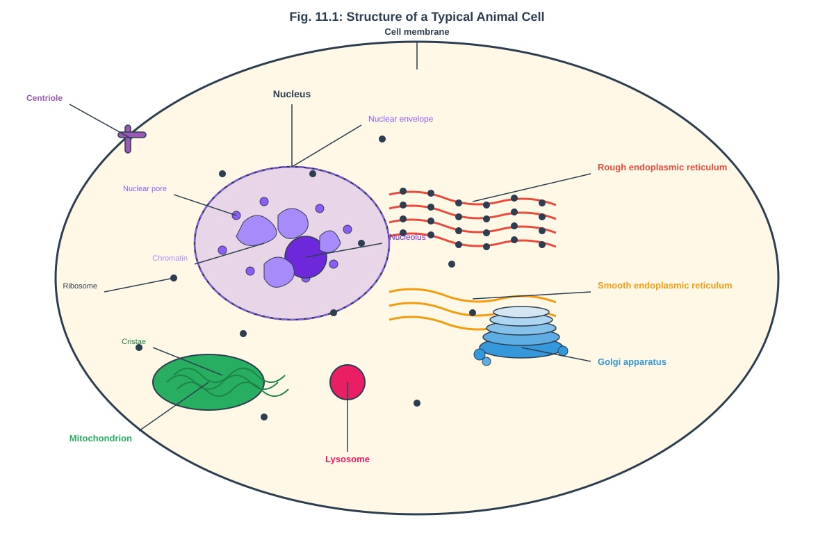
Generated diagram for Q11.
(a) Identify the organelle labelled X (pointing to the Golgi apparatus) and state two of its functions. [2]
(b) With reference to Fig. 11.1, state one structural difference between the rough endoplasmic reticulum and the smooth endoplasmic reticulum. [1]
(c) Explain why the mitochondrion is described as the "powerhouse of the cell." [2]
12. Fig. 12.1 shows the effect of temperature on the rate of an enzyme-catalysed reaction.
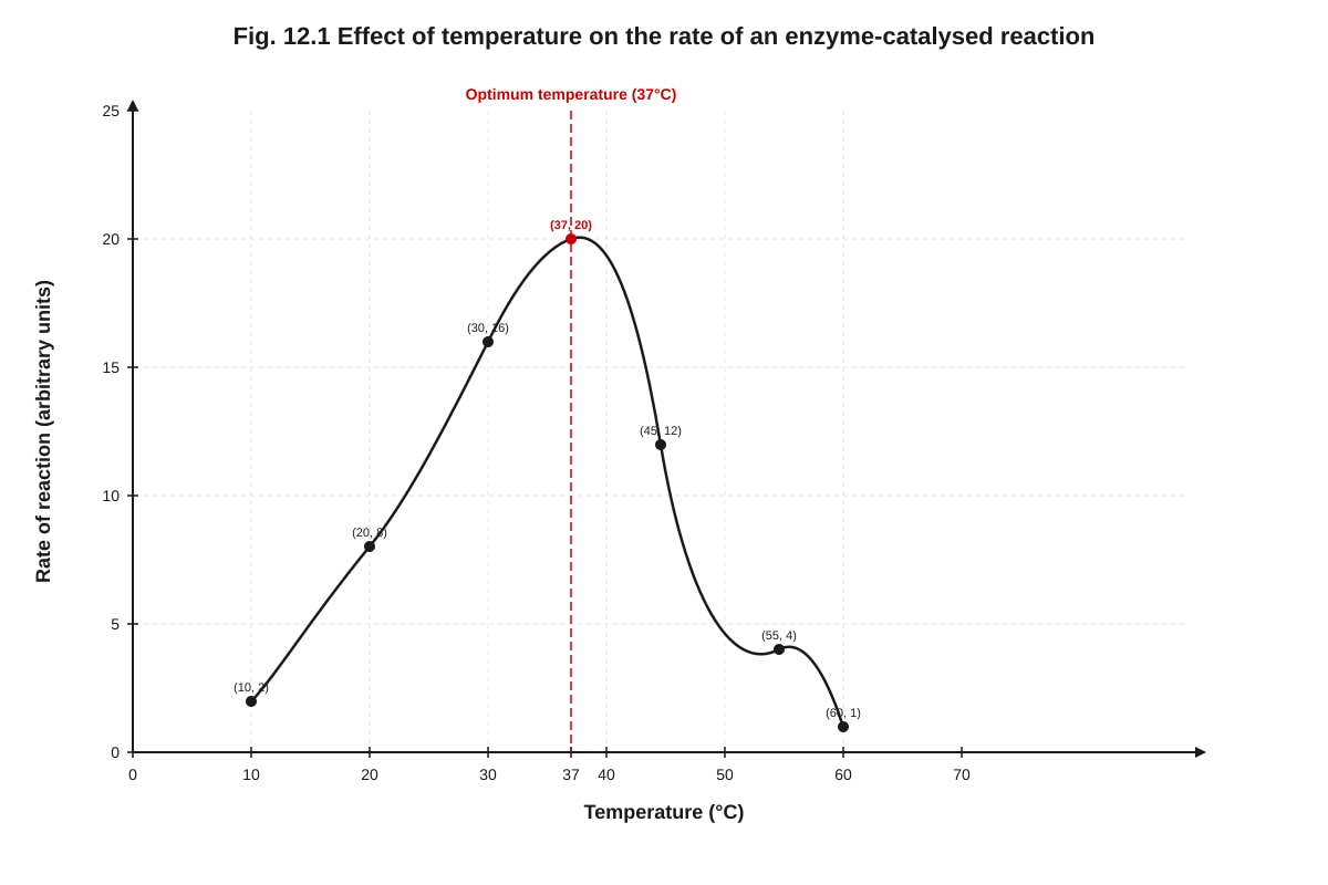
Generated graph for Q12.
(a) With reference to Fig. 12.1, state the optimum temperature for this enzyme. [1]
(b) Explain the shape of the curve between 10°C and 37°C. [2]
(c) Explain the sharp decrease in the rate of reaction above 37°C. [2]
(d) A student repeated the experiment using the same enzyme from a thermophilic bacterium. Predict and explain how the curve would differ. [2]
13. Table 13.1 shows the results of tests carried out on four food samples using different biochemical reagents.
| Food Sample | Benedict's Test | Iodine Test | Biuret Test | Ethanol Emulsion Test |
|---|---|---|---|---|
| A | Blue | Blue-black | Pale blue | Cloudy white |
| B | Brick-red | Blue | Pale blue | Clear |
| C | Blue | Blue | Violet | Cloudy white |
| D | Brick-red | Blue-black | Violet | Cloudy white |
(a) Which food sample contains starch but no reducing sugar? Explain your answer. [2]
(b) Which food sample contains all three types of biological molecules tested? Identify the sample and explain your reasoning. [2]
(c) Describe how the Biuret test is carried out and explain the chemical basis for a positive result. [2]
14. Fig. 14.1 shows a section through a cell surface membrane based on the fluid mosaic model.
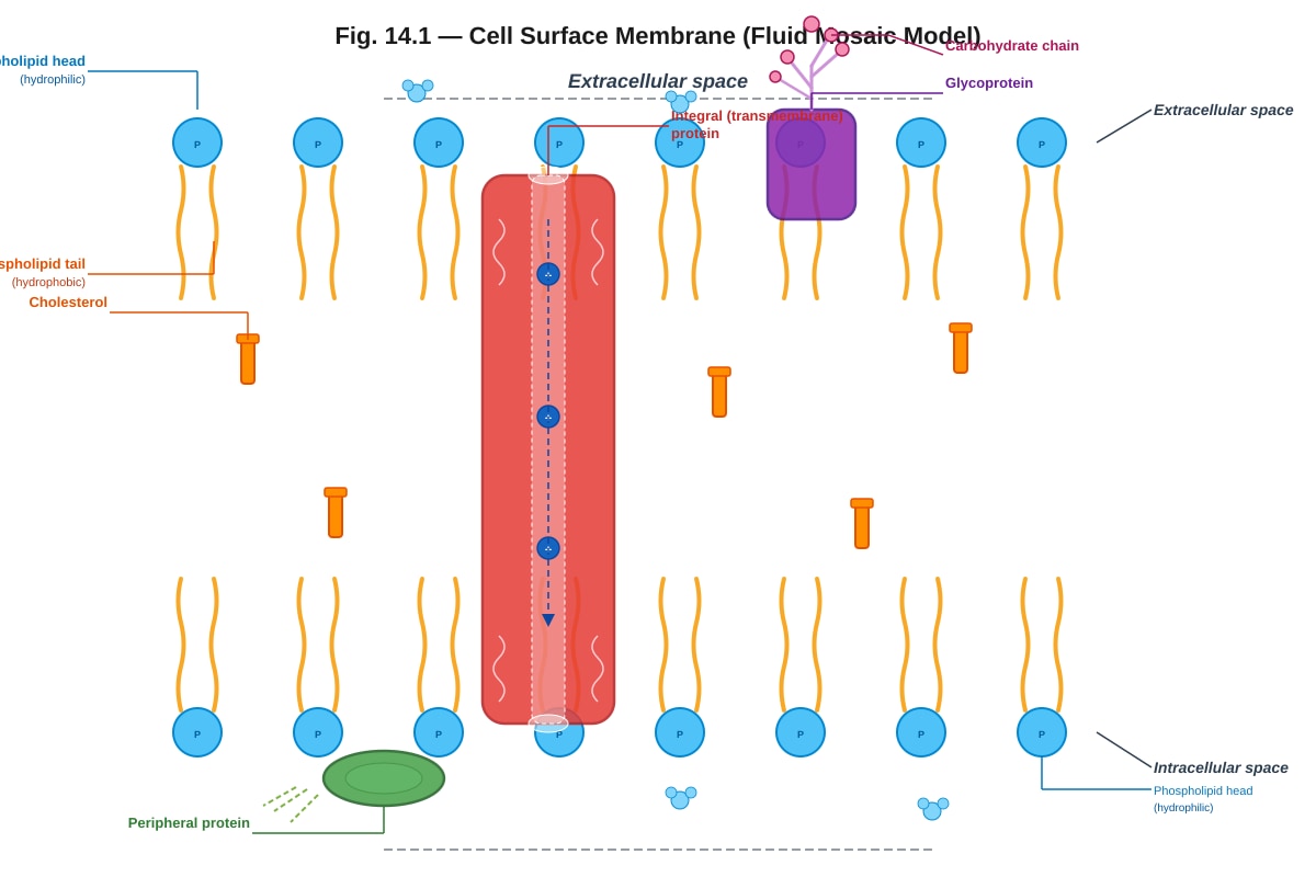
Generated diagram for Q14.
(a) With reference to Fig. 14.1, explain why the phospholipid bilayer is described as a "fluid" mosaic. [2]
(b) State two functions of cholesterol in the cell membrane. [2]
(c) Explain how the structure of the transmembrane protein enables it to function as a channel for the transport of ions across the membrane. [2]
Section C: Free Response [20 marks]
Answer two questions from this section. Each question carries 10 marks.
15. (a) Describe the structure of DNA, including the arrangement of nucleotides, the nature of the sugar-phosphate backbone, and the role of hydrogen bonding in maintaining the double helix. [5]
(b) Explain how the structure of DNA enables it to carry out two of the following functions: (i) accurate replication during cell division (ii) coding for the sequence of amino acids in a protein (iii) undergoing mutations that contribute to genetic variation [5]
16. (a) Describe the process of protein synthesis, including transcription and translation. In your answer, refer to the roles of mRNA, tRNA, ribosomes, and the genetic code. [6]
(b) Explain how a single gene mutation (a base substitution) can lead to a non-functional protein. Use a named example to illustrate your answer. [4]
17. (a) Compare and contrast the processes of facilitated diffusion and active transport across cell membranes. In your answer, refer to the role of membrane proteins, the direction of movement relative to the concentration gradient, and the requirement for energy. [6]
(b) Explain how the processes described in (a) contribute to the maintenance of a stable internal environment in a multicellular organism. [4]
End of Paper
Answers
TuitionGoWhere Practice Paper — Biology H2 A-Level
Answer Key & Marking Scheme
Paper: Practice Paper — Cells & Biomolecules Total Marks: 50
Section A: Multiple Choice [10 marks]
1. C — Ribosome [1]
- Explanation: Ribosomes are the only organelle listed that are found in both prokaryotic and eukaryotic cells. Prokaryotes lack a true nucleus (A), mitochondria (B), and endoplasmic reticulum (D). Ribosomes are the universal site of protein synthesis in all cells. Note that prokaryotic ribosomes (70S) are smaller than eukaryotic ribosomes (80S), but both types are present.
2. C — The hydrophobic tails face inward and the hydrophilic heads face outward. [1]
- Explanation: Phospholipids are amphipathic molecules — they have a hydrophilic (water-loving) phosphate head and two hydrophobic (water-fearing) fatty acid tails. In an aqueous environment, they spontaneously arrange into a bilayer to shield the hydrophobic tails from water while exposing the hydrophilic heads to the aqueous surroundings on both sides. This is the fundamental principle underlying membrane formation.
3. C — Hydrogen bonds [1]
- Explanation: The two antiparallel strands of DNA are held together by hydrogen bonds between complementary base pairs: adenine forms two hydrogen bonds with thymine, and guanine forms three hydrogen bonds with cytosine. Covalent bonds (A) form the sugar-phosphate backbone within each strand. Ionic bonds (B) are not involved in DNA structure. Phosphodiester bonds (D) are covalent bonds linking adjacent nucleotides within a single strand.
4. B — All active sites of the enzyme molecules are saturated with substrate. [1]
- Explanation: At very high substrate concentrations, every enzyme molecule has its active site occupied by a substrate molecule at any given moment. The enzyme is working at its maximum capacity (Vmax), so adding more substrate cannot increase the rate further. The enzyme is not denatured (A) — denaturation involves structural damage, typically from heat or pH extremes. Substrate inhibition (C) is a specific phenomenon not described here. The activation energy (D) is a fixed property of the reaction pathway and does not change with substrate concentration.
5. B — Lipid synthesis and detoxification [1]
- Explanation: The smooth endoplasmic reticulum (SER) lacks ribosomes and is involved in lipid synthesis (including phospholipids and steroids), carbohydrate metabolism, and detoxification of drugs and poisons. Protein synthesis (A) occurs on the rough endoplasmic reticulum. Packaging of proteins (C) is a function of the Golgi apparatus. ATP production (D) occurs in mitochondria.
6. B — A brick-red precipitate forms. [1]
- Explanation: Benedict's test detects reducing sugars (e.g., glucose, maltose, fructose). Benedict's reagent is blue due to copper(II) sulfate. In the presence of reducing sugars, the copper(II) ions (Cu²⁺) are reduced to copper(I) oxide (Cu₂O), which is insoluble and appears as a brick-red precipitate. Blue-black (A) is the positive result for the iodine test (starch). Violet (C) is the positive result for the Biuret test (protein). A purple ring (D) describes the Molisch test for carbohydrates.
7. B — S phase [1]
- Explanation: The S phase (Synthesis phase) of interphase is when DNA replication occurs, resulting in each chromosome being duplicated into two sister chromatids. G1 phase is a growth phase before DNA replication. G2 phase is a growth phase after DNA replication, where the cell prepares for mitosis. M phase (mitosis) is when the already-replicated chromosomes are separated into daughter cells.
8. C — ATP [1]
- Explanation: ATP (adenosine triphosphate) is the universal energy currency of the cell. The hydrolysis of ATP to ADP + Pᵢ releases energy that directly powers cellular processes such as active transport, muscle contraction, and biosynthesis. Glucose (A) is a fuel molecule that must be broken down to produce ATP. NADH (B) and FADH₂ (D) are electron carriers that donate electrons to the electron transport chain to generate ATP, but they are not the immediate source of energy for most cellular work.
9. C — Glycoproteins [1]
- Explanation: Glycoproteins (proteins with attached carbohydrate chains) on the cell surface act as recognition markers, enabling cells to identify each other. This is crucial for immune recognition, tissue formation, and cell signalling. Phospholipids (A) form the bilayer structure. Cholesterol (B) regulates membrane fluidity. Peripheral proteins (D) are involved in structural support and enzymatic functions but not primarily in cell-cell recognition.
10. C — Water is a medium in which enzymes and substrates can move and interact. [1]
- Explanation: Water provides the aqueous environment necessary for enzymes and substrates to diffuse, collide, and interact. Enzymes function in aqueous solution, and their three-dimensional structure (essential for activity) is maintained by interactions with water molecules. Water is not always a product (A) — it is a reactant in hydrolysis reactions and a product in condensation reactions. Water does not provide energy (B). Water does not directly participate in all active sites (D) — only in reactions where water is a substrate.
Section B: Structured Questions [20 marks]
11.
(a) Golgi apparatus (body / complex). [1 for identification]
- Two functions (any two): [1 — ½ mark each, max 1]
- Modifies proteins (e.g., glycosylation — adding carbohydrate groups to proteins to form glycoproteins)
- Packages proteins into vesicles for transport to their destinations (e.g., secretion via exocytosis, delivery to lysosomes)
- Sorts and labels proteins for delivery to the correct cellular location
- Produces lysosomes
- Involved in the processing and secretion of lipids
(b) The rough endoplasmic reticulum has ribosomes attached to its cytoplasmic surface, while the smooth endoplasmic reticulum lacks ribosomes. [1]
- Teaching note: This is the defining structural difference. The presence of ribosomes gives the RER its "rough" appearance under the electron microscope and is directly related to its role in protein synthesis.
(c) The mitochondrion is called the "powerhouse of the cell" because it is the site of aerobic cellular respiration, where energy stored in organic molecules (primarily glucose) is converted into ATP. [1]
- Specifically, the electron transport chain (located on the inner mitochondrial membrane / cristae) generates the majority of ATP through oxidative phosphorylation. [1]
- The highly folded cristae increase the surface area for the electron transport chain and ATP synthase enzymes, maximising ATP production. [1 — max 2 marks]
12.
(a) 37°C [1]
- Read directly from the graph — the peak of the curve occurs at 37°C.
(b) As temperature increases from 10°C to 37°C, the kinetic energy of both enzyme and substrate molecules increases. [1]
- This results in more frequent and more energetic collisions between enzyme and substrate molecules, leading to more enzyme-substrate complexes being formed per unit time, hence the rate of reaction increases. [1]
- Teaching note: Students should link temperature → kinetic energy → collision frequency/energy → rate. Simply stating "the rate increases" without explanation is insufficient for 2 marks.
(c) Above 37°C, the enzyme molecules gain excessive kinetic energy that disrupts the hydrogen bonds and other weak interactions (e.g., hydrophobic interactions, ionic bonds) that maintain the enzyme's three-dimensional tertiary structure. [1]
- This causes the enzyme to denature — the active site changes shape so that the substrate can no longer bind effectively, and the rate of reaction decreases sharply. [1]
- Common mistake: Students often say "the enzyme is killed" — enzymes are not living organisms and cannot be "killed." The correct term is "denatured."
(d) The curve would be shifted to the right, with the optimum temperature at a higher temperature (e.g., 60–80°C). [1]
- This is because enzymes from thermophilic bacteria have evolved to function at high temperatures — they have more stable tertiary structures (e.g., more disulfide bonds, stronger hydrophobic interactions, more compact folding) that resist denaturation at elevated temperatures. [1]
- Teaching note: The student should not simply say "the rate is higher" — they must explain the shift in optimum temperature and the structural basis for thermostability.
13.
(a) Sample C [1]
- Sample C gives a blue result with Benedict's test (no reducing sugar present) and a blue-black result with the iodine test (starch present). [1]
- Teaching note: Benedict's test is blue in the absence of reducing sugar. Iodine turns blue-black in the presence of starch.
(b) Sample D [1]
- Sample D gives a brick-red precipitate with Benedict's test (reducing sugar present), a blue-black result with the iodine test (starch present), a violet result with the Biuret test (protein present), and a cloudy white emulsion with the ethanol emulsion test (lipid present). [1]
- Therefore, Sample D contains reducing sugar, starch, protein, and lipid — all four biological molecules tested.
(c) Procedure: Add sodium hydroxide (NaOH) solution to the test sample, then add a few drops of copper(II) sulfate (CuSO₄) solution. [1]
- Chemical basis: In alkaline conditions, peptide bonds in proteins form a violet-coloured complex with copper(II) ions (Cu²⁺). The violet colour indicates the presence of peptide bonds and therefore protein. [1]
- Common mistake: Students sometimes confuse the Biuret test with Benedict's test. Benedict's test requires heating and detects reducing sugars, while the Biuret test detects peptide bonds and does not require heating.
14.
(a) "Fluid" — The phospholipids and proteins are not static; they can move laterally within the bilayer, giving the membrane a flexible, fluid character. [1]
- "Mosaic" — The membrane is composed of a variety of different components (phospholipids, proteins, cholesterol, glycoproteins) scattered throughout like tiles in a mosaic pattern. [1]
- Teaching note: The term "fluid mosaic" was proposed by Singer and Nicolson in 1972. Students should address both parts of the term separately.
(b) Two functions of cholesterol: [1 each, max 2]
- Regulates membrane fluidity — at high temperatures, cholesterol reduces fluidity by restraining phospholipid movement; at low temperatures, it prevents the membrane from becoming too rigid by preventing close packing of phospholipid tails.
- Increases mechanical stability of the membrane.
- Reduces permeability to small polar molecules and ions.
- Helps maintain the shape of animal cells (which lack cell walls).
(c) The transmembrane channel protein spans the entire phospholipid bilayer. [1]
- The hydrophobic exterior of the protein interacts with the hydrophobic fatty acid tails of the phospholipids, anchoring the protein in the membrane. [1]
- The interior of the channel is hydrophilic, creating a pore through which hydrophilic ions can pass across the otherwise impermeable hydrophobic core of the bilayer. [1 — max 2 marks]
- Teaching note: Students should link the protein's structure (hydrophobic exterior, hydrophilic interior) to its function (allowing ion transport across the hydrophobic membrane core).
Section C: Free Response [20 marks]
Answer two questions. Each question carries 10 marks.
15.
(a) Description of DNA structure [5 marks]
- DNA is a double helix composed of two antiparallel polynucleotide strands wound around a common axis. [1]
- Each nucleotide consists of three components: a deoxyribose sugar, a phosphate group, and a nitrogenous base (adenine, thymine, guanine, or cytosine). [1]
- The sugar-phosphate backbone is formed by covalent phosphodiester bonds linking the 3' carbon of one deoxyribose to the 5' carbon of the next, providing structural stability. [1]
- The two strands are held together by hydrogen bonds between complementary base pairs: A=T (two hydrogen bonds) and G≡C (three hydrogen bonds). [1]
- The strands run in antiparallel directions — one runs 5'→3' and the other 3'→5'. [1]
(b) Explanation of how DNA structure enables two functions [5 marks — 2.5 per function, or 3+2]
(i) Accurate replication:
- Complementary base pairing (A-T, G-C) means that each strand serves as a template for the synthesis of a new complementary strand. [1]
- During replication, the double helix is unwound by helicase, and DNA polymerase adds nucleotides to the new strand according to base-pairing rules, ensuring that the sequence of the new strand is precisely determined by the template strand. [1]
- This semi-conservative mechanism ensures genetic fidelity — each daughter DNA molecule contains one original and one new strand. [½]
(ii) Coding for amino acid sequences:
- The sequence of bases along a DNA strand constitutes the genetic code. [1]
- Each amino acid is specified by a triplet code (codon) of three consecutive bases. During transcription, the DNA sequence is copied into mRNA, which is then translated into a polypeptide by ribosomes. [1]
- The linear sequence of codons determines the linear sequence of amino acids in the protein, which in turn determines the protein's structure and function. [½]
(iii) Mutations contributing to genetic variation:
- Changes in the DNA base sequence (e.g., base substitutions, insertions, deletions) can alter the amino acid sequence of the encoded protein. [1]
- These mutations are a source of new alleles, which contribute to genetic variation within a population. [1]
- If the mutation confers a selective advantage, it may be favoured by natural selection and increase in frequency in the population over generations. [½]
16.
(a) Description of protein synthesis [6 marks]
Transcription (in the nucleus):
- The gene encoding the protein is transcribed. RNA polymerase binds to the promoter region of the gene and unwinds the DNA double helix. [1]
- RNA polymerase synthesises a complementary mRNA strand using one DNA strand as the template, following base-pairing rules (A-U, T-A, G-C, C-G), in the 5'→3' direction. [1]
- The pre-mRNA undergoes processing (5' capping, 3' polyadenylation, splicing to remove introns) before mature mRNA exits the nucleus via nuclear pores. [½]
Translation (at ribosomes in the cytoplasm):
- The mRNA binds to a ribosome. The ribosome reads the mRNA sequence in triplets called codons. [1]
- tRNA molecules, each carrying a specific amino acid, have anticodons that are complementary to the mRNA codons. The tRNA anticodon base-pairs with the mRNA codon at the ribosome's A site. [1]
- A peptide bond forms between the amino acids at the A site and P site, catalysed by peptidyl transferase (a ribozyme). The ribosome translocates along the mRNA, and the process continues until a stop codon is reached. [1]
- The polypeptide chain is released and folds into its functional three-dimensional structure. [½]
(b) Explanation of how a base substitution can lead to a non-functional protein [4 marks]
- A base substitution is a point mutation where one nucleotide is replaced by another in the DNA sequence. [1]
- This changes the codon in the mRNA, which may code for a different amino acid (missense mutation). If the substituted amino acid is in a critical region of the protein (e.g., the active site of an enzyme), the protein's three-dimensional structure and function may be severely disrupted. [1]
- Example: Sickle cell anaemia — a single base substitution (GAG→GTG) in the gene for the β-globin chain of haemoglobin changes the codon from GAG (glutamic acid) to GTG (valine). [1]
- This single amino acid change causes haemoglobin molecules to polymerise under low oxygen conditions, distorting red blood cells into a sickle shape, leading to blockage of blood vessels and reduced oxygen transport. The protein is effectively non-functional in its oxygen-carrying role under these conditions. [1]
- Alternative example: A base substitution could also create a nonsense mutation (premature stop codon), resulting in a truncated, non-functional protein.
17.
(a) Comparison of facilitated diffusion and active transport [6 marks]
| Feature | Facilitated Diffusion | Active Transport |
|---|---|---|
| Direction | Down the concentration gradient (high → low) | Against the concentration gradient (low → high) |
| Energy requirement | No energy (ATP) required | Requires energy (ATP) |
| Membrane proteins | Uses channel proteins or carrier proteins | Uses carrier proteins (pumps) |
| Specificity | Specific to particular molecules/ions | Specific to particular molecules/ions |
| Saturation | Rate plateaus at high concentrations (all proteins occupied) | Rate plateaus at high concentrations (all pumps occupied) |
- Facilitated diffusion relies on channel proteins (which form hydrophilic pores for ions) or carrier proteins (which change shape to transport molecules such as glucose). The substance moves passively down its concentration gradient — no metabolic energy is required. [1]
- Active transport uses carrier proteins (e.g., the sodium-potassium pump) that use energy from ATP hydrolysis to change conformation and pump substances against their concentration gradient. [1]
- Both processes are specific — the shape and binding site of the transport protein determines which substance is transported. [1]
- Both show saturation kinetics — at high solute concentrations, all transport proteins are occupied, and the rate reaches a maximum. [1]
- Key difference: Active transport requires energy input (directly from ATP or indirectly via an electrochemical gradient), while facilitated diffusion does not. [1]
- Key difference: Active transport can move substances against their concentration gradient, while facilitated diffusion cannot. [1]
(b) Contribution to maintaining a stable internal environment [4 marks]
- Active transport enables cells to maintain internal concentrations of ions and molecules that differ from their surroundings. For example, the sodium-potassium pump (Na⁺/K⁺-ATPase) maintains a high intracellular K⁺ concentration and low intracellular Na⁺ concentration, which is essential for nerve impulse transmission, muscle contraction, and osmotic balance. [1]
- Facilitated diffusion allows rapid uptake of essential nutrients (e.g., glucose via GLUT transporters) when extracellular concentrations are high, ensuring a constant supply of substrates for cellular respiration. [1]
- Together, these processes enable homeostasis — the maintenance of a stable internal environment despite changes in external conditions. For example, cells can regulate their volume by controlling ion concentrations through a combination of active transport and facilitated diffusion. [1]
- In multicellular organisms, these transport processes at the cellular level contribute to the regulation of blood glucose levels, pH balance, and electrolyte concentrations at the organism level. [1]
End of Answer Key
Mark Summary:
| Section | Marks |
|---|---|
| A: Multiple Choice (Q1–10) | 10 |
| B: Structured Questions (Q11–14) | 20 |
| C: Free Response (Q15–17, best 2) | 20 |
| Total | 50 |
Free quiz and exam paper access
Enter your details to view this paper
Your access is remembered on this device.