AI Generated Exam Paper
A Level H2 Biology Practice Paper 5
Free A Level H2 Biology Practice Paper 5, DeepSeek AI version, with questions, answers, and A Level-style practice for Singapore students.
These static practice materials are generated from the site's syllabus and paper-generation workflow, with source and model context shown so students and parents can evaluate the material before use.
Questions
TuitionGoWhere Practice Paper - Biology H2 A-Level
TuitionGoWhere Practice Paper (AI)
Subject: Biology H2 (9477) Level: A-Level Paper: Practice Paper 5 (Cells & Biomolecules) Duration: 2 hours Total Marks: 75
Name: _________________________ Class: _________________________ Date: _________________________
Instructions to Candidates
- This paper consists of three sections: Section A, Section B, and Section C.
- Answer all questions in Section A and Section B.
- Section C contains one essay question. Answer the question.
- Write your answers in the spaces provided.
- The number of marks is given in brackets [ ] at the end of each question or part question.
- You are advised to spend no more than 50 minutes on Section A, 50 minutes on Section B, and 20 minutes on Section C.
Section A: Structured Questions (30 marks)
Answer all questions in this section.
1. Figure 1.1 shows a transmission electron micrograph of a eukaryotic cell.
(a) Identify the organelles labelled P, Q, and R. [3]
P: _________________________ Q: _________________________ R: _________________________
(b) Organelle P is described as being "semi-autonomous". Explain what this term means and provide one piece of evidence that supports this description. [2]
(c) Compare and contrast the structure of organelle Q with that of a typical prokaryotic ribosome. [2]
2. A student investigated the effect of temperature on the rate of diffusion of potassium permanganate crystals in agar jelly. The results are shown in Table 2.1.
Table 2.1
| Temperature / °C | Distance diffused after 30 minutes / mm |
|---|---|
| 10 | 4.2 |
| 20 | 6.8 |
| 30 | 9.5 |
| 40 | 12.1 |
| 50 | 14.3 |
(a) Describe the relationship between temperature and the distance diffused. [1]
(b) Explain the effect of temperature on the rate of diffusion, with reference to the kinetic energy of particles. [2]
(c) The student suggested that the rate of diffusion would continue to increase indefinitely with temperature. Evaluate this suggestion. [2]
3. Figure 3.1 represents the fluid mosaic model of the cell surface membrane.
(a) Name the structures labelled X, Y, and Z. [3]
X: _________________________ Y: _________________________ Z: _________________________
(b) Explain how the structure of a phospholipid molecule enables it to form a bilayer in an aqueous environment. [2]
(c) Glycoproteins and glycolipids are components of the cell surface membrane. Outline two functions of these molecules. [2]
4. A biologist measured the water potential of potato tuber cells using a range of sucrose solutions. The results are shown in Table 4.1.
Table 4.1
| Sucrose concentration / mol dm⁻³ | Percentage change in mass of potato cylinders |
|---|---|
| 0.0 | +18.5 |
| 0.2 | +10.2 |
| 0.4 | +3.1 |
| 0.6 | -4.8 |
| 0.8 | -12.6 |
| 1.0 | -19.3 |
(a) Using the data in Table 4.1, estimate the water potential of the potato tuber cells. Explain your reasoning. [2]
(b) Explain why the potato cylinders gained mass when placed in 0.0 mol dm⁻³ sucrose solution. [2]
(c) A student suggested that placing the potato cylinders in a 1.5 mol dm⁻³ sucrose solution would result in a greater percentage loss in mass. Discuss whether this prediction is likely to be correct. [2]
5. Figure 5.1 shows the molecular structure of a disaccharide.
(a) Name the disaccharide shown in Figure 5.1. [1]
(b) Identify the type of glycosidic bond present in this disaccharide. [1]
(c) Describe how this disaccharide is formed from its constituent monosaccharides. [2]
(d) Explain why this disaccharide is classified as a reducing sugar. [2]
Section B: Data-Based and Extended Response Questions (30 marks)
Answer all questions in this section.
6. Amylase is an enzyme that catalyses the hydrolysis of starch to maltose. A student investigated the effect of pH on the activity of amylase extracted from two different organisms: a bacterium found in hot springs (Bacillus sp.) and a fungus found in soil (Aspergillus sp.).
The results are shown in Figure 6.1.
Figure 6.1
| pH | Bacillus amylase activity / arbitrary units | Aspergillus amylase activity / arbitrary units |
|---|---|---|
| 3.0 | 2 | 18 |
| 4.0 | 8 | 42 |
| 5.0 | 22 | 68 |
| 6.0 | 48 | 85 |
| 7.0 | 72 | 72 |
| 8.0 | 88 | 38 |
| 9.0 | 65 | 12 |
| 10.0 | 28 | 3 |
(a) Compare the pH optima of the two amylase enzymes. Use data from Figure 6.1 to support your answer. [2]
(b) Explain why enzyme activity decreases at pH values above and below the optimum. [3]
(c) The bacterium Bacillus sp. lives in hot springs with temperatures around 70°C and pH 8.0. Suggest and explain how the structure of Bacillus amylase is adapted to function in this environment. [3]
7. Mitochondria are the site of aerobic respiration in eukaryotic cells. Figure 7.1 is a diagram of a mitochondrion.
(a) Name the structures labelled A, B, and C. [3]
A: _________________________ B: _________________________ C: _________________________
(b) Explain how the structure of the inner mitochondrial membrane is adapted for its function in oxidative phosphorylation. [3]
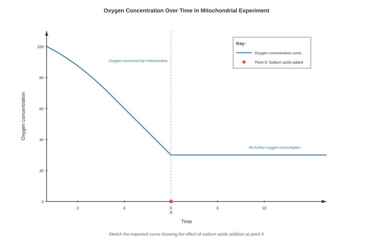
(c) An experiment was carried out using isolated mitochondria in a buffer containing ADP and inorganic phosphate (Pi). The oxygen concentration in the buffer was monitored over time. At point X, sodium azide (an inhibitor of cytochrome c oxidase) was added.
Sketch the expected oxygen concentration curve on the axes below, showing the effect of adding sodium azide at point X. Label your graph clearly. [2]
Generated graph for this question.
(d) Explain the effect of sodium azide on oxygen consumption by the mitochondria. [2]
8. Collagen is a fibrous protein found in connective tissues such as tendons, ligaments, and skin.
(a) Describe the primary, secondary, and tertiary structure of a collagen molecule. [4]
(b) Explain how the structure of collagen is related to its function as a structural protein in tendons. [3]
(c) Scurvy is a disease caused by vitamin C deficiency. Vitamin C is required for the hydroxylation of proline and lysine residues in collagen. Suggest and explain why individuals with scurvy have weakened connective tissues. [3]
9. Figure 9.1 shows the structure of a nucleotide.
(a) Name the components labelled D, E, and F. [3]
D: _________________________ E: _________________________ F: _________________________
(b) This nucleotide is a monomer of RNA. Identify one structural difference between this nucleotide and a DNA nucleotide. [1]
(c) Describe how nucleotides are joined together to form a polynucleotide strand. [2]
Section C: Essay Question (15 marks)
Answer one question from this section.
Write your answer in the space provided. You are advised to spend approximately 20 minutes on this section.
10. Discuss the importance of hydrogen bonding in determining the structure and properties of biological molecules. In your answer, you should refer to at least three different types of biological molecules.
[15]
END OF PAPER
© TuitionGoWhere 2025. This is an AI-generated practice paper intended for educational use. It is not derived from past-year examination papers.
Answers
TuitionGoWhere Practice Paper - Biology H2 A-Level
Answer Key and Marking Scheme
Subject: Biology H2 (9477) Paper: Practice Paper 5 (Cells & Biomolecules) Total Marks: 75
Section A: Structured Questions (30 marks)
Question 1
(a) [3 marks]
- P: Mitochondrion / Mitochondria [1]
- Q: Rough endoplasmic reticulum / RER [1]
- R: Golgi apparatus / Golgi body / Golgi complex [1]
(b) [2 marks]
- Semi-autonomous means the organelle can replicate independently of the cell cycle / contains its own genetic material and ribosomes / can synthesise some of its own proteins [1].
- Evidence: Mitochondria contain their own circular DNA (mtDNA) / mitochondria contain 70S ribosomes similar to prokaryotes / mitochondria divide by binary fission [1].
(c) [2 marks]
- Similarity: Both are composed of rRNA and proteins / both have a large and small subunit [1].
- Difference: Organelle Q (RER ribosomes) are 80S ribosomes, while prokaryotic ribosomes are 70S / RER ribosomes are larger than prokaryotic ribosomes [1].
Question 2
(a) [1 mark]
- As temperature increases, the distance diffused increases / positive correlation between temperature and distance diffused [1].
(b) [2 marks]
- At higher temperatures, particles have greater kinetic energy [1].
- Particles move faster, so they diffuse more rapidly / collide more frequently, spreading further in the same time period [1].
(c) [2 marks]
- The suggestion is incorrect / unlikely to be correct [1].
- At very high temperatures, the agar jelly would melt / the protein structure of the agar would denature / the medium would break down, so diffusion cannot be measured / at extremely high temperatures, the crystals may decompose [1].
Question 3
(a) [3 marks]
- X: Phospholipid / phospholipid bilayer [1]
- Y: Integral protein / intrinsic protein / channel protein / carrier protein [1]
- Z: Cholesterol [1]
(b) [2 marks]
- Phospholipids have a hydrophilic (polar) phosphate head and two hydrophobic (non-polar) fatty acid tails [1].
- In an aqueous environment, the hydrophilic heads face outwards towards the water, while the hydrophobic tails face inwards away from the water, forming a bilayer [1].
(c) [2 marks]
- Any two from:
- Cell recognition / cell-to-cell recognition [1]
- Cell adhesion / binding cells together [1]
- Receptor sites for hormones / neurotransmitters / signalling molecules [1]
- Antigens / involved in immune response [1]
- Forming the glycocalyx [1]
Question 4
(a) [2 marks]
- The water potential of the potato cells is approximately equal to the water potential of the sucrose solution where there is no net change in mass / where the percentage change is zero [1].
- From the graph/interpolation, this occurs at approximately 0.45–0.50 mol dm⁻³ sucrose / between 0.4 and 0.6 mol dm⁻³ where the line crosses zero [1].
(b) [2 marks]
- The 0.0 mol dm⁻³ sucrose solution (distilled water) has a higher water potential (less negative / zero) than the potato cells [1].
- Water enters the cells by osmosis down the water potential gradient, causing the cells to swell and gain mass [1].
(c) [2 marks]
- The prediction is likely to be correct because the trend shows increasing mass loss with increasing sucrose concentration [1].
- However, the relationship may not be linear at very high concentrations / the cells may become fully plasmolysed and mass loss may plateau / at very high concentrations, the water potential gradient may not increase proportionally [1].
Question 5
(a) [1 mark]
- Maltose [1]
(b) [1 mark]
- α-1,4-glycosidic bond / alpha-1,4-glycosidic bond [1]
(c) [2 marks]
- Maltose is formed by a condensation reaction between two α-glucose molecules [1].
- A molecule of water is removed, and a glycosidic bond forms between carbon-1 of one glucose and carbon-4 of the other [1].
(d) [2 marks]
- Maltose has a free anomeric carbon / free carbon-1 on one of the glucose residues that is not involved in the glycosidic bond [1].
- This free anomeric carbon can open to form an aldehyde group, which can reduce Benedict's reagent / act as a reducing agent [1].
Section B: Data-Based and Extended Response Questions (30 marks)
Question 6
(a) [2 marks]
- Bacillus amylase has an optimum pH of approximately 8.0 (activity = 88 arbitrary units) [1].
- Aspergillus amylase has an optimum pH of approximately 6.0 (activity = 85 arbitrary units) / Aspergillus amylase has a lower pH optimum than Bacillus amylase [1].
(b) [3 marks]
- At pH values above and below the optimum, the charges on the amino acid side chains in the active site are altered [1].
- This disrupts the ionic and hydrogen bonds that maintain the specific three-dimensional shape (tertiary structure) of the enzyme [1].
- The active site is no longer complementary to the substrate, so the enzyme-substrate complex cannot form / the enzyme is denatured at extreme pH [1].
(c) [3 marks]
- The enzyme has a high proportion of disulfide bonds / more ionic bonds / more hydrophobic interactions that stabilise its tertiary structure at high temperatures [1].
- The enzyme's amino acid composition results in an active site with charges that are optimal at pH 8.0 [1].
- The enzyme is adapted to the hot spring environment, so it is not denatured at 70°C and functions most efficiently at pH 8.0, allowing the bacterium to digest starch in its habitat [1].
Question 7
(a) [3 marks]
- A: Outer mitochondrial membrane [1]
- B: Crista / cristae [1]
- C: Matrix [1]
(b) [3 marks]
- The inner membrane is highly folded into cristae, which greatly increases the surface area for the electron transport chain and ATP synthase [1].
- The membrane contains the protein complexes of the electron transport chain (NADH dehydrogenase, cytochrome b-c1 complex, cytochrome c oxidase) for electron transfer and proton pumping [1].
- The membrane contains ATP synthase / stalked particles, which use the proton gradient to synthesise ATP via chemiosmosis [1].
- The membrane is impermeable to protons (H⁺), allowing a proton gradient to be established [1].
(c) [2 marks]
- Graph should show:
- A steady decrease in oxygen concentration before point X (indicating oxygen consumption by respiring mitochondria) [1].
- After point X, the rate of oxygen consumption decreases significantly / the line becomes almost flat (indicating inhibition of the electron transport chain) [1].
(d) [2 marks]
- Sodium azide inhibits cytochrome c oxidase (Complex IV), the final enzyme in the electron transport chain [1].
- This prevents the transfer of electrons to oxygen (the final electron acceptor), so oxygen is no longer reduced to water, and oxygen consumption stops or decreases dramatically [1].
Question 8
(a) [4 marks]
- Primary structure: The repeating amino acid sequence Gly-X-Y, where X is often proline and Y is often hydroxyproline / a specific sequence rich in glycine, proline, and hydroxyproline [1].
- Secondary structure: Each polypeptide chain forms a left-handed helix / a tight, extended helix (not an α-helix) [1].
- Tertiary structure: Three polypeptide chains wind around each other to form a right-handed triple helix / tropocollagen molecule [1].
- The structure is stabilised by hydrogen bonds between the chains and covalent cross-links between lysine and hydroxylysine residues [1].
(b) [3 marks]
- Collagen molecules assemble into fibrils, which are further bundled into fibres, providing high tensile strength [1].
- The triple helix structure and covalent cross-links between molecules make collagen strong and resistant to stretching [1].
- This allows tendons to transmit the force of muscle contraction to bones without breaking / collagen provides structural support in tendons [1].
(c) [3 marks]
- Vitamin C is a cofactor for the enzymes prolyl hydroxylase and lysyl hydroxylase, which catalyse the hydroxylation of proline and lysine residues in collagen [1].
- Without hydroxylation, fewer hydrogen bonds form between the three polypeptide chains, so the triple helix is less stable [1].
- The collagen molecules are weaker and cannot form strong fibres, leading to weakened connective tissues, fragile blood vessels, and poor wound healing [1].
Question 9
(a) [3 marks]
- D: Phosphate group [1]
- E: Ribose / pentose sugar [1]
- F: Nitrogenous base / adenine / guanine / cytosine / uracil [1]
(b) [1 mark]
- The sugar is ribose (in RNA) instead of deoxyribose (in DNA) / RNA contains uracil instead of thymine [1].
(c) [2 marks]
- Nucleotides are joined by phosphodiester bonds formed through condensation reactions [1].
- The phosphate group of one nucleotide bonds to the hydroxyl group on carbon-3' of the sugar of the next nucleotide, forming a sugar-phosphate backbone [1].
Section C: Essay Question (15 marks)
Question 10
Marking scheme for essay question:
The essay should demonstrate a comprehensive understanding of the role of hydrogen bonding in biological molecules. Marks are allocated for content (maximum 12 marks) and for the quality of written communication and organisation (maximum 3 marks).
Content marks (maximum 12):
Candidates should refer to at least three different types of biological molecules. Award marks for accurate, detailed explanations of the role of hydrogen bonding in each.
1. Water (up to 4 marks):
- Water molecules are polar, with δ+ on hydrogen atoms and δ- on oxygen atom [1].
- Hydrogen bonds form between the δ+ hydrogen of one water molecule and the δ- oxygen of another [1].
- Hydrogen bonding gives water its properties: high specific heat capacity (many H-bonds absorb energy before temperature rises), high latent heat of vaporisation (H-bonds must be broken for evaporation), cohesion and surface tension (H-bonds between water molecules), and solvent properties (H-bonds with polar solutes) [up to 2 marks for explaining at least two properties].
2. Proteins (up to 4 marks):
- Hydrogen bonds form between the C=O and N-H groups of amino acids in the polypeptide backbone [1].
- In secondary structure: H-bonds stabilise α-helices (between every fourth amino acid) and β-pleated sheets (between adjacent polypeptide strands) [1].
- In tertiary structure: H-bonds form between R-groups of amino acids (e.g., between serine, threonine, tyrosine residues) [1].
- Hydrogen bonding is essential for maintaining the specific three-dimensional shape of proteins, which determines their function (e.g., enzyme active sites, antibody binding sites) [1].
3. DNA (up to 4 marks):
- Hydrogen bonds form between complementary nitrogenous bases on the two antiparallel polynucleotide strands [1].
- Adenine forms two hydrogen bonds with thymine; guanine forms three hydrogen bonds with cytosine [1].
- Hydrogen bonding holds the two strands together in the double helix, but allows them to separate during replication and transcription [1].
- The specificity of base pairing (complementary) ensures accurate replication and transmission of genetic information [1].
4. Polysaccharides / Carbohydrates (up to 3 marks):
- Hydrogen bonds form between adjacent cellulose chains in plant cell walls [1].
- These H-bonds cross-link cellulose microfibrils, giving cellulose high tensile strength [1].
- This enables plant cell walls to withstand turgor pressure and provide structural support [1].
5. Collagen (up to 3 marks):
- Hydrogen bonds form between the three polypeptide chains in the collagen triple helix [1].
- These H-bonds, along with covalent cross-links, stabilise the tropocollagen structure [1].
- This contributes to the high tensile strength of collagen fibres in connective tissues [1].
Quality of written communication (maximum 3 marks):
- 3 marks: Answer is well-structured, uses appropriate scientific terminology accurately, and presents a logical and coherent argument with clear links between structure and function.
- 2 marks: Answer is mostly well-organised with some use of scientific terminology; ideas are generally clear but may lack full coherence.
- 1 mark: Answer has some relevant content but lacks organisation; limited or inaccurate use of scientific terminology.
- 0 marks: Answer is poorly organised with little or no relevant scientific content.
END OF ANSWER KEY
© TuitionGoWhere 2025. This is an AI-generated answer key intended for educational use.
Free quiz and exam paper access
Enter your details to view this paper
Your access is remembered on this device.