AI Generated Exam Paper
A Level H2 Biology Practice Paper 4
Free A Level H2 Biology Practice Paper 4, LongCat AI version, with questions, answers, and A Level-style practice for Singapore students.
These static practice materials are generated from the site's syllabus and paper-generation workflow, with source and model context shown so students and parents can evaluate the material before use.
Questions
TuitionGoWhere Practice Paper - Biology H2 A-Level
TuitionGoWhere Practice Paper (AI)
Subject: Biology
Level: A-Level H2
Paper: Practice Paper — Cells & Biomolecules
Version: 4 of 5
Duration: 1 hour 30 minutes
Total Marks: 60
Name: ___________________________
Class: ___________________________
Date: ___________________________
Instructions
- Answer all questions in the spaces provided.
- Write in dark blue or black pen.
- You may use a pencil for any diagrams or graphs.
- Do not use correction fluid.
- The number of marks for each question or part question is shown in brackets [ ].
- The total marks for this paper is 60.
- You are advised to spend no more than 90 minutes on this paper.
Section A: Structured Questions (30 marks)
Answer all questions 1–10.
1. Fig. 1.1 shows the structure of a typical eukaryotic animal cell as seen under an electron microscope.
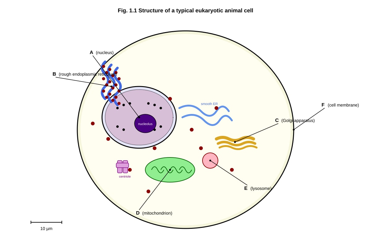
Generated diagram for Q1.
(a) State the function of the organelle labelled C.
___________________________________________________________________________ [1]
(b) Organelle D is the site of aerobic respiration. Describe two structural features of organelle D that are visible in Fig. 1.1 and explain how each feature is adapted to its function.
___________________________________________________________________________ [4]
(c) Suggest why organelle E contains hydrolytic enzymes and explain the consequence if these enzymes were released into the cytoplasm.
___________________________________________________________________________ [2]
[Total: 7 marks]
2. Table 2.1 shows the relative concentrations of ions inside and outside a typical mammalian cell.
| Ion | Intracellular concentration (mmol dm⁻³) | Extracellular concentration (mmol dm⁻³) |
|---|---|---|
| Na⁺ | 15 | 145 |
| K⁺ | 150 | 5 |
| Ca²⁺ | 0.0001 | 2.5 |
| Cl⁻ | 10 | 110 |
Table 2.1
(a) With reference to Table 2.1, identify the ion with the steepest concentration gradient across the cell membrane. Show your reasoning.
___________________________________________________________________________ [2]
(b) Explain how the sodium-potassium pump maintains the ion distribution shown in Table 2.1.
___________________________________________________________________________ [3]
[Total: 5 marks]
3. Fig. 3.1 shows the effect of temperature on the rate of an enzyme-catalysed reaction.
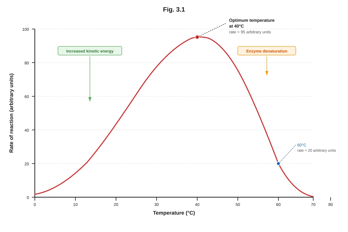
Generated graph for Q3.
(a) With reference to Fig. 3.1, describe the effect of temperature on the rate of this enzyme-catalysed reaction between 0 °C and 40 °C.
___________________________________________________________________________ [2]
(b) Explain the decrease in the rate of reaction above 40 °C.
___________________________________________________________________________ [2]
(c) A student claims that doubling the enzyme concentration at 60 °C would restore the rate of reaction to its maximum. Evaluate this claim.
___________________________________________________________________________ [2]
[Total: 6 marks]
4. Fig. 4.1 shows a phospholipid bilayer forming part of a cell surface membrane.
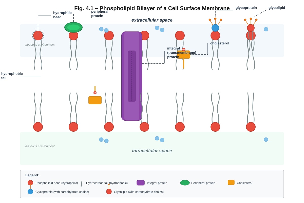
Generated diagram for Q4.
(a) With reference to Fig. 4.1, explain why phospholipids spontaneously arrange themselves into a bilayer in an aqueous environment.
___________________________________________________________________________ [2]
(b) Explain the role of cholesterol in the cell surface membrane.
___________________________________________________________________________ [2]
(c) State two functions of glycoproteins in the cell surface membrane.
-
- _________________________________________________________________________ [2]
[Total: 6 marks]
5. Describe the process of mitosis and explain how it results in the production of two genetically identical daughter cells.
___________________________________________________________________________ [5]
[Total: 5 marks]
6. A student carried out an experiment to investigate the effect of pH on the activity of the enzyme catalase, which breaks down hydrogen peroxide into water and oxygen. The volume of oxygen produced in the first 2 minutes was measured at different pH values. The results are shown in Table 6.1.
| pH | Volume of O₂ produced in 2 min / cm³ |
|---|---|
| 3 | 2.1 |
| 5 | 8.4 |
| 7 | 18.6 |
| 9 | 12.3 |
| 11 | 3.7 |
Table 6.1
(a) State the optimum pH for catalase activity based on the data in Table 6.1.
___________________________________________________________________________ [1]
(b) Explain why the volume of oxygen produced decreases at pH values above and below the optimum.
___________________________________________________________________________ [3]
(c) Identify two variables that should be kept constant in this experiment to ensure a fair test.
-
- _________________________________________________________________________ [2]
[Total: 6 marks]
7. Fig. 7.1 shows the structure of a nucleotide found in DNA.
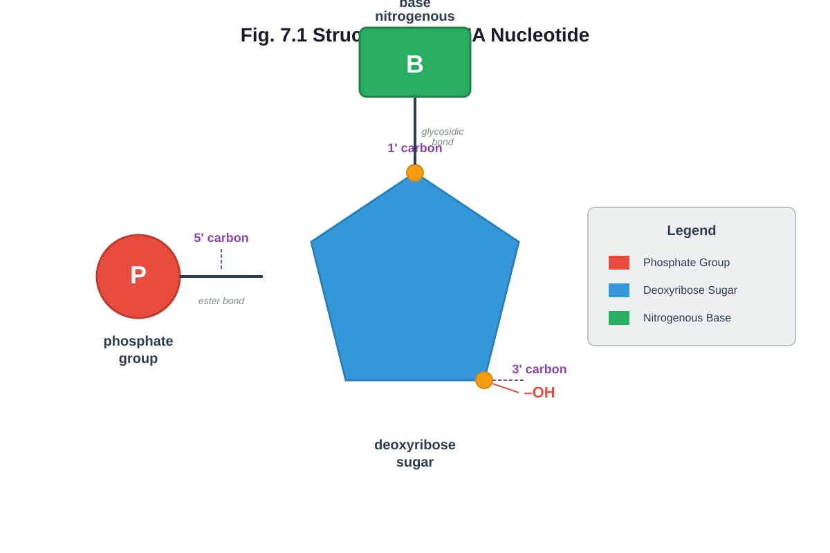
Generated diagram for Q7.
(a) With reference to Fig. 7.1, name the bond that joins the nitrogenous base to the deoxyribose sugar.
___________________________________________________________________________ [1]
(b) Describe how nucleotides are joined together to form a polynucleotide chain.
___________________________________________________________________________ [2]
(c) Explain how the structure of DNA allows it to carry genetic information.
___________________________________________________________________________ [3]
[Total: 6 marks]
8. Distinguish between prokaryotic and eukaryotic cells by completing Table 8.1. For each feature, write the correct description in the appropriate column.
| Feature | Prokaryotic cell | Eukaryotic cell |
|---|---|---|
| Size | ||
| DNA organisation | ||
| Ribosome type | ||
| Membrane-bound organelles |
Table 8.1
[4]
[Total: 4 marks]
9. Fig. 9.1 shows the fluid mosaic model of the cell surface membrane with a transmembrane protein.
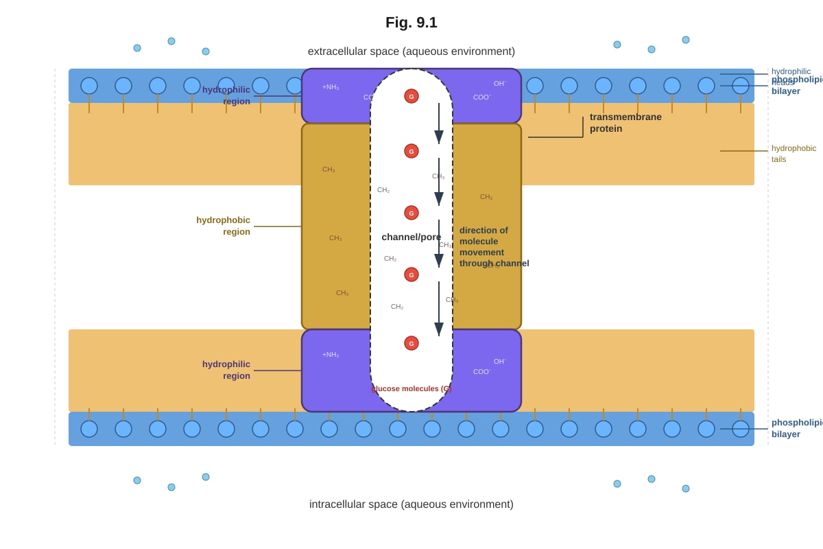
Generated diagram for Q9.
(a) With reference to Fig. 9.1, explain how the structure of the transmembrane protein is related to its position within the phospholipid bilayer.
___________________________________________________________________________ [2]
(b) Explain how this transmembrane protein could facilitate the transport of glucose molecules across the cell membrane.
___________________________________________________________________________ [3]
[Total: 5 marks]
10. A solution of starch was incubated with amylase at 37 °C. Samples were taken at regular intervals and tested with iodine solution. The results are shown in Table 10.1.
| Time / min | Colour with iodine solution |
|---|---|
| 0 | Blue-black |
| 2 | Blue-black |
| 4 | Brown |
| 6 | Brown |
| 8 | Yellow-brown (no colour change) |
| 10 | Yellow-brown (no colour change) |
Table 10.1
(a) Explain the colour change observed in this experiment.
___________________________________________________________________________ [2]
(b) State the product of starch digestion by amylase that gives a yellow-brown colour with iodine.
___________________________________________________________________________ [1]
(c) Explain why the reaction was carried out at 37 °C rather than at 60 °C.
___________________________________________________________________________ [2]
[Total: 5 marks]
Section B: Data-Based Question (15 marks)
Answer all questions 11–13.
11. Read the following passage and answer the questions that follow.
The Discovery and Significance of Aquaporins
In 1992, Peter Agre discovered a family of integral membrane proteins called aquaporins that facilitate the rapid movement of water across cell membranes. Before this discovery, scientists believed that water simply diffused through the phospholipid bilayer. Aquaporins form tetrameric channels in the membrane, with each monomer containing a narrow pore that allows only water molecules to pass through in single file.
The pore of an aquaporin contains a conserved NPA (asparagine-proline-alanine) motif that creates an electrostatic barrier, preventing the passage of protons (H⁺) while allowing water molecules to pass. This is critical because the movement of protons would dissipate the proton gradient across the membrane, which is essential for ATP synthesis in mitochondria.
Aquaporins are found in many tissues, including the kidneys, where aquaporin-2 (AQP2) plays a vital role in water reabsorption in the collecting ducts. The hormone antidiuretic hormone (ADH) triggers the insertion of AQP2 into the apical membrane of collecting duct cells, increasing water permeability and reducing water loss in urine.
Mutations in the AQP2 gene can cause nephrogenic diabetes insipidus, a condition in which the kidneys are unable to concentrate urine, leading to the production of large volumes of dilute urine.
(a) Explain why the discovery of aquaporins changed the understanding of water movement across cell membranes.
___________________________________________________________________________ [2]
(b) With reference to the passage, explain how the NPA motif in aquaporins prevents the passage of protons while allowing water molecules to pass.
___________________________________________________________________________ [3]
(c) Explain the role of ADH in regulating water reabsorption in the kidney collecting duct.
___________________________________________________________________________ [3]
(d) Suggest how a mutation in the AQP2 gene could lead to the production of large volumes of dilute urine.
___________________________________________________________________________ [2]
[Total: 10 marks]
12. Fig. 12.1 shows the effect of substrate concentration on the rate of reaction of enzyme X in the absence and presence of substance Y.
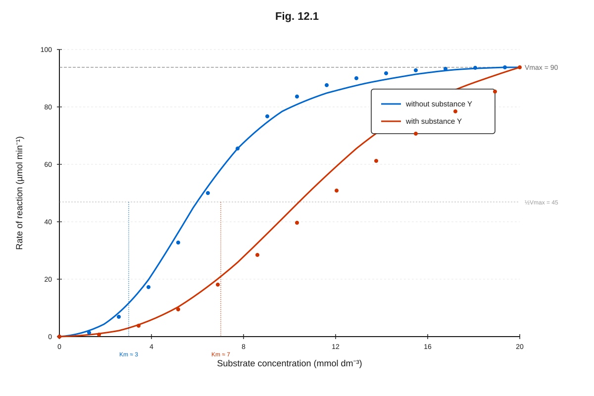
Generated graph for Q12.
(a) With reference to Fig. 12.1, describe the effect of substance Y on the rate of reaction of enzyme X.
___________________________________________________________________________ [2]
(b) Identify the type of inhibition caused by substance Y. Explain your answer using evidence from Fig. 12.1.
___________________________________________________________________________ [2]
(c) Explain how substance Y affects the Km of enzyme X.
___________________________________________________________________________ [1]
[Total: 5 marks]
Section C: Extended Response (15 marks)
Answer all questions 14–15.
14. Describe the structure of the cell surface membrane according to the fluid mosaic model and explain how its structure enables it to control the movement of substances into and out of the cell.
___________________________________________________________________________ [10]
[Total: 10 marks]
15. Explain how the structure of biological molecules determines their function. In your answer, refer to at least two different types of biological molecules.
___________________________________________________________________________ [5]
[Total: 5 marks]
End of Paper
Section A: 30 marks | Section B: 15 marks | Section C: 15 marks | Total: 60 marks
Answers
TuitionGoWhere Practice Paper — Biology H2 A-Level
Answer Key & Marking Scheme
Version 4 of 5 | Cells & Biomolecules | Total: 60 marks
Section A
Question 1 (7 marks)
(a) The Golgi apparatus (organelle C) modifies, sorts, and packages proteins (and lipids) for secretion or delivery to other organelles. [1]
Marking note: Award 1 mark for a correct function. Acceptable answers include: "modifies proteins," "packages proteins into vesicles," "sorts and transports proteins," "forms lysosomes," or "adds carbohydrate groups to proteins (glycosylation)." Do not accept vague answers like "transport" without specifying what is being transported.
(b) Four marks total — 2 marks per feature (1 mark for identifying the feature + 1 mark for explaining how it is adapted to its function).
-
Double membrane / inner membrane folded into cristae: The cristae (folds of the inner membrane) increase the surface area available for the attachment of electron carriers and ATP synthase, which are involved in oxidative phosphorylation during aerobic respiration. [2]
-
Matrix containing enzymes: The mitochondrial matrix contains enzymes for the Krebs cycle (e.g., citrate synthase, isocitrate dehydrogenase) and fatty acid oxidation, providing the necessary catalytic environment for these metabolic reactions. [2]
Alternative acceptable features:
- Small circular DNA / ribosomes in matrix: Allows the mitochondrion to synthesise some of its own proteins independently, enabling rapid production of respiratory chain proteins.
- Intermembrane space: The small volume of the intermembrane space allows a high concentration of protons (H⁺) to be accumulated quickly, creating a steep proton gradient for chemiosmosis.
Common mistakes: Students often state that the mitochondrion is the "site of aerobic respiration" without linking a specific visible structure to its function. Both the feature and the functional link are required for full marks.
(c) Lysosomes contain hydrolytic enzymes (e.g., proteases, lipases, nucleases) to digest worn-out organelles, engulfed pathogens, and macromolecules through hydrolysis. [1] If these enzymes were released into the cytoplasm, they would digest the cell's own organelles and macromolecules, leading to autolysis (self-digestion) and cell death. [1]
Teaching note: Lysosomal enzymes have an acidic pH optimum (pH ~5), maintained by proton pumps in the lysosomal membrane. The cytoplasm has a near-neutral pH (~7.2), so the enzymes would have reduced activity if released, but they would still cause significant damage over time. This is why lysosomal membrane integrity is critical.
Question 2 (5 marks)
(a) Ca²⁺ has the steepest concentration gradient. [1] The ratio of extracellular to intracellular concentration for Ca²⁺ is 2.5 / 0.0001 = 25,000:1, which is much greater than for Na⁺ (145/15 ≈ 9.7:1), K⁺ (5/150 ≈ 0.03:1, i.e., 150/5 = 30:1 inward), or Cl⁻ (110/10 = 11:1). [1]
Marking note: Award 1 mark for identifying Ca²⁺ and 1 mark for showing the calculation or comparative reasoning. Students may express the gradient as a ratio or simply state that the difference is greatest for Ca²⁺.
(b) The sodium-potassium pump (Na⁺/K⁺-ATPase) is an integral membrane protein that uses energy from ATP hydrolysis to actively transport 3 Na⁺ ions out of the cell and 2 K⁺ ions into the cell per cycle. [1] This maintains the high intracellular K⁺ concentration and high extracellular Na⁺ concentration shown in Table 2.1. [1] The pump works against the concentration gradients of both ions, which is why it requires energy (ATP). [1]
Teaching note: The Na⁺/K⁺-ATPase is an example of active transport. It is electrogenic because it moves 3 positive charges out for every 2 positive charges in, creating a net movement of one positive charge out of the cell, which contributes to the resting membrane potential. Students should understand that without this pump, the concentration gradients would eventually equilibrate through passive transport.
Question 3 (6 marks)
(a) As temperature increases from 0 °C to 40 °C, the rate of reaction increases. [1] This is because increasing temperature increases the kinetic energy of both enzyme and substrate molecules, leading to more frequent and more energetic collisions, resulting in more enzyme-substrate complexes being formed per unit time. [1]
Marking note: Award 1 mark for describing the trend (rate increases) and 1 mark for the explanation involving kinetic energy and collision frequency/energy.
(b) Above 40 °C, the rate of reaction decreases sharply because the enzyme molecules begin to denature. [1] The high temperature disrupts the hydrogen bonds, ionic bonds, and hydrophobic interactions that maintain the enzyme's tertiary structure, causing the active site to change shape so that the substrate can no longer bind effectively. [1]
Common mistake: Students often say "the enzyme is killed" — enzymes are not living organisms and cannot be "killed." The correct term is "denatured."
(c) The student's claim is incorrect. [1] At 60 °C, the enzyme is already denatured (as shown by the low rate of reaction at this temperature). Denaturation is a structural change to the protein, so adding more enzyme would simply add more enzyme that would also be denatured. The rate would not be restored to the maximum because the problem is not a lack of enzyme but the loss of enzyme function due to denaturation. [1]
Teaching note: This question tests understanding of the difference between temperature effects below the optimum (increased kinetic energy) and above the optimum (denaturation). Students must recognise that denaturation is irreversible under normal conditions and that increasing enzyme concentration cannot overcome it.
Question 4 (6 marks)
(a) Phospholipid molecules have hydrophilic (water-attracting) phosphate heads and hydrophobic (water-repelling) hydrocarbon tails. [1] In an aqueous environment, the hydrophilic heads orientate towards the water on both sides of the bilayer, while the hydrophobic tails orientate away from the water, facing each other in the interior. This arrangement is thermodynamically favourable and forms spontaneously. [1]
(b) Cholesterol molecules are located between the phospholipid tails in the bilayer. [1] At high temperatures, cholesterol restricts the movement of phospholipid fatty acid tails, reducing membrane fluidity and preventing the membrane from becoming too permeable. At low temperatures, cholesterol prevents the phospholipids from packing too closely together, maintaining membrane fluidity and preventing the membrane from becoming too rigid. [1]
Marking note: Award 1 mark for the location of cholesterol and 1 mark for explaining its dual role in regulating fluidity at both high and low temperatures. Students who only mention one temperature condition should receive only partial credit.
(c) Two functions of glycoproteins:
- Cell recognition / cell signalling — glycoproteins act as receptor molecules for hormones, neurotransmitters, and other signalling molecules. [1]
- Cell adhesion / cell-to-cell recognition — glycoproteins enable cells to recognise each other, which is important for immune responses and tissue formation. [1]
Alternative acceptable answers: Protection of the cell surface (glycocalyx), acting as antigens for immune recognition, or acting as binding sites for pathogens (which can also be exploited by viruses).
Question 5 (5 marks)
Marking scheme (5 marks):
-
Prophase: Chromatin condenses and becomes visible as chromosomes, each consisting of two sister chromatids joined at the centromere. The nuclear envelope breaks down. The spindle apparatus forms from the centrioles. [1]
-
Metaphase: Chromosomes align at the equator (metaphase plate) of the cell. Spindle fibres from opposite poles attach to the centromere of each chromosome. [1]
-
Anaphase: The centromeres split, and the sister chromatids are pulled apart to opposite poles of the cell by the shortening of spindle fibres. Each chromatid is now considered an individual chromosome. [1]
-
Telophase: The chromosomes arrive at opposite poles and begin to decondense. Nuclear envelopes reform around each set of chromosomes. The spindle breaks down. [1]
-
Cytokinesis and genetic identity: Cytokinesis divides the cytoplasm, producing two daughter cells. Because DNA replication during S phase produces identical copies of each chromosome, and mitosis ensures that one copy of each chromosome is distributed to each daughter cell, the two daughter cells are genetically identical to each other and to the parent cell. [1]
Common mistakes: Students often confuse mitosis with meiosis, mentioning homologous pairs or crossing over. Mitosis involves the separation of sister chromatids, not homologous chromosomes. Also, students should note that DNA replication occurs during interphase (S phase), not during mitosis itself.
Question 6 (6 marks)
(a) pH 7 [1]
(b) At the optimum pH (pH 7), the enzyme's active site has the correct shape and charge distribution to bind the substrate effectively, and the rate of reaction is highest. [1] At pH values below the optimum (acidic conditions), excess H⁺ ions disrupt the ionic bonds and hydrogen bonds that maintain the enzyme's tertiary structure, causing the active site to change shape (denaturation at extreme pH), so the substrate can no longer bind effectively. [1] At pH values above the optimum (alkaline conditions), excess OH⁻ ions similarly disrupt the bonds maintaining the enzyme's tertiary structure, altering the active site shape and reducing enzyme activity. [1]
Marking note: Award 1 mark for the optimum pH explanation, 1 mark for the acidic pH explanation, and 1 mark for the alkaline pH explanation. Students must mention the disruption of bonds/tertiary structure/active site shape change for the non-optimum pH values.
(c) Two variables to keep constant:
- Temperature — to ensure that only pH affects the rate of reaction. [1]
- Concentration (or volume) of hydrogen peroxide (substrate) — to ensure that substrate concentration is not a limiting factor. [1]
Alternative acceptable answers: Concentration/volume of catalase enzyme, volume of buffer solution, or surface area of any solid catalyst used.
Question 7 (6 marks)
(a) Glycosidic bond [1]
(b) Nucleotides are joined together by phosphodiester bonds formed between the phosphate group of one nucleotide and the 3' hydroxyl group of the deoxyribose sugar of the next nucleotide. [1] This condensation reaction forms a sugar-phosphate backbone with the nitrogenous bases projecting sideways. The polynucleotide chain has a 5' end (with a free phosphate group) and a 3' end (with a free hydroxyl group), giving the chain directionality. [1]
(c) DNA carries genetic information in the sequence of its four nitrogenous bases (adenine, thymine, guanine, cytosine). [1] The specific sequence of bases along a gene codes for the sequence of amino acids in a protein. [1] The double-helical structure, with complementary base pairing (A-T and G-C), ensures that genetic information can be accurately copied during DNA replication and transmitted to daughter cells. [1]
Marking note: Award 1 mark for the base sequence coding concept, 1 mark for the link to protein synthesis (amino acid sequence), and 1 mark for the role of complementary base pairing in accurate replication/information storage.
Question 8 (4 marks)
| Feature | Prokaryotic cell | Eukaryotic cell |
|---|---|---|
| Size | Smaller (typically 1–5 µm) | Larger (typically 10–100 µm) |
| DNA organisation | Circular DNA in nucleoid region, not associated with histones (in most prokaryotes) | Linear DNA in nucleus, associated with histone proteins to form chromosomes |
| Ribosome type | 70S ribosomes | 80S ribosomes |
| Membrane-bound organelles | Absent | Present (e.g., mitochondria, ER, Golgi) |
Marking note: Award 1 mark per correct row (all entries in the row must be correct for the mark). Acceptable alternatives: for DNA organisation, "no nuclear envelope" for prokaryotic and "enclosed within nuclear envelope" for eukaryotic; for ribosome type, "smaller" for prokaryotic and "larger" for eukaryotic.
Question 9 (5 marks)
(a) The transmembrane protein has hydrophobic amino acid residues in the region that spans the interior of the phospholipid bilayer, where it interacts with the hydrophobic fatty acid tails. [1] The regions of the protein exposed to the aqueous extracellular and intracellular environments have hydrophilic amino acid residues, which interact favourably with water. [1] This arrangement of hydrophobic and hydrophilic regions anchors the protein stably within the bilayer.
(b) The transmembrane protein acts as a carrier protein (or channel protein) for glucose. [1] Glucose is a large, polar molecule that cannot diffuse through the hydrophobic core of the phospholipid bilayer. [1] The transmembrane protein provides a hydrophilic channel (or undergoes conformational changes as a carrier) that allows glucose to cross the membrane down its concentration gradient via facilitated diffusion. [1]
Marking note: Students should specify that glucose transport via a transmembrane protein is facilitated diffusion (passive, down the concentration gradient). If the question context suggested active transport, that would also be acceptable with appropriate explanation.
Question 10 (5 marks)
(a) At the start of the experiment, starch gives a blue-black colour with iodine. [1] As amylase breaks down starch into maltose (and other smaller sugars), the starch concentration decreases, so the colour becomes less intense (brown) until no starch remains, at which point iodine gives its original yellow-brown colour. [1]
(b) Maltose [1]
(c) 37 °C is close to the optimum temperature for amylase (human amylase optimum is approximately 37 °C). [1] At 60 °C, the enzyme would be denatured (as it is well above the optimum), and the reaction would proceed much more slowly or not at all, making it impossible to observe the progressive digestion of starch over time. [1]
Teaching note: This experiment demonstrates the progressive nature of enzyme-catalysed reactions. The iodine test is used because it specifically detects starch (blue-black) but not its breakdown products (maltose gives no colour change with iodine, so the solution appears yellow-brown, the natural colour of iodine).
Section B
Question 11 (10 marks)
(a) Before the discovery of aquaporins, scientists believed that water moved across cell membranes by simple diffusion through the phospholipid bilayer. [1] The discovery of aquaporins showed that water can also move rapidly across membranes through specific protein channels, which explains the much higher rate of water transport observed in many cell types than could be accounted for by simple diffusion alone. [1]
(b) The NPA (asparagine-proline-alanine) motif creates an electrostatic barrier within the aquaporin pore. [1] The positive charges associated with the NPA motif repel protons (H⁺ ions), preventing them from passing through the channel. [1] Water molecules, being uncharged and small, can pass through the narrow pore in single file without being repelled by this electrostatic barrier. [1]
Marking note: Award 1 mark for the electrostatic barrier concept, 1 mark for the repulsion of H⁺, and 1 mark for explaining why water can pass (uncharged, small size, single file).
(c) When the body is dehydrated, the hypothalamus detects increased blood osmolarity and signals the posterior pituitary to release ADH into the bloodstream. [1] ADH binds to receptors on the basal membrane of collecting duct cells in the kidney, triggering a signalling cascade that causes vesicles containing AQP2 to fuse with the apical membrane. [1] This increases the number of aquaporin-2 channels in the apical membrane, increasing water permeability and allowing more water to be reabsorbed from the filtrate back into the blood by osmosis, thus concentrating the urine. [1]
(d) A mutation in the AQP2 gene could result in a non-functional aquaporin-2 protein, or a protein that cannot be correctly inserted into the apical membrane of collecting duct cells. [1] Without functional AQP2 channels, the collecting duct remains impermeable to water, so water cannot be reabsorbed from the filtrate. This results in the production of large volumes of dilute urine, as seen in nephrogenic diabetes insipidus. [1]
Question 12 (5 marks)
(a) In the presence of substance Y, the rate of reaction at lower substrate concentrations is reduced compared to the reaction without substance Y. [1] However, at high substrate concentrations, the maximum rate of reaction (Vmax) is the same with or without substance Y. [1]
Marking note: Award 1 mark for stating that the rate is reduced at low substrate concentrations and 1 mark for stating that Vmax is unchanged.
(b) Substance Y causes competitive inhibition. [1] This is because the Vmax is the same in the presence and absence of substance Y, but a higher substrate concentration is required to reach Vmax when substance Y is present (the apparent Km is increased). This indicates that substance Y competes with the substrate for the active site of the enzyme. [1]
Teaching note: Competitive inhibitors structurally resemble the substrate and bind reversibly to the active site. At sufficiently high substrate concentrations, the substrate outcompetes the inhibitor, and Vmax can still be reached. This is a key distinguishing feature from non-competitive inhibition, where Vmax is reduced.
(c) Substance Y increases the apparent Km of enzyme X. [1] This is because a higher substrate concentration is required to reach half of Vmax in the presence of the competitive inhibitor, indicating that the enzyme's apparent affinity for the substrate is reduced.
Section C
Question 14 (10 marks)
Marking scheme (10 marks):
Structure of the cell surface membrane (fluid mosaic model):
-
The membrane is based on a phospholipid bilayer, with hydrophilic phosphate heads facing the aqueous environments on both sides and hydrophobic fatty acid tails facing inward. [1]
-
Proteins are embedded in the bilayer in two main forms: integral (transmembrane) proteins that span the entire bilayer, and peripheral proteins that are attached to the surface. [1]
-
Cholesterol molecules are found between the phospholipid tails, helping to regulate membrane fluidity. [1]
-
Glycoproteins and glycolipids are found on the extracellular surface, with carbohydrate chains extending outward. [1]
-
The term "fluid" refers to the fact that phospholipids and proteins can move laterally within the bilayer, giving the membrane flexibility. The term "mosaic" refers to the scattered arrangement of proteins within the bilayer, like tiles in a mosaic. [1]
How structure enables control of substance movement:
-
The hydrophobic core of the phospholipid bilayer acts as a barrier to the passage of large polar molecules and ions, preventing them from freely crossing the membrane. [1]
-
Channel proteins provide hydrophilic pores that allow specific ions and small polar molecules to pass through by facilitated diffusion. [1]
-
Carrier proteins bind to specific molecules (e.g., glucose, amino acids) and undergo conformational changes to transport them across the membrane, either by facilitated diffusion or active transport. [1]
-
The sodium-potassium pump (an example of a carrier protein) uses ATP to actively transport Na⁺ and K⁺ against their concentration gradients, maintaining the cell's electrochemical gradient. [1]
-
The selective permeability of the membrane, determined by the combination of the phospholipid bilayer and specific transport proteins, allows the cell to control which substances enter and exit, maintaining homeostasis. [1]
Marking descriptors:
- 8–10 marks: Comprehensive description of membrane structure with accurate terminology, clear explanation of how at least three structural features enable control of substance movement, well-organised response.
- 5–7 marks: Good description of membrane structure, explanation of at least two features enabling control of movement, some use of correct terminology.
- 3–4 marks: Basic description of membrane structure, limited explanation of how structure relates to function.
- 1–2 marks: Very limited or inaccurate description of membrane structure.
Question 15 (5 marks)
Marking scheme (5 marks):
Students must refer to at least two different types of biological molecules. Award marks as follows:
For each biological molecule (up to 2 marks per molecule, maximum 4 marks):
Example 1: Proteins
- Proteins are made of amino acids linked by peptide bonds. The sequence of amino acids (primary structure) determines how the protein folds into its secondary (α-helix, β-pleated sheet), tertiary (3D shape), and quaternary structures. [1]
- The specific 3D shape of a protein, particularly the shape of its active site (in enzymes) or binding site (in receptors), determines its function. For example, the complementary shape of an enzyme's active site to its substrate allows specific catalysis. [1]
Example 2: Nucleic acids (DNA)
- DNA is made of nucleotides containing deoxyribose sugar, a phosphate group, and one of four nitrogenous bases (A, T, G, C). The sequence of bases along the DNA strand encodes genetic information. [1]
- The double-helical structure, held together by complementary base pairing (A-T, G-C) via hydrogen bonds, allows DNA to be accurately replicated and to transmit genetic information. [1]
Example 3: Carbohydrates
- Monosaccharides (e.g., glucose) are linked by glycosidic bonds to form disaccharides (e.g., maltose) and polysaccharides (e.g., starch, glycogen, cellulose). [1]
- Starch and glycogen are compact, branched molecules used for energy storage, while cellulose forms straight, strong fibres held together by hydrogen bonds, providing structural support in plant cell walls. The type of glycosidic bond (α-1,4 vs β-1,4) determines the shape and function of the polysaccharide. [1]
Example 4: Lipids (triglycerides)
- Triglycerides are made of one glycerol molecule bonded to three fatty acid chains by ester bonds. [1]
- The long hydrocarbon chains of fatty acids contain many C-H bonds, which release large amounts of energy when broken during respiration, making lipids efficient energy storage molecules. The hydrophobic nature of lipids also makes them suitable for forming the phospholipid bilayer of cell membranes. [1]
Quality of communication: [1 mark]
- Award 1 mark for a well-organised, coherent response with correct use of scientific terminology.
Marking descriptors:
- 5 marks: Detailed explanation of structure-function relationship for at least two biological molecules with accurate scientific terminology and clear reasoning.
- 3–4 marks: Explanation of structure-function relationship for two molecules, but with some gaps in detail or terminology.
- 1–2 marks: Limited explanation, possibly only one molecule addressed, or significant inaccuracies.
End of Answer Key
Section A: 30 marks | Section B: 15 marks | Section C: 15 marks | Total: 60 marks
Free quiz and exam paper access
Enter your details to view this paper
Your access is remembered on this device.