AI Generated Exam Paper
A Level H2 Biology Practice Paper 3
Free A Level H2 Biology Practice Paper 3, LongCat AI version, with questions, answers, and A Level-style practice for Singapore students.
These static practice materials are generated from the site's syllabus and paper-generation workflow, with source and model context shown so students and parents can evaluate the material before use.
Questions
TuitionGoWhere Practice Paper - Biology H2 A-Level
TuitionGoWhere Practice Paper (AI)
Subject: Biology Level: A-Level H2 Paper: Practice Paper — Cells & Biomolecules Duration: 1 hour 30 minutes Total Marks: 50
Name: ___________________________ Class: ___________________________ Date: ___________________________
Instructions
- Answer all questions in the spaces provided.
- Write in dark blue or black pen.
- You may use a pencil for any diagrams or graphs.
- Do not use correction fluid.
- The number of marks for each question is shown in brackets [ ].
- The total marks for this paper is 50.
- You are advised to spend no more than 1 hour 30 minutes on this paper.
Section A: Multiple Choice [10 marks]
Questions 1–10. Each question is worth 1 mark. Choose the one best answer.
1. Which of the following organelles is present in both prokaryotic and eukaryotic cells?
A. Nucleus B. Mitochondrion C. Ribosome D. Endoplasmic reticulum
[1]
2. A student observed a cell under an electron microscope and noted the presence of a double membrane with pores, a nucleolus, and chromatin. Which structure was the student observing?
A. Golgi apparatus B. Nucleus C. Mitochondrion D. Chloroplast
[1]
3. Which property of water is most directly responsible for its role as a universal solvent in biological systems?
A. High specific heat capacity B. Cohesion and surface tension C. Polarity of the water molecule D. High latent heat of vaporisation
[1]
4. The diagram below shows a phospholipid bilayer.
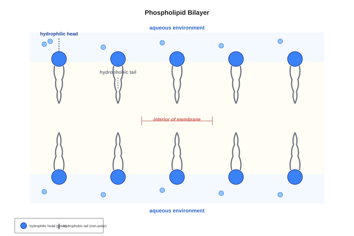
Generated diagram for Q4.
Which statement about the phospholipid bilayer is correct?
A. The hydrophobic heads face outward to interact with water. B. The hydrophilic tails face inward to avoid water. C. The hydrophobic tails face inward, away from the aqueous environment. D. The hydrophilic heads face inward to form a non-polar core.
[1]
5. Which of the following is a function of the smooth endoplasmic reticulum?
A. Protein synthesis B. Lipid synthesis and detoxification C. Packaging and modification of proteins D. ATP production
[1]
6. A polypeptide has the amino acid sequence: Met–Leu–Asp–Val–Arg. How many peptide bonds are present in this polypeptide?
A. 3 B. 4 C. 5 D. 6
[1]
7. Which level of protein structure is determined by hydrogen bonding between the peptide backbone to form α-helices and β-pleated sheets?
A. Primary structure B. Secondary structure C. Tertiary structure D. Quaternary structure
[1]
8. An enzyme-catalysed reaction was carried out at 35 °C. When the temperature was increased to 70 °C, the rate of reaction decreased significantly. What is the best explanation for this observation?
A. The enzyme was denatured, losing its three-dimensional active site shape. B. The substrate molecules moved too fast to bind to the active site. C. The activation energy of the reaction increased. D. The enzyme was inhibited by the products of the reaction.
[1]
9. Which of the following molecules contains a β-1,4-glycosidic bond?
A. Maltose B. Sucrose C. Lactose D. Fructose
[1]
10. During which phase of mitosis do sister chromatids separate and move to opposite poles of the cell?
A. Prophase B. Metaphase C. Anaphase D. Telophase
[1]
Section B: Structured Questions [25 marks]
Questions 11–15. Answer all questions. Show your working where appropriate.
11. Fig. 11.1 shows the structure of a typical animal cell as seen under an electron microscope.
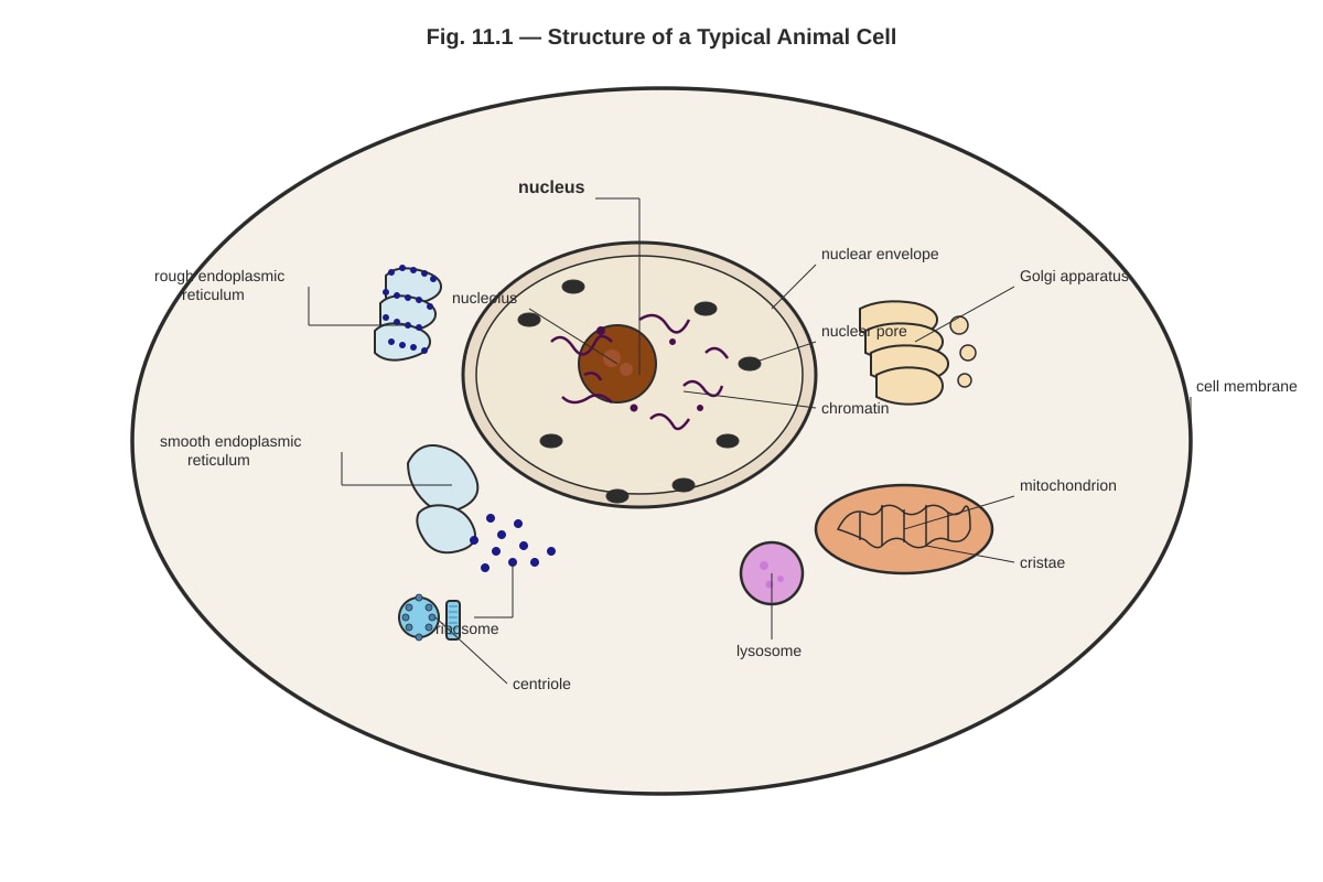
Generated diagram for Q11.
(a) Identify the organelle labelled X (pointing to the Golgi apparatus) and state two of its functions. [2]
.......................................................................................................................
.......................................................................................................................
.......................................................................................................................
(b) With reference to Fig. 11.1, describe one structural feature of the mitochondrion that is adapted for its role in aerobic respiration. [1]
.......................................................................................................................
.......................................................................................................................
(c) Explain why the rough endoplasmic reticulum appears "rough" under the electron microscope. [1]
.......................................................................................................................................................................
(d) State the function of the lysosome. [1]
.......................................................................................................................
[5]
12. Fig. 12.1 shows the effect of substrate concentration on the rate of an enzyme-catalysed reaction, with and without the presence of substance X.
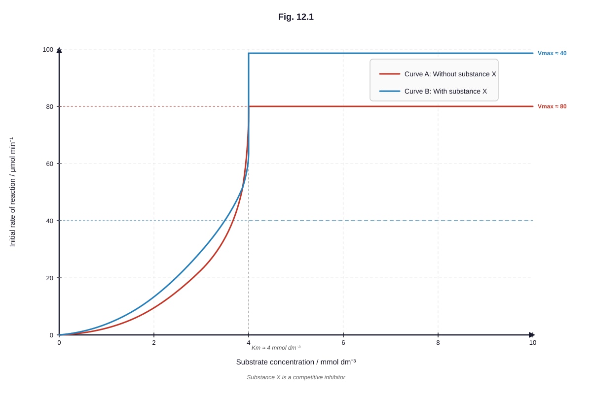
Generated graph for Q12.
(a) With reference to Fig. 12.1, describe the effect of increasing substrate concentration on the initial rate of reaction for Curve A (without substance X). [2]
.......................................................................................................................
.......................................................................................................................
.......................................................................................................................
(b) Compare the curves for reactions with and without substance X. Suggest what type of inhibition substance X causes, giving a reason for your answer. [3]
.......................................................................................................................
.......................................................................................................................................................................
.......................................................................................................................
.......................................................................................................................
.......................................................................................................................
(c) Explain why the rate of reaction plateaus at high substrate concentrations. [2]
.......................................................................................................................................................................
.......................................................................................................................
.......................................................................................................................
[7]
13. A student carried out an experiment to test for the presence of biological molecules in three unknown solutions, P, Q, and R. The results are shown in Table 13.1.
Table 13.1
| Test | Solution P | Solution Q | Solution R |
|---|---|---|---|
| Biuret test | Blue | Purple/violet | Blue |
| Benedict's test (after heating) | Blue | Blue | Brick-red precipitate |
| Ethanol emulsion test | Cloudy white emulsion | No change | No change |
| Iodine test | No change | No change | No change |
(a) Identify the biological molecule(s) present in each of solutions P, Q, and R. [3]
Solution P: ..........................................................................................................
Solution Q: ..........................................................................................................
Solution R: ..........................................................................................................
(b) Explain why the Benedict's test requires heating in a water bath. [1]
.......................................................................................................................
.......................................................................................................................
(c) Describe the chemical bonding that holds the primary structure of a protein together. [1]
.......................................................................................................................
.......................................................................................................................
[5]
14. Fig. 14.1 shows a section through a cell surface membrane.
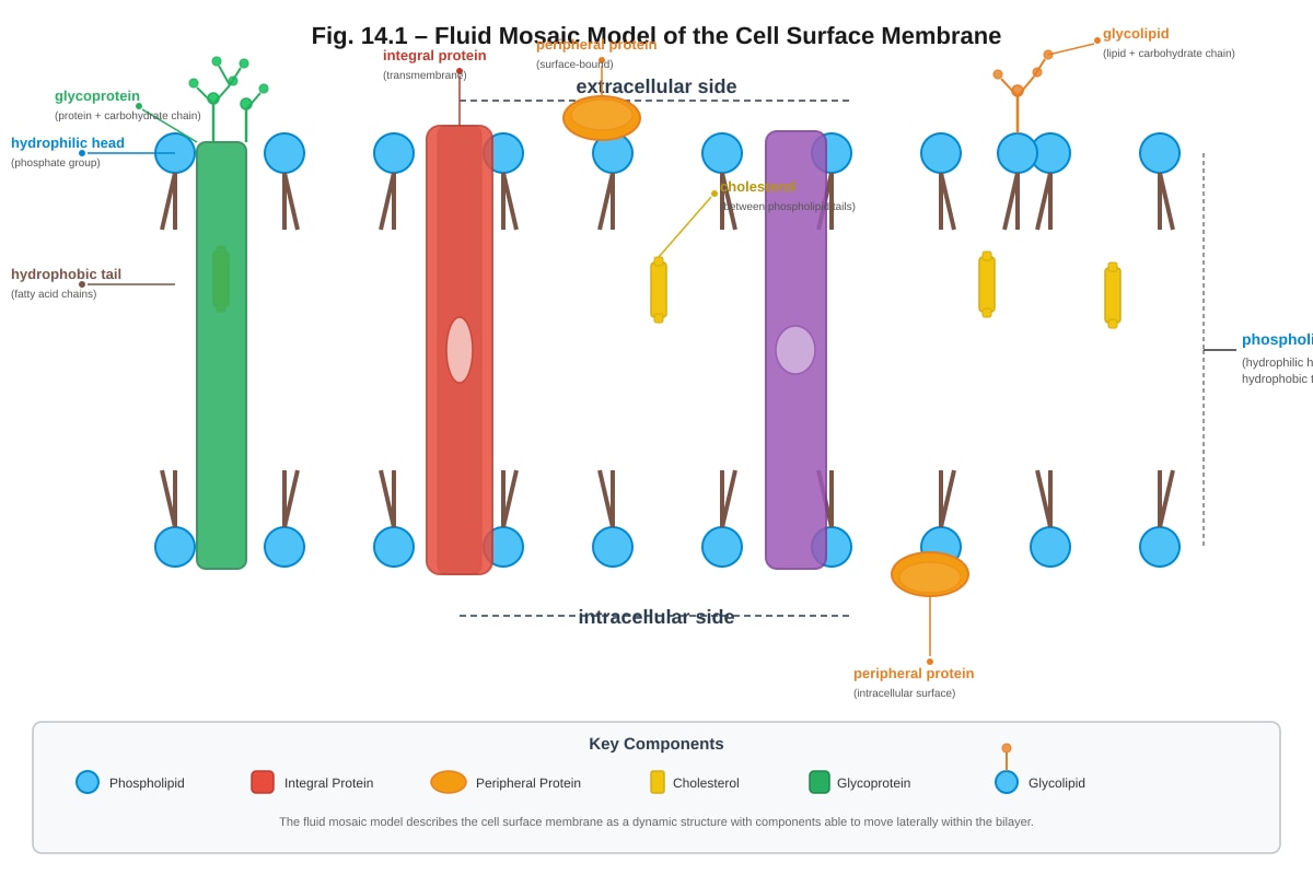
Generated diagram for Q14.
(a) With reference to Fig. 14.1, explain how the structure of the phospholipid bilayer provides a barrier to the movement of ions across the membrane. [2]
.......................................................................................................................
.......................................................................................................................
.......................................................................................................................
(b) State two functions of cholesterol in the cell surface membrane. [2]
.......................................................................................................................
.......................................................................................................................
(c) Explain the role of glycoproteins in cell signalling. [2]
.......................................................................................................................
.......................................................................................................................
.......................................................................................................................
[6]
15. Describe the role of the following in protein synthesis:
(a) Messenger RNA (mRNA) [2]
.......................................................................................................................
.......................................................................................................................
.......................................................................................................................
(b) Transfer RNA (tRNA) [2]
.......................................................................................................................
.......................................................................................................................
.......................................................................................................................
(c) Ribosomes [2]
.......................................................................................................................
.......................................................................................................................
.......................................................................................................................
[6]
Section C: Free Response [15 marks]
Answer any two of the three questions. Each question is worth 7 or 8 marks. Write your answers in continuous prose in the spaces provided.
16. (a) Describe the process of mitosis and explain how it results in the production of two genetically identical daughter cells. [5]
.......................................................................................................................
.......................................................................................................................
.......................................................................................................................
.......................................................................................................................
.......................................................................................................................
.......................................................................................................................
.......................................................................................................................
.......................................................................................................................
.......................................................................................................................
.......................................................................................................................
(b) Explain why mitosis is important in the growth and repair of multicellular organisms. [2]
.......................................................................................................................
.......................................................................................................................
.......................................................................................................................
.......................................................................................................................
.......................................................................................................................
.......................................................................................................................
[7]
17. (a) Describe the structure of DNA and explain how its structure enables it to carry genetic information and replicate itself. [5]
.......................................................................................................................
.......................................................................................................................
.......................................................................................................................
.......................................................................................................................
.......................................................................................................................
.......................................................................................................................
.......................................................................................................................
.......................................................................................................................
.......................................................................................................................
.......................................................................................................................
(b) Explain the significance of complementary base pairing in DNA replication. [3]
.......................................................................................................................
.......................................................................................................................
.......................................................................................................................
.......................................................................................................................
.......................................................................................................................
.......................................................................................................................
.......................................................................................................................
[8]
18. (a) Explain how the structure of an enzyme relates to its function, including the induced-fit model of enzyme action. [4]
.......................................................................................................................
.......................................................................................................................
.......................................................................................................................
.......................................................................................................................
.......................................................................................................................
.......................................................................................................................
.......................................................................................................................
.......................................................................................................................
(b) Describe how two factors affect the rate of an enzyme-catalysed reaction. For each factor, explain the effect at the molecular level. [4]
.......................................................................................................................
.......................................................................................................................
.......................................................................................................................
.......................................................................................................................
.......................................................................................................................
.......................................................................................................................
.......................................................................................................................
.......................................................................................................................
.......................................................................................................................
.......................................................................................................................
.......................................................................................................................
.......................................................................................................................
[8]
End of Paper
Total Marks: 50
Answers
TuitionGoWhere Practice Paper — Biology H2 A-Level
Answer Key & Marking Scheme
Paper: Practice Paper — Cells & Biomolecules Total Marks: 50
Section A: Multiple Choice [10 marks]
1. C — Ribosome [1]
- Explanation: Ribosomes are the only organelle listed that are found in both prokaryotic and eukaryotic cells. Prokaryotes lack a true nucleus (A), mitochondria (B), and endoplasmic reticulum (D). Ribosomes are essential for protein synthesis in all cells. Prokaryotic ribosomes are 70S while eukaryotic ribosomes are 80S, but both types possess them.
2. B — Nucleus [1]
- Explanation: The nucleus is the only organelle with a double membrane (nuclear envelope) containing nuclear pores, a nucleolus, and chromatin. The Golgi apparatus (A) is a stack of cisternae without a double membrane or nucleolus. Mitochondria (C) have a double membrane but no nucleolus or chromatin. Chloroplasts (D) have a double membrane but contain thylakoids, not chromatin or a nucleolus.
3. C — Polarity of the water molecule [1]
- Explanation: Water is a polar molecule due to the unequal sharing of electrons between oxygen and hydrogen atoms (oxygen is more electronegative). This polarity allows water to surround and dissolve ions and polar molecules, making it an excellent solvent. High specific heat capacity (A) relates to temperature buffering. Cohesion (B) relates to surface tension and water transport. High latent heat of vaporisation (D) relates to cooling by evaporation.
4. C — The hydrophobic tails face inward, away from the aqueous environment. [1]
- Explanation: In a phospholipid bilayer, the hydrophilic (water-loving) phosphate heads face outward toward the aqueous environments (both extracellular and intracellular), while the hydrophobic (water-fearing) fatty acid tails face inward, shielded from water. This arrangement is driven by the amphipathic nature of phospholipids and is the fundamental basis of membrane structure. Options A, B, and D incorrectly describe the orientation.
5. B — Lipid synthesis and detoxification [1]
- Explanation: The smooth endoplasmic reticulum (SER) lacks ribosomes and is involved in lipid synthesis (including phospholipids and steroids) and detoxification of drugs and poisons. Protein synthesis (A) occurs on the rough ER. Packaging and modification of proteins (C) is a function of the Golgi apparatus. ATP production (D) occurs in mitochondria.
6. B — 4 [1]
- Explanation: A peptide bond forms between the carboxyl group (–COOH) of one amino acid and the amino group (–NH₂) of the next amino acid. In a polypeptide of n amino acids, there are always (n − 1) peptide bonds. Here, 5 amino acids → 5 − 1 = 4 peptide bonds. Common mistake: students may choose 5 (C) by counting the number of amino acids instead of bonds.
7. B — Secondary structure [1]
- Explanation: Secondary structure refers to local folding patterns (α-helices and β-pleated sheets) stabilised by hydrogen bonds between the C=O and N–H groups of the peptide backbone (not the R groups). Primary structure (A) is the sequence of amino acids. Tertiary structure (C) involves interactions between R groups (hydrophobic interactions, disulfide bonds, ionic bonds, hydrogen bonds). Quaternary structure (D) involves the assembly of multiple polypeptide subunits.
8. A — The enzyme was denatured, losing its three-dimensional active site shape. [1]
- Explanation: Most human enzymes have an optimum temperature around 37 °C. At 70 °C, the enzyme's tertiary structure is disrupted (denatured) — hydrogen bonds and other weak interactions break, causing the active site to lose its specific shape. Substrate molecules can no longer bind effectively. Option B is incorrect because faster molecular motion would increase collisions, not decrease rate. Option C is incorrect because activation energy is a property of the reaction, not temperature. Option D is not supported by the information given.
9. C — Lactose [1]
- Explanation: Lactose is a disaccharide formed from glucose and galactose linked by a β-1,4-glycosidic bond. Maltose (A) contains an α-1,4-glycosidic bond. Sucrose (B) contains an α-1,2-glycosidic bond. Fructose (D) is a monosaccharide and contains no glycosidic bonds.
10. C — Anaphase [1]
- Explanation: During anaphase, the centromeres holding sister chromatids together divide, and the spindle fibres shorten, pulling the sister chromatids apart to opposite poles of the cell. In prophase (A), chromosomes condense and the spindle forms. In metaphase (B), chromosomes align at the cell equator. In telophase (D), the nuclear envelope reforms around the separated chromosomes.
Section B: Structured Questions [25 marks]
11.
(a) [2 marks]
- Organelle X: Golgi apparatus (also called Golgi body or Golgi complex) [1]
- Two functions (any two of the following): [1 — ½ mark each, max 1]
- Modifies proteins (e.g., by adding carbohydrate groups to form glycoproteins)
- Packages proteins into vesicles for transport
- Sorts and directs proteins to their correct destinations (e.g., secretion, lysosomes, cell membrane)
- Produces lysosomes
- Processes proteins received from the rough endoplasmic reticulum
(b) [1 mark]
- The mitochondrion has cristae (folds of the inner membrane) which increase the surface area for the attachment of enzymes and electron carriers involved in aerobic respiration / the electron transport chain / oxidative phosphorylation. [1]
- Accept: Large surface area of inner membrane for ATP synthesis; presence of matrix containing enzymes for the Krebs cycle.
(c) [1 mark]
- The rough endoplasmic reticulum appears "rough" because ribosomes are attached to its cytoplasmic surface. [1]
(d) [1 mark]
- Lysosomes contain hydrolytic (digestive) enzymes that break down worn-out organelles, engulfed pathogens, or cellular waste materials. [1]
- Accept: Digestion of foreign materials / autophagy / autolysis.
[5]
12.
(a) [2 marks]
- As substrate concentration increases, the initial rate of reaction increases rapidly at first [1], then the rate of increase gradually slows down until it reaches a maximum (plateau) [1].
- Marking note: Both points needed for 2 marks. "Increases" alone is insufficient — must describe the changing rate of increase (steep then levelling off).
(b) [3 marks]
- Substance X is a non-competitive inhibitor. [1]
- Reason: The maximum rate of reaction (Vmax) is lower in the presence of substance X (Curve B plateaus at ~40 compared to ~80 without), but the substrate concentration at which the reaction reaches half Vmax (Km) appears similar. [1]
- Alternative acceptable reasoning: Substance X binds to a site other than the active site (allosteric site), changing the shape of the enzyme's active site so that substrate cannot bind effectively. This reduces the effective enzyme concentration, lowering Vmax. Increasing substrate concentration cannot overcome this type of inhibition. [1]
- Common mistake: Students may incorrectly identify this as competitive inhibition. Competitive inhibition would show the same Vmax but a higher apparent Km (the curve would plateau at the same height but require higher substrate concentration to reach it).
(c) [2 marks]
- At high substrate concentrations, all active sites of the enzyme molecules are occupied (saturated) with substrate. [1]
- The rate of reaction is limited by the turnover rate of the enzyme (how quickly each enzyme molecule can convert substrate to product and release it) / the total enzyme concentration, so adding more substrate does not increase the rate. [1]
[7]
13.
(a) [3 marks — 1 mark each]
- Solution P: Lipid (fats/oils) [1] — positive ethanol emulsion test (cloudy white emulsion), negative for protein, reducing sugar, and starch.
- Solution Q: Protein [1] — positive Biuret test (purple/violet colour), negative for reducing sugar, lipid, and starch.
- Solution R: Reducing sugar (e.g., glucose) [1] — positive Benedict's test (brick-red precipitate), negative for protein, lipid, and starch.
(b) [1 mark]
- Heating provides the activation energy needed for the redox reaction between the reducing sugar and copper(II) sulfate in Benedict's reagent / heating is necessary for the reduction of blue Cu²⁺ ions (in CuSO₄) to red Cu⁺ ions (as Cu₂O precipitate). [1]
(c) [1 mark]
- The primary structure of a protein is held together by peptide bonds (covalent bonds) formed between the carboxyl group (–COOH) of one amino acid and the amino group (–NH₂) of the next amino acid, with the loss of a water molecule (condensation reaction). [1]
- Accept: Covalent peptide bonds linking amino acids in a specific sequence.
[5]
14.
(a) [2 marks]
- The hydrophobic interior (fatty acid tails) of the phospholipid bilayer creates a non-polar core that acts as a barrier to the passage of ions (which are charged/polar). [1]
- Ions cannot dissolve in / pass through the hydrophobic interior because they are hydrophilic / charged and are repelled by the non-polar fatty acid tails. [1]
- Marking note: Must mention both the hydrophobic nature of the interior AND why this prevents ion passage.
(b) [2 marks — 1 mark each, any two of:]
- Regulates membrane fluidity — at high temperatures, cholesterol reduces fluidity by restricting phospholipid movement; at low temperatures, it prevents the membrane from becoming too rigid by preventing close packing of phospholipids. [1]
- Increases mechanical stability of the membrane. [1]
- Reduces permeability of the membrane to small polar molecules and ions. [1]
- Helps maintain the shape of animal cells (which lack a cell wall). [1]
(c) [2 marks]
- Glycoproteins have carbohydrate chains (oligosaccharides) on the extracellular surface of the membrane that act as cell surface antigens / recognition sites. [1]
- These carbohydrate chains are involved in cell-cell recognition (e.g., immune cells recognising self vs. non-self) and cell signalling (e.g., binding of hormones or signalling molecules to trigger a response in the target cell). [1]
- Accept: Specific examples such as receptor binding, immune recognition, or cell adhesion.
[6]
15.
(a) mRNA [2 marks]
- mRNA is transcribed from a DNA template in the nucleus and carries the genetic code (as a sequence of codons, each consisting of three bases) from the DNA to the ribosome in the cytoplasm. [1]
- Each codon on the mRNA specifies a particular amino acid, so the mRNA determines the sequence of amino acids in the polypeptide being synthesised. [1]
(b) tRNA [2 marks]
- tRNA molecules have an anticodon (a sequence of three bases) that is complementary to a specific codon on the mRNA. [1]
- Each tRNA molecule carries a specific amino acid corresponding to its anticodon, delivering it to the ribosome for incorporation into the growing polypeptide chain. [1]
- Accept: tRNA acts as an adaptor molecule linking the mRNA codon to the correct amino acid.
(c) Ribosomes [2 marks]
- Ribosomes provide the site (platform) for translation / protein synthesis, where mRNA and tRNA interact. [1]
- Ribosomes catalyse the formation of peptide bonds between adjacent amino acids (via peptidyl transferase activity in the large subunit) and move along the mRNA, reading each codon in sequence. [1]
- Accept: Ribosomes have two subunits (large and small) that come together during translation; they contain binding sites for mRNA and tRNA (A site, P site, E site).
[6]
Section C: Free Response [15 marks]
Answer any two questions.
16.
(a) Describe the process of mitosis and explain how it results in the production of two genetically identical daughter cells. [5 marks]
Marking scheme:
-
Prophase: Chromosomes condense (shorten and thicken) and become visible; each chromosome consists of two sister chromatids joined at the centromere. The nuclear envelope breaks down. The mitotic spindle forms from the centrioles (which have moved to opposite poles). [1]
-
Metaphase: Chromosomes align at the metaphase plate (equator) of the cell. Spindle fibres from opposite poles attach to the centromere of each chromosome. [1]
-
Anaphase: The centromeres divide, and the spindle fibres shorten, pulling sister chromatids apart to opposite poles of the cell. Each chromatid is now considered an individual chromosome. [1]
-
Telophase: Chromosomes arrive at opposite poles and begin to decondense. Nuclear envelopes reform around each set of chromosomes. The spindle breaks down. [1]
-
Cytokinesis: The cytoplasm divides (in animal cells, by cleavage furrow; in plant cells, by cell plate formation), resulting in two daughter cells, each with the same number and type of chromosomes as the parent cell. Because DNA replication during S phase produced identical sister chromatids, and these were separated equally during anaphase, the daughter cells are genetically identical to each other and to the parent cell. [1]
Marking descriptors:
- 5 marks: All four phases described accurately with key details; clear explanation of genetic identity.
- 3–4 marks: Most phases described with some detail; explanation of genetic identity present but incomplete.
- 1–2 marks: Limited description of phases; little or no explanation of genetic identity.
(b) Explain why mitosis is important in the growth and repair of multicellular organisms. [2 marks]
- Growth: Mitosis produces new cells, increasing the total number of cells in the organism, enabling the organism to grow larger. Each new cell has the same genetic information, ensuring all cells carry the complete genome. [1]
- Repair: When tissues are damaged (e.g., a cut or wound), mitosis produces new cells to replace the damaged or dead cells, restoring the structure and function of the tissue. [1]
- Accept: Asexual reproduction in some organisms; maintenance of chromosome number across cell generations.
[7]
17.
(a) Describe the structure of DNA and explain how its structure enables it to carry genetic information and replicate itself. [5 marks]
Marking scheme:
-
DNA is a double helix composed of two polynucleotide strands (chains) running antiparallel to each other (one runs 5'→3', the other 3'→5'). [1]
-
Each nucleotide consists of a deoxyribose sugar, a phosphate group, and a nitrogenous base (adenine, thymine, guanine, or cytosine). Nucleotides are linked by phosphodiester bonds between the sugar of one nucleotide and the phosphate of the next, forming the sugar-phosphate backbone. [1]
-
The two strands are held together by hydrogen bonds between complementary base pairs: A pairs with T (2 hydrogen bonds) and G pairs with C (3 hydrogen bonds). This is called complementary base pairing. [1]
-
Carrying genetic information: The sequence of bases along the DNA strand encodes genetic information. The vast number of possible base sequences allows DNA to store enormous amounts of information. Genes are specific sequences of bases that code for specific proteins. [1]
-
Self-replication: Because of complementary base pairing, each strand can serve as a template for the synthesis of a new complementary strand. During replication, the double helix is unwound and each strand is used to build a new partner strand, producing two identical DNA molecules. [1]
Marking descriptors:
- 5 marks: Comprehensive description of double helix, nucleotides, base pairing, and clear explanation of both information storage and replication.
- 3–4 marks: Good description with most structural features; explanations present but may lack detail.
- 1–2 marks: Basic description; limited explanation of function.
(b) Explain the significance of complementary base pairing in DNA replication. [3 marks]
-
Complementary base pairing ensures that each strand of the original DNA molecule can act as a template for the synthesis of a new strand. [1]
-
During replication, the two strands separate (catalysed by helicase), and free DNA nucleotides pair with their complementary bases on each template strand (A with T, G with C). [1]
-
This ensures that the two resulting DNA molecules are identical to the original and to each other, because the base sequence of each new strand is determined precisely by the template strand. This is essential for accurate transmission of genetic information from parent cell to daughter cells during cell division. [1]
Marking descriptors:
- 3 marks: Clear explanation of template role, mechanism, and significance for fidelity of replication.
- 2 marks: Explanation covers template role and accuracy but lacks detail on mechanism.
- 1 mark: Basic statement about complementary pairing without clear explanation.
[8]
18.
(a) Explain how the structure of an enzyme relates to its function, including the induced-fit model of enzyme action. [4 marks]
Marking scheme:
-
Enzymes are globular proteins with a specific three-dimensional shape determined by their tertiary (and sometimes quaternary) structure. [1]
-
The active site is a specific region on the enzyme surface with a unique shape and chemical properties (due to the arrangement of amino acid R groups) that is complementary to the shape and chemistry of the substrate. This gives enzymes their specificity — each enzyme catalyses only one type of reaction (or a specific substrate). [1]
-
Induced-fit model: When the substrate binds to the active site, the enzyme's active site changes shape slightly to fit the substrate more closely. This is an improvement over the older "lock and key" model. The conformational change puts strain on the substrate bonds, lowering the activation energy and facilitating the reaction. [1]
-
The enzyme-substrate complex forms, the reaction occurs, and products are released. The enzyme is unchanged and can be reused. The active site returns to its original shape after products leave. [1]
Marking descriptors:
- 4 marks: Clear explanation of enzyme structure, active site specificity, induced-fit model, and enzyme reusability.
- 2–3 marks: Good understanding but may miss induced-fit detail or enzyme reusability.
- 1 mark: Basic description of enzyme-substrate interaction only.
(b) Describe how two factors affect the rate of an enzyme-catalysed reaction. For each factor, explain the effect at the molecular level. [4 marks]
Factor 1: Temperature [2 marks]
- As temperature increases, both enzyme and substrate molecules gain kinetic energy, move faster, and collide more frequently, increasing the rate of formation of enzyme-substrate complexes and thus increasing the reaction rate. [1]
- Above the optimum temperature, the enzyme's tertiary structure is disrupted (denatured) — hydrogen bonds and other weak interactions break, the active site loses its specific shape, and the substrate can no longer bind. The rate of reaction decreases sharply. [1]
Factor 2: pH [2 marks]
- Enzymes have an optimum pH at which the rate of reaction is highest. At the optimum pH, the charges on the amino acid residues in the active site are correct for substrate binding and catalysis. [1]
- Changes in pH alter the ionisation of amino acid R groups in the active site and throughout the enzyme, disrupting ionic bonds and hydrogen bonds that maintain the enzyme's three-dimensional shape. This changes the shape of the active site, reducing substrate binding and catalytic efficiency. Extreme pH causes denaturation. [1]
Alternative acceptable factors:
- Substrate concentration: At low substrate concentration, rate increases with concentration because more substrate molecules are available to bind to active enzyme sites. At high concentration, all active sites are saturated and rate plateaus at Vmax.
- Enzyme concentration: Increasing enzyme concentration increases the number of active sites available, increasing the rate of reaction (provided substrate is in excess).
- Inhibitors: Competitive inhibitors compete with substrate for the active site; non-competitive inhibitors bind elsewhere and change the active site shape.
Marking descriptors:
- 4 marks: Two factors described accurately with clear molecular-level explanations for both.
- 2–3 marks: Two factors described but molecular explanations may be incomplete for one or both.
- 1 mark: Only one factor described, or descriptions lack molecular-level explanation.
[8]
End of Answer Key
Mark Summary:
| Section | Marks |
|---|---|
| A: Multiple Choice (Q1–10) | 10 |
| B: Structured Questions (Q11–15) | 25 |
| C: Free Response (any 2 of Q16–18) | 15 |
| Total | 50 |
Free quiz and exam paper access
Enter your details to view this paper
Your access is remembered on this device.