AI Generated Exam Paper
A Level H2 Biology Practice Paper 2
Free A Level H2 Biology Practice Paper 2, LongCat AI version, with questions, answers, and A Level-style practice for Singapore students.
These static practice materials are generated from the site's syllabus and paper-generation workflow, with source and model context shown so students and parents can evaluate the material before use.
Questions
TuitionGoWhere Practice Paper - Biology H2 A-Level
TuitionGoWhere Practice Paper (AI)
Subject: Biology Level: A-Level H2 Paper: Practice Paper — Cells & Biomolecules Duration: 1 hour 30 minutes Total Marks: 50
Name: ___________________________ Class: ___________________________ Date: ___________________________
Instructions
- Answer all questions in the spaces provided.
- Write your answers in the blank spaces or on lined pages as indicated.
- The number of marks for each question or part-question is shown in brackets [ ].
- You are advised to spend no more than 20 minutes on Section A, 30 minutes on Section B, and 40 minutes on Section C.
- Credit will be given for the correct use of biological terminology and for clear, well-organised answers.
- Where a question requires explanation or description, answers should be written in continuous prose where appropriate.
Section A: Multiple Choice [10 marks]
Questions 1–10: Each question is worth 1 mark. Choose the single best answer.
1. Which of the following organelles is present in both prokaryotic and eukaryotic cells?
A. Nucleus B. Mitochondrion C. Ribosome D. Endoplasmic reticulum
Answer: ______________ [1]
2. A student observed a cell under an electron microscope and noted an organelle consisting of a stack of flattened membrane-bound cisternae. This organelle is most likely the
A. rough endoplasmic reticulum. B. smooth endoplasmic reticulum. C. Golgi apparatus. D. lysosome.
Answer: ______________ [1]
3. Which property of water is most directly responsible for its role as a solvent for ionic biological molecules?
A. High specific heat capacity B. High latent heat of vaporisation C. Polarity of the water molecule D. Cohesion between water molecules
Answer: ______________ [1]
4. The diagram below shows a section of a phospholipid bilayer.
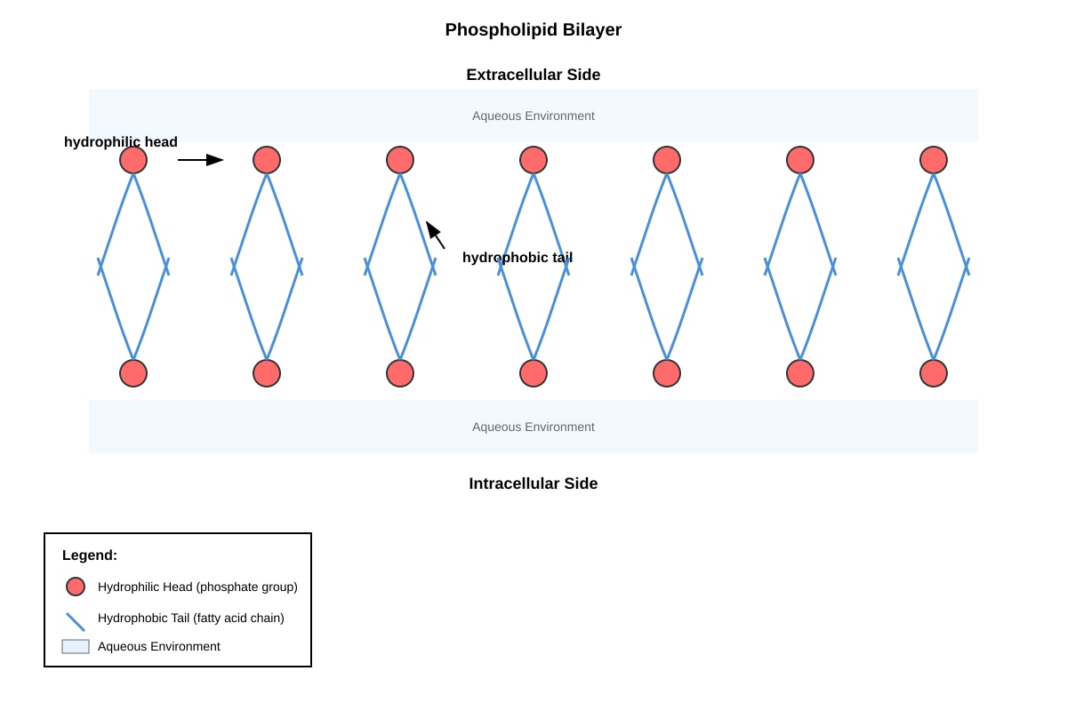
Generated diagram for Q4.
Which statement about the phospholipid bilayer shown is correct?
A. The hydrophobic heads face the aqueous environment. B. The hydrophobic tails interact with water molecules. C. The bilayer is permeable to large polar molecules. D. The bilayer forms a barrier to ions and large polar molecules.
Answer: ______________ [1]
5. Which of the following bonds is responsible for linking amino acids together in a polypeptide chain?
A. Hydrogen bond B. Ionic bond C. Peptide bond D. Disulfide bond
Answer: ______________ [1]
6. An enzyme-catalysed reaction was carried out at 35 °C. When the temperature was increased to 70 °C, the rate of reaction decreased sharply. The best explanation is that
A. the enzyme was denatured and lost its three-dimensional active site conformation. B. the substrate molecules moved too slowly to bind to the enzyme. C. the activation energy of the reaction increased. D. the enzyme-substrate complex became more stable.
Answer: ______________ [1]
7. Which of the following is a function of cholesterol in animal cell membranes?
A. It increases membrane fluidity at all temperatures. B. It acts as a receptor for signalling molecules. C. It reduces membrane fluidity at high temperatures and increases fluidity at low temperatures. D. It provides energy for active transport across the membrane.
Answer: ______________ [1]
8. During which phase of the cell cycle does DNA replication occur?
A. G1 phase B. S phase C. G2 phase D. M phase
Answer: ______________ [1]
9. A molecule of glucose and a molecule of maltose are compared. Which statement is correct?
A. Both are monosaccharides. B. Glucose is a monosaccharide and maltose is a disaccharide. C. Both are reducing sugars but only glucose gives a positive Benedict's test. D. Maltose is formed by a glycosidic bond between two fructose molecules.
Answer: ______________ [1]
10. Which level of protein structure is directly determined by the sequence of amino acids in the polypeptide chain?
A. Primary structure B. Secondary structure C. Tertiary structure D. Quaternary structure
Answer: ______________ [1]
Section B: Structured Questions [20 marks]
Questions 11–15: Answer all questions. Show your working where applicable.
11. Fig. 11.1 shows the structure of a typical animal cell as seen under an electron microscope.
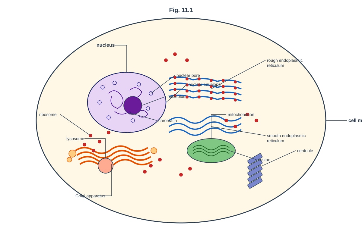
Generated diagram for Q11.
(a) State two structural features visible in Fig. 11.1 that distinguish this as an animal cell rather than a plant cell. [2]
(b) With reference to two organelles visible in Fig. 11.1, describe how their structures are related to their functions. [4]
(c) Explain why the nucleus is described as the control centre of the cell. [2]
[Total: 8 marks]
12. Fig. 12.1 shows the effect of substrate concentration on the rate of an enzyme-catalysed reaction, both in the absence and presence of substance X.
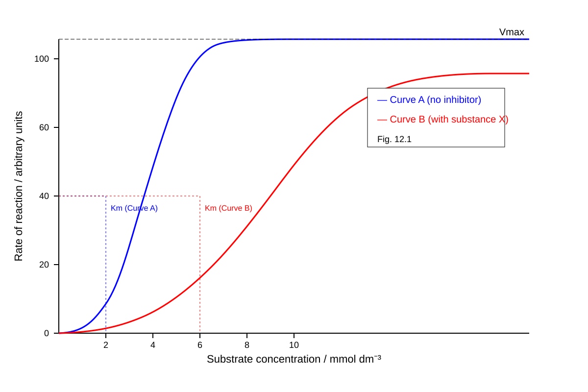
Generated graph for Q12.
(a) With reference to Fig. 12.1, describe the effect of increasing substrate concentration on the rate of reaction in the absence of substance X. [2]
(b) Compare the curves for the reaction with and without substance X. [2]
(c) Suggest the type of inhibition caused by substance X. Explain your answer with reference to Fig. 12.1. [3]
[Total: 7 marks]
13. Table 13.1 shows the results of a food test carried out on a sample of food.
| Test | Reagent used | Observation |
|---|---|---|
| Test 1 | Iodine solution | Blue-black colour |
| Test 2 | Benedict's solution (heated in water bath) | Orange-red precipitate |
| Test 3 | Biuret solution | Violet colour |
| Test 4 | Ethanol emulsion test | Milky white emulsion |
Table 13.1
(a) State the biological molecules that are present in the food sample. [2]
(b) For one of the positive tests in Table 13.1, describe the biochemical basis of the colour change observed. [2]
(c) Explain why Benedict's test requires heating in a water bath rather than direct heating with a Bunsen burner. [1]
[Total: 5 marks]
Section C: Free Response [20 marks]
Questions 14–16: Answer all questions. Write your answers in continuous prose where appropriate. Use clearly labelled diagrams where they support your answer.
14. Fig. 14.1 shows a simplified diagram of the fluid mosaic model of the cell surface membrane.
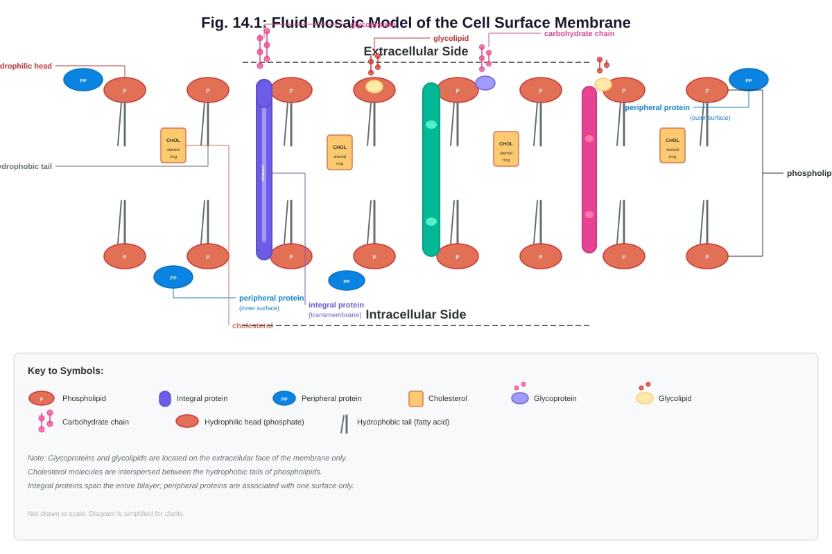
Generated diagram for Q14.
(a) With reference to Fig. 14.1, describe the roles of cholesterol and glycoproteins in the cell surface membrane. [4]
(b) Explain how the structure of the phospholipid bilayer provides a partially permeable barrier. [3]
(c) A cell was placed in a hypertonic solution. Describe and explain what would happen to the cell. [3]
[Total: 10 marks]
15. Fig. 15.1 shows a polypeptide consisting of five amino acids.
Image pending generation: diagram for Q15.
(a) With reference to Fig. 15.1, state the number of peptide bonds in the polypeptide. [1]
(b) Name the type of reaction that joins amino acids together to form a polypeptide. [1]
(c) Explain how the structure of the R-group of cysteine allows it to contribute to the tertiary structure of a protein. [2]
(d) Describe four types of bond or interaction that stabilise the tertiary structure of a protein. For each, state where in the protein it is likely to occur. [4]
[Total: 8 marks]
16. Describe the process of mitosis and explain its significance in living organisms. [2]
[Total: 2 marks]
END OF PAPER
Total Marks: 50
Answers
TuitionGoWhere Practice Paper — Biology H2 A-Level
Answer Key & Marking Scheme
Paper: Practice Paper — Cells & Biomolecules Total Marks: 50
Section A: Multiple Choice [10 marks]
1. C [1]
- Explanation: Ribosomes are the only organelles listed that are found in both prokaryotic and eukaryotic cells. Prokaryotes lack a nucleus, mitochondria, and endoplasmic reticulum. Ribosomes in prokaryotes are 70S (smaller) compared to 80S in eukaryotes, but they are present in both cell types and are essential for protein synthesis.
2. C [1]
- Explanation: The Golgi apparatus is characterised by a stack of flattened membrane-bound cisternae. The rough endoplasmic reticulum is a network of tubules and flattened sacs studded with ribosomes, not a discrete stack. The smooth ER lacks ribosomes and is tubular. Lysosomes are spherical vesicles, not stacks of cisternae.
3. C [1]
- Explanation: The polarity of the water molecule (partial positive charge on hydrogen atoms and partial negative charge on oxygen) allows water to surround and solvate ions and polar molecules, making it an excellent solvent for ionic biological substances. High specific heat capacity relates to temperature buffering, latent heat of vaporisation relates to cooling, and cohesion relates to surface tension and water transport.
4. D [1]
- Explanation: The hydrophobic interior of the phospholipid bilayer acts as a barrier to ions and large polar molecules, which cannot easily pass through the non-polar fatty acid tails. Hydrophilic heads (not tails) face the aqueous environment. The bilayer is impermeable to large polar molecules without the aid of transport proteins.
5. C [1]
- Explanation: Amino acids are linked by peptide bonds, which form between the carboxyl group (–COOH) of one amino acid and the amino group (–NH₂) of another in a condensation reaction. Hydrogen bonds stabilise secondary structure. Disulfide bonds form between cysteine residues in tertiary structure. Ionic bonds can also contribute to tertiary structure.
6. A [1]
- Explanation: At 70 °C, the enzyme's three-dimensional structure is disrupted (denatured) due to the breaking of hydrogen bonds, ionic interactions, and other weak bonds that maintain the tertiary structure. This alters the shape of the active site so that the substrate can no longer bind effectively. Increasing temperature initially increases reaction rate, but beyond the optimum, denaturation causes a sharp decline.
7. C [1]
- Explanation: Cholesterol modulates membrane fluidity in a dual manner: at high temperatures, it restricts phospholipid movement, reducing fluidity; at low temperatures, it prevents tight packing of phospholipids, maintaining fluidity. This buffering effect helps maintain membrane integrity across a range of temperatures.
8. B [1]
- Explanation: DNA replication occurs during the S (synthesis) phase of interphase. G1 is a growth phase before DNA replication, G2 is a growth phase after DNA replication in preparation for mitosis, and M phase is mitosis itself (nuclear division).
9. B [1]
- Explanation: Glucose is a monosaccharide (C₆H₁₂O₆), while maltose is a disaccharide formed from two glucose molecules joined by a α-1,4-glycosidic bond. Both glucose and maltose are reducing sugars and both give a positive Benedict's test. Maltose is formed from two glucose units, not fructose.
10. A [1]
- Explanation: The primary structure is the linear sequence of amino acids in a polypeptide chain, which is directly encoded by the gene. Secondary structure (α-helix, β-pleated sheet) arises from hydrogen bonding between the peptide backbone. Tertiary structure results from interactions between R-groups. Quaternary structure involves the assembly of multiple polypeptide subunits.
Section B: Structured Questions [20 marks]
11. (a) [2]
Marking scheme — 1 mark each, max 2:
- Absence of a cell wall
- Absence of chloroplasts
- Absence of a large permanent central vacuole
- Presence of centrioles
Teaching note: Plant cells have a rigid cell wall made of cellulose, chloroplasts for photosynthesis, and a large central vacuole for turgor and storage. Animal cells lack all three. Centrioles are typically found in animal cells (though not all plant cells lack them — lower plants may have them). The most reliable distinguishing features are the absence of a cell wall and chloroplasts.
11. (b) [4]
Marking scheme — 2 marks per organelle (1 mark for structure + 1 mark for link to function), max 4:
Example answer 1: Mitochondrion
- Structure: Has a double membrane; the inner membrane is highly folded into cristae; contains a matrix.
- Function link: The cristae provide a large surface area for the attachment of enzymes and electron carriers involved in aerobic respiration (oxidative phosphorylation/electron transport chain). The matrix contains enzymes for the Krebs cycle.
Example answer 2: Rough endoplasmic reticulum (RER)
- Structure: Membrane-bound flattened sacs (cisternae) studded with ribosomes on the cytoplasmic surface.
- Function link: The ribosomes on the RER synthesise proteins (particularly secretory and membrane proteins). The cisternae provide a transport pathway and allow proteins to be folded and modified within the lumen.
Other acceptable organelles:
- Golgi apparatus: Stack of flattened cisternae; modifies, sorts, and packages proteins into vesicles for secretion or delivery to other organelles.
- Lysosome: Single membrane-bound vesicle containing hydrolytic enzymes; the membrane isolates the enzymes from the rest of the cell to prevent unwanted digestion.
- Nucleus: Double membrane (nuclear envelope) with nuclear pores; houses DNA and controls gene expression; nuclear pores regulate the transport of molecules between nucleus and cytoplasm.
11. (c) [2]
Marking scheme — 2 marks:
- The nucleus contains DNA / chromosomes / genetic material [1]
- DNA carries the genes / genetic code that control protein synthesis, which determines cell structure and function [1]
Teaching note: The nucleus is called the control centre because it houses the cell's genetic information. Genes on DNA are transcribed into mRNA, which is translated into proteins. Proteins determine virtually all cellular structures and functions, including enzymes that catalyse metabolic reactions. Without the nucleus, the cell cannot produce new proteins and cannot regulate its activities.
12. (a) [2]
Marking scheme — 2 marks:
- As substrate concentration increases, the rate of reaction increases [1]
- The rate eventually plateaus / reaches a maximum (Vmax) when all enzyme active sites are saturated / occupied [1]
Teaching note: At low substrate concentrations, there are many free active sites, so increasing substrate leads to more enzyme-substrate complexes forming per unit time. At high substrate concentrations, all active sites are occupied at any given moment, so further increases in substrate do not increase the rate — the enzyme is working at maximum capacity.
12. (b) [2]
Marking scheme — 2 marks:
- Both curves reach the same Vmax [1]
- The curve with substance X requires a higher substrate concentration to reach half Vmax / has a higher apparent Km [1]
Teaching note: The key observation is that Vmax is unchanged but the curve is shifted to the right, indicating a higher Km. This is the hallmark of competitive inhibition.
12. (c) [3]
Marking scheme — 3 marks:
- Competitive inhibition [1]
- Substance X / the inhibitor has a similar structure to the substrate and competes for the active site of the enzyme [1]
- The inhibition can be overcome by increasing substrate concentration, as shown by the same Vmax being reached [1]
Teaching note: In competitive inhibition, the inhibitor binds reversibly to the active site, preventing substrate binding. Because the inhibitor and substrate compete, a sufficiently high substrate concentration can outcompete the inhibitor, restoring the reaction rate to the same Vmax. The apparent Km increases because a higher substrate concentration is needed to reach half Vmax. Classic examples include malonate inhibiting succinate dehydrogenase (malonate resembles succinate) and statins inhibiting HMG-CoA reductase.
13. (a) [2]
Marking scheme — ½ mark per correct molecule, max 2:
- Starch (from iodine test — blue-black)
- Reducing sugar (from Benedict's test — orange-red precipitate)
- Protein (from Biuret test — violet colour)
- Lipid (from ethanol emulsion test — milky white emulsion)
Teaching note: The food sample contains all four major classes of biological molecules. The orange-red precipitate in Benedict's test indicates a moderate to high concentration of reducing sugar (green = low, yellow = moderate, orange = high, red = very high).
13. (b) [2]
Marking scheme — 2 marks for a clear biochemical explanation:
Example: Benedict's test
- Benedict's solution contains copper(II) sulfate (Cu²⁺ ions in alkaline solution) [1]
- Reducing sugars have a free aldehyde or ketone group that reduces Cu²⁺ (blue) to Cu⁺, forming insoluble copper(I) oxide (Cu₂O), which appears as a coloured precipitate (green → yellow → orange → red depending on concentration) [1]
Alternative: Biuret test
- Biuret solution contains copper(II) sulfate in alkaline (NaOH) solution [1]
- The Cu²⁺ ions form a violet-coloured complex with peptide bonds (specifically, the nitrogen atoms in the –CO–NH– groups) in alkaline conditions [1]
13. (c) [1]
Marking scheme — 1 mark:
- A water bath provides gentle, uniform heating / prevents the solution from boiling too vigorously / ensures the temperature does not exceed 100 °C, which could degrade the reagents or cause the solution to spatter [1]
Teaching note: Direct heating with a Bunsen burner can cause uneven heating, localised overheating, and the risk of the test tube cracking or the contents spattering. A water bath ensures controlled, even heating to the required temperature (typically around 80–100 °C for Benedict's test).
Section C: Free Response [20 marks]
14. (a) [4]
Marking scheme — 2 marks per component:
Cholesterol: [2]
- Cholesterol molecules are located between the phospholipid tails in the bilayer [1]
- They regulate membrane fluidity — at high temperatures they restrict phospholipid movement (reducing fluidity), and at low temperatures they prevent tight packing of phospholipids (maintaining fluidity). They also help maintain membrane mechanical stability [1]
Glycoproteins: [2]
- Glycoproteins are proteins with carbohydrate chains attached, located on the extracellular surface of the membrane [1]
- They function in cell-cell recognition (e.g., immune recognition, blood group antigens), act as receptors for signalling molecules (e.g., hormones), and help protect the cell surface [1]
14. (b) [3]
Marking scheme — 3 marks:
- The phospholipid bilayer has a hydrophilic (polar) phosphate head and two hydrophobic (non-polar) fatty acid tails [1]
- The hydrophobic interior of the bilayer acts as a barrier to the passage of ions, large polar molecules, and hydrophilic substances, which cannot dissolve in the non-polar core [1]
- Small non-polar molecules (e.g., O₂, CO₂) and small uncharged polar molecules (e.g., water) can pass through the bilayer by simple diffusion, making the membrane partially/permeable [1]
Teaching note: The term "partially permeable" (or "selectively permeable") means that the membrane allows some substances to pass through freely while restricting others. This is essential for maintaining different intracellular and extracellular environments.
14. (c) [3]
Marking scheme — 3 marks:
- A hypertonic solution has a higher solute concentration (lower water potential) than the cell cytoplasm [1]
- Water molecules move out of the cell by osmosis, from a region of higher water potential (inside the cell) to a region of lower water potential (the solution), across the partially permeable membrane [1]
- The cell will shrink / lose volume. In an animal cell, the cell membrane will pull away from the cell wall (if present) — in plant cells this is called plasmolysis; in animal cells, the cell becomes crenated [1]
Teaching note: Osmosis is the net movement of water molecules from a region of higher water potential to a region of lower water potential across a partially permeable membrane. In plant cells, plasmolysis can be observed as the cell membrane detaching from the cell wall. In animal cells (e.g., red blood cells), the cell shrinks and becomes crenated.
15. (a) [1]
Answer: 4 [1]
Teaching note: A polypeptide of n amino acids has (n – 1) peptide bonds. With 5 amino acids, there are 4 peptide bonds linking them in a chain.
15. (b) [1]
Answer: Condensation reaction (or dehydration synthesis) [1]
Teaching note: A condensation reaction joins two monomers by removing a water molecule. In this case, the –OH from the carboxyl group of one amino acid and the –H from the amino group of another combine to form H₂O, creating a peptide bond (–CO–NH–). The reverse reaction (breaking a peptide bond by adding water) is hydrolysis.
15. (c) [2]
Marking scheme — 2 marks:
- Cysteine contains a sulfhydryl group (–SH) in its R-group [1]
- Two cysteine residues can form a disulfide bond (–S–S–) through oxidation, which covalently links different parts of the polypeptide chain, stabilising the tertiary structure [1]
Teaching note: Disulfide bonds are strong covalent bonds that form between the –SH groups of two cysteine residues. They are particularly important in extracellular proteins (e.g., antibodies, insulin) where they help maintain the protein's three-dimensional shape in the extracellular environment. The formation of disulfide bonds is an oxidation reaction (2 R–SH → R–S–S–R + 2H⁺ + 2e⁻).
15. (d) [4]
Marking scheme — 1 mark per bond/interaction + location, max 4:
| Bond/Interaction | Where it occurs | Mark |
|---|---|---|
| Hydrogen bonds | Between polar R-groups (e.g., –OH, –NH₂, –COOH) or between backbone groups forming secondary structures | [1] |
| Ionic bonds (salt bridges) | Between positively charged R-groups (e.g., lysine –NH₃⁺) and negatively charged R-groups (e.g., aspartate –COO⁻) | [1] |
| Disulfide bonds | Between the –SH groups of two cysteine residues, forming covalent –S–S– bridges | [1] |
| Hydrophobic interactions | Between non-polar / hydrophobic R-groups (e.g., leucine, valine, phenylalanine), which cluster together in the interior of the protein away from water | [1] |
Teaching note: The tertiary structure is the overall three-dimensional shape of a single polypeptide chain, stabilised by interactions between R-groups. Hydrogen bonds are weak individually but numerous. Ionic bonds are strong but can be disrupted by changes in pH. Disulfide bonds are the strongest and are covalent. Hydrophobic interactions drive non-polar residues to the protein interior, which is a major stabilising force. Van der Waals forces between closely packed non-polar groups also contribute.
16. [2]
Marking scheme — 2 marks:
- Mitosis is the process of nuclear division that results in the production of two genetically identical daughter nuclei, each with the same number of chromosomes as the parent nucleus [1]
- Significance: It is important for growth (increasing cell number), repair/replacement of damaged or dead cells, and asexual reproduction. It ensures genetic consistency — each daughter cell receives an exact copy of the genetic material [1]
Teaching note: Mitosis consists of four main stages: prophase (chromosomes condense, nuclear envelope breaks down), metaphase (chromosomes align at the metaphase plate), anaphase (sister chromatids separate and move to opposite poles), and telophase (nuclear envelopes reform, chromosomes decondense). This is followed by cytokinesis (division of the cytoplasm). Mitosis ensures that each daughter cell is genetically identical to the parent cell, which is essential for maintaining genetic stability in multicellular organisms.
END OF ANSWER KEY
Mark Summary:
| Section | Marks |
|---|---|
| A: Multiple Choice (Q1–10) | 10 |
| B: Structured Questions (Q11–13) | 20 |
| C: Free Response (Q14–16) | 20 |
| Total | 50 |
Free quiz and exam paper access
Enter your details to view this paper
Your access is remembered on this device.