From Real Exams Exam Paper
A Level H2 Biology Practice Paper 5
Free A Level H2 Biology Practice Paper 5, LongCat Exam version, with questions, answers, and A Level-style practice for Singapore students.
These static practice materials are generated from the site's syllabus and paper-generation workflow, with source and model context shown so students and parents can evaluate the material before use.
Questions
TuitionGoWhere Practice Paper — Biology H2 A-Level
TuitionGoWhere Secondary School (AI)
| Subject: | Biology H2 |
| Level: | A-Level |
| Paper: | Practice Paper (Cells & Biomolecules) |
| Version: | 5 of 5 |
| Duration: | 1 hour 15 minutes |
| Total Marks: | 60 |
| Name: | ________________________ |
| Class: | ________________________ |
| Date: | ________________________ |
Instructions
- Answer all questions in the spaces provided.
- Write in dark blue or black pen.
- You may use a pencil for any diagrams or graphs.
- No calculators are permitted.
- The total mark for this paper is 60.
- The number of marks for each question or part question is shown in brackets [ ].
- You are advised to spend no more than 1 hour 15 minutes on this paper.
Section A: Multiple Choice Questions (10 marks)
Questions 1–10: Choose the single best answer.
1. Which of the following organelles is present in both prokaryotic and eukaryotic cells?
A. Golgi apparatus
B. Mitochondrion
C. Ribosome
D. Endoplasmic reticulum
[1 mark]
2. A student analysed the chemical composition of a membrane. Which component would be most abundant in a typical eukaryotic plasma membrane?
A. Cholesterol
B. Glycolipid
C. Phospholipid
D. Integral protein
[1 mark]
3. Which property of water is most important in enabling it to act as a solvent for ionic biological molecules?
A. High specific heat capacity
B. High latent heat of vaporisation
C. Cohesion between water molecules
D. Polarity of the water molecule
[1 mark]
4. During an enzyme-catalysed reaction, the rate of reaction decreases as the temperature rises above the optimum. Which statement best explains this?
A. The substrate molecules have too much kinetic energy to bind to the active site.
B. The enzyme is denatured, so the tertiary structure of the active site is altered.
C. The activation energy increases at higher temperatures.
D. The enzyme-substrate complex becomes more stable at higher temperatures.
[1 mark]
5. Which of the following bonds is responsible for holding the two strands of the DNA double helix together?
A. Covalent bonds
B. Ionic bonds
C. Hydrogen bonds
D. Phosphodiester bonds
[1 mark]
6. A polypeptide has the amino acid sequence: Met–Leu–Asp–Val–Arg. A mutation changes the codon for Asp to a codon for Glu. What type of mutation is this?
A. Nonsense mutation
B. Frameshift mutation
C. Silent mutation
D. Missense mutation
[1 mark]
7. Which of the following is a function of the smooth endoplasmic reticulum?
A. Synthesis of ribosomal RNA
B. Synthesis of lipids and steroids
C. Packaging of proteins into vesicles
D. Translation of mRNA into polypeptides
[1 mark]
8. A reducing sugar test was performed on four unknown solutions. Which observation indicates a positive result with Benedict's reagent?
A. A blue solution remains blue.
B. A blue solution turns green, then yellow, then orange-red precipitate.
C. A blue solution turns violet.
D. A colourless solution turns blue-black.
[1 mark]
9. Which level of protein structure is directly determined by the sequence of amino acids in the polypeptide chain?
A. Secondary structure
B. Tertiary structure
C. Quaternary structure
D. Primary structure
[1 mark]
10. In an experiment, a cell was placed in a hypertonic solution. Which of the following best describes what would happen to an animal cell?
A. The cell would swell and burst.
B. The cell would become turgid.
C. The cell would shrink and become crenated.
D. The cell would remain unchanged.
[1 mark]
Section B: Structured Questions (35 marks)
Question 11.
Image pending generation: diagram for Q11.
Fig. 11.1 shows a diagram of a typical animal cell as seen under an electron microscope.
(a) Identify the organelles labelled A, B, C, and D in Fig. 11.1.
A: ________________________ [1]
B: ________________________ [1]
C: ________________________ [1]
D: ________________________ [1]
(b) Describe two structural features of the organelle labelled C that are related to its function.
- _____________________________________________________________ [1]
- _____________________________________________________________ [1]
(c) Explain why organelle B is abundant in cells that secrete large quantities of protein.
_____________________________________________________________ [2]
[Total: 7 marks]
Question 12.
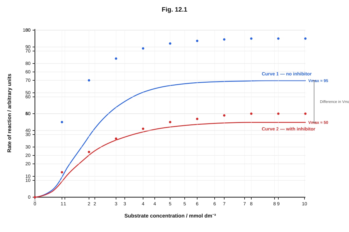
Generated graph for Q12.
Fig. 12.1 shows the effect of substrate concentration on the rate of an enzyme-catalysed reaction, with and without the presence of an inhibitor.
(a) Describe the effect of increasing substrate concentration on the rate of reaction in the absence of the inhibitor (Curve 1).
_____________________________________________________________ [2]
(b) With reference to Fig. 12.1, explain the effect of the inhibitor on the enzyme.
_____________________________________________________________ [3]
(c) Suggest, with a reason, whether the inhibitor shown is competitive or non-competitive.
_____________________________________________________________ [1]
[Total: 6 marks]
Question 13.
The table below shows the results of food tests carried out on three different food samples, P, Q, and R.
| Test | Food sample P | Food sample Q | Food sample R |
|---|---|---|---|
| Benedict's test | Blue solution | Orange-red precipitate | Blue solution |
| Biuret test | Pale blue solution | Pale blue solution | Violet solution |
| Ethanol emulsion test | Clear solution | Cloudy white emulsion | Clear solution |
| Iodine test | Blue-black colour | Brown solution | Brown solution |
(a) Identify the main biological molecule(s) present in each food sample.
P: ____________________________________________________________ [1]
Q: ____________________________________________________________ [1]
R: ____________________________________________________________ [1]
(b) Describe how the Biuret test is carried out.
_____________________________________________________________ [2]
(c) Explain why food sample P gives a positive result with the iodine test.
_____________________________________________________________ [2]
[Total: 7 marks]
Question 14.
Generated diagram for Q14.
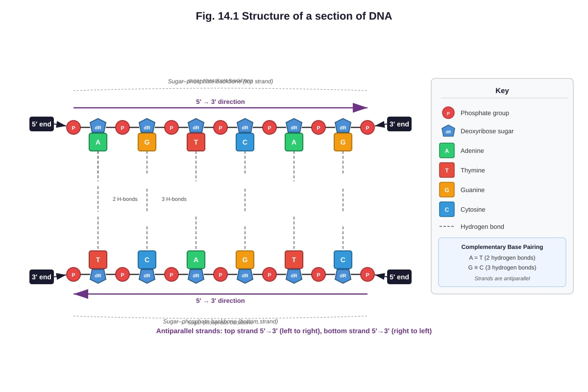
Fig. 14.1 shows a short section of a DNA molecule.
(a) With reference to Fig. 14.1, state two structural features of DNA that enable it to carry out its function of storing genetic information.
- _____________________________________________________________ [1]
- _____________________________________________________________ [1]
(b) Explain how the structure of DNA allows it to replicate itself accurately.
_____________________________________________________________ [3]
(c) Name the type of bond labelled X that holds the two strands of DNA together.
_____________________________________________________________ [1]
[Total: 6 marks]
Question 15.
(a) Describe the role of the following organelles in the production and secretion of a protein such as insulin.
(i) Ribosome:
_____________________________________________________________ [1]
(ii) Golgi apparatus:
_____________________________________________________________ [1]
(b) Explain the significance of the fluid mosaic model of the plasma membrane for the process of exocytosis.
_____________________________________________________________ [3]
[Total: 5 marks]
Question 16.
A student carried out an experiment to investigate the effect of temperature on the permeability of beetroot cell membranes. Beetroot cylinders of equal size were placed in distilled water at different temperatures for 10 minutes. The colour of the surrounding water was then measured using a colorimeter.
The results are shown in the table below.
| Temperature / °C | Absorbance of solution (arbitrary units) |
|---|---|
| 10 | 0.05 |
| 20 | 0.08 |
| 30 | 0.12 |
| 40 | 0.25 |
| 50 | 0.58 |
| 60 | 0.89 |
| 70 | 0.92 |
(a) Describe the trend shown in the results.
_____________________________________________________________ [2]
(b) Explain the results obtained at 60 °C and 70 °C.
_____________________________________________________________ [3]
(c) State one variable that should be kept constant in this experiment, other than the size of the beetroot cylinders.
_____________________________________________________________ [1]
[Total: 6 marks]
Section C: Free Response (15 marks)
Question 17.
Generated diagram for Q17.
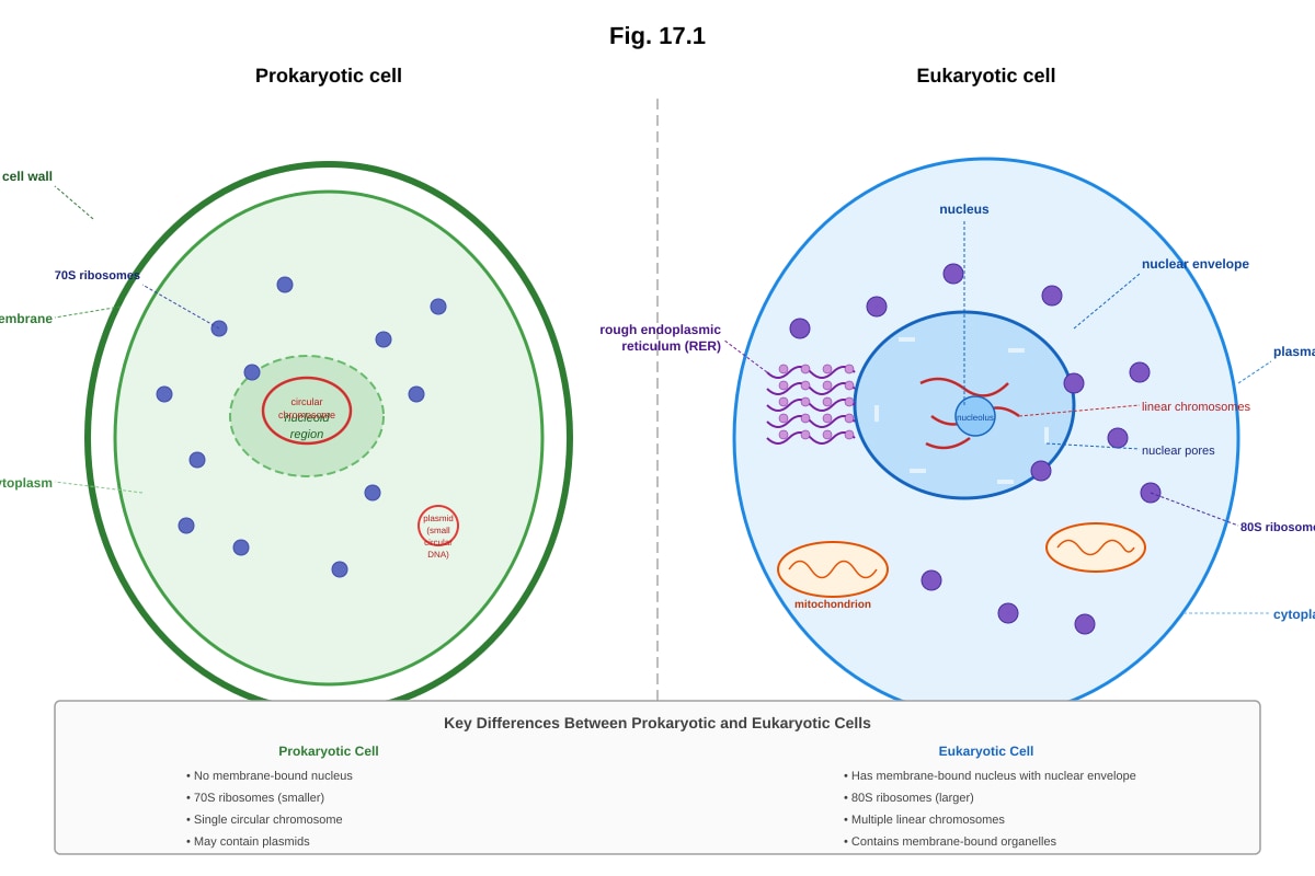
Fig. 17.1 shows a comparison between a prokaryotic cell and a eukaryotic cell.
(a) State three structural differences between a prokaryotic cell and a eukaryotic cell, other than the presence or absence of a nucleus.
- _____________________________________________________________ [1]
- _____________________________________________________________ [1]
- _____________________________________________________________ [1]
(b) Explain how the structural differences between prokaryotic and eukaryotic cells affect the way gene expression occurs in each type of cell.
_____________________________________________________________ [4]
[Total: 7 marks]
Question 18.
Haemoglobin is a globular protein with quaternary structure. It is made up of four polypeptide chains, each containing a haem group with an iron(II) ion (Fe2+) at its centre.
(a) Explain what is meant by the quaternary structure of a protein.
_____________________________________________________________ [2]
(b) Describe how the structure of haemoglobin is related to its function of transporting oxygen.
_____________________________________________________________ [4]
(c) Explain how a change in the primary structure of one polypeptide chain in haemoglobin could affect its ability to transport oxygen.
_____________________________________________________________ [2]
**[Total: 8 marks]]
End of Paper
| Section | Marks |
|---|---|
| A: Multiple Choice (Q1–10) | 10 |
| B: Structured Questions (Q11–16) | 35 |
| C: Free Response (Q17–18) | 15 |
| Total | 60 |
Answers
TuitionGoWhere Practice Paper — Biology H2 A-Level
Answer Key — Version 5 of 5
Total Marks: 60
Section A: Multiple Choice Questions (10 marks)
1. C — Ribosome [1]
Teaching note: Ribosomes are the only organelle listed that are found in both prokaryotic and eukaryotic cells. They are non-membrane-bound and essential for protein synthesis in all living cells. The Golgi apparatus, mitochondrion, and endoplasmic reticulum are all membrane-bound organelles found only in eukaryotic cells.
2. C — Phospholipid [1]
Teaching note: Phospholipids are the most abundant component of the plasma membrane, forming the basic bilayer structure. While proteins are also abundant by mass, phospholipids form the continuous structural framework. Cholesterol and glycolipids are present in smaller quantities.
3. D — Polarity of the water molecule [1]
Teaching note: The polarity of water (partial negative charge on oxygen, partial positive charges on hydrogen atoms) allows water molecules to surround and stabilise ions through electrostatic interactions, enabling dissolution of ionic compounds. High specific heat capacity, latent heat, and cohesion are important properties but not directly related to dissolving ionic substances.
4. B — The enzyme is denatured, so the tertiary structure of the active site is altered. [1]
Teaching note: Above the optimum temperature, the increased kinetic energy disrupts the hydrogen bonds, ionic bonds, and hydrophobic interactions that maintain the enzyme's tertiary structure. This changes the shape of the active site so that the substrate can no longer bind effectively. The activation energy does not increase — the enzyme's ability to lower it is reduced.
5. C — Hydrogen bonds [1]
Teaching note: Hydrogen bonds between complementary base pairs (A–T with 2 bonds, G–C with 3 bonds) hold the two antiparallel strands of DNA together. Covalent bonds form the sugar-phosphate backbone within each strand. Phosphodiester bonds are the specific covalent bonds linking nucleotides in a strand. Ionic bonds are not involved in DNA strand association.
6. D — Missense mutation [1]
Teaching note: A missense mutation results in one amino acid being replaced by another. Here, aspartic acid (Asp) is replaced by glutamic acid (Glu) — both are different amino acids, so this is a substitution that changes the amino acid. A nonsense mutation introduces a stop codon. A frameshift mutation involves insertion or deletion of nucleotides. A silent mutation does not change the amino acid.
7. B — Synthesis of lipids and steroids [1]
Teaching note: The smooth endoplasmic reticulum (SER) lacks ribosomes and is involved in lipid synthesis, steroid hormone production, and detoxification. rRNA synthesis occurs in the nucleolus. Protein packaging is carried out by the Golgi apparatus. Translation occurs on ribosomes.
8. B — A blue solution turns green, then yellow, then orange-red precipitate. [1]
Teaching note: Benedict's reagent is blue due to copper(II) sulfate. In the presence of a reducing sugar, the Cu2+ ions are reduced to Cu+ ions, forming copper(I) oxide (Cu2O), which is an orange-red precipitate. The colour change progresses through green and yellow as the concentration of reducing sugar increases. Option C describes a positive Biuret test, and option D describes a positive iodine test.
9. D — Primary structure [1]
Teaching note: The primary structure is the linear sequence of amino acids linked by peptide bonds. This sequence is directly encoded by the gene (DNA). The secondary, tertiary, and quaternary structures all arise as consequences of the primary structure — the amino acid sequence determines how the polypeptide folds.
10. C — The cell would shrink and become crenated. [1]
Teaching note: In a hypertonic solution, the water potential outside the cell is lower than inside the cell. Water moves out of the cell by osmosis, causing the cell to shrink. In animal cells, this is called crenation. Plant cells undergo plasmolysis in the same conditions, but the cell wall prevents them from shrinking as dramatically. Option A describes what happens in a hypotonic solution. Option B describes a plant cell in a hypotonic solution.
Section B: Structured Questions (35 marks)
Question 11 (7 marks)
(a) Identification of organelles:
- A: Nucleus [1]
- B: Rough endoplasmic reticulum (RER) [1]
- C: Mitochondrion [1]
- D: Golgi apparatus [1]
Marking note: Accept "rough ER" for B. Accept "mitochondria" for C. Spelling must be reasonably accurate.
(b) Two structural features of the mitochondrion related to its function: [2]
-
The inner membrane is folded into cristae — this increases the surface area for the attachment of electron carriers and ATP synthase enzymes involved in oxidative phosphorylation / the electron transport chain. [1]
-
The matrix contains enzymes for the Krebs cycle / link reaction / the matrix is the site of the Krebs cycle. [1]
Alternative acceptable answers:
- The mitochondrion has a double membrane — the inner membrane is the site of the electron transport chain and chemiosmosis.
- The matrix contains mitochondrial DNA and ribosomes — allowing the mitochondrion to synthesise some of its own proteins.
- The intermembrane space is small — allowing a proton gradient to build up quickly across the inner membrane.
Marking note: Each mark requires a structural feature linked to its function. Structure alone or function alone scores 0 for that point.
(c) Explanation of why RER is abundant in cells that secrete large quantities of protein: [2]
- The RER has ribosomes attached to its surface [1] which synthesise proteins / polypeptides that are destined for secretion. The RER provides a large surface area for protein synthesis and the lumen of the RER provides a pathway for transport of these proteins to the Golgi apparatus for processing and packaging [1].
Marking note: Award 1 mark for mentioning ribosomes on RER synthesising proteins. Award 1 mark for linking this to the secretory pathway (transport to Golgi / packaging for secretion). Simply stating "RER makes proteins" without linking to secretion scores only 1 mark.
Question 12 (6 marks)
(a) Description of the effect of increasing substrate concentration (Curve 1): [2]
- At low substrate concentrations, the rate of reaction increases rapidly / almost linearly as substrate concentration increases [1]. At higher substrate concentrations, the rate of reaction levels off / reaches a maximum / plateaus because all active sites are occupied / saturated [1].
Marking note: Award 1 mark for describing the initial increase and 1 mark for describing the plateau. Simply stating "rate increases" without mentioning the plateau scores only 1 mark.
(b) Explanation of the effect of the inhibitor: [3]
- In the presence of the inhibitor, the maximum rate of reaction (Vmax) is lower than without the inhibitor [1]. At any given substrate concentration, the rate of reaction is lower with the inhibitor present [1]. This is because the inhibitor binds to the enzyme, reducing the number of functional enzyme molecules available to catalyse the reaction / reducing the rate of formation of enzyme-substrate complexes [1].
Marking note: Award 1 mark for identifying the reduced Vmax. Award 1 mark for noting the lower rate at given substrate concentrations. Award 1 mark for explaining the mechanism (fewer functional enzymes / fewer E-S complexes).
(c) Type of inhibitor with reason: [1]
- Non-competitive inhibitor [1] because the Vmax is reduced / the maximum rate of reaction is lower in the presence of the inhibitor.
Teaching note: In competitive inhibition, Vmax remains the same (the inhibitor can be overcome by increasing substrate concentration), but a higher substrate concentration is needed to reach Vmax. In non-competitive inhibition, the inhibitor binds to a site other than the active site, reducing the number of functional enzyme molecules, so Vmax is reduced. Since Curve 2 shows a lower Vmax than Curve 1, this is non-competitive inhibition.
Question 13 (7 marks)
(a) Identification of main biological molecules: [3]
-
P: Starch [1] — positive iodine test (blue-black) indicates starch; negative Benedict's test rules out reducing sugars; negative Biuret test rules out protein; negative ethanol emulsion test rules out lipids.
-
Q: Lipid and reducing sugar [1] — positive Benedict's test (orange-red precipitate) indicates reducing sugar; positive ethanol emulsion test (cloudy white emulsion) indicates lipid; negative Biuret test rules out protein; negative iodine test rules out starch.
-
R: Protein [1] — positive Biuret test (violet) indicates protein; negative Benedict's test rules out reducing sugars; negative ethanol emulsion test rules out lipids; negative iodine test rules out starch.
(b) Description of how the Biuret test is carried out: [2]
- Add sodium hydroxide solution (NaOH) / alkali to the sample [1], then add a few drops of copper(II) sulfate solution (CuSO4) [1]. A violet / purple colour indicates the presence of protein / peptide bonds.
Marking note: Both steps required for full marks. The order matters — NaOH must be added first to create alkaline conditions. Simply stating "add Biuret reagent" without describing the two components scores 1 mark.
(c) Explanation for positive iodine test with food sample P: [2]
- Food sample P contains starch [1]. Starch has a helical / coiled structure (amylose) that traps iodine molecules, forming a starch-iodine complex which appears blue-black [1].
Marking note: Award 1 mark for identifying starch. Award 1 mark for explaining the mechanism (helical structure trapping iodine / formation of starch-iodine complex). Simply stating "starch turns iodine blue-black" without explaining why scores only 1 mark.
Question 14 (6 marks)
(a) Two structural features of DNA enabling it to store genetic information: [2]
-
The sequence of bases along the strand can vary — the four bases (A, T, G, C) can be arranged in any order, allowing a vast amount of genetic information to be encoded. [1]
-
Complementary base pairing (A–T and G–C) — ensures that the genetic information is stored redundantly / in a stable manner on both strands, allowing accurate replication and repair. [1]
Alternative acceptable answers:
- The sugar-phosphate backbone is strong and stable, protecting the genetic information stored in the bases.
- The double-stranded structure provides a template for replication and repair.
- Hydrogen bonds between base pairs allow the strands to be separated for replication and transcription.
(b) Explanation of how DNA structure allows accurate replication: [4]
- The two strands of DNA are held together by hydrogen bonds between complementary base pairs [1]. During replication, the double helix is unwound and the hydrogen bonds between the strands are broken by helicase, separating the two strands [1]. Each separated strand acts as a template for the synthesis of a new complementary strand [1]. Free DNA nucleotides bind to their complementary bases on each template strand (A with T, G with C) via hydrogen bonding, and DNA polymerase catalyses the formation of phosphodiester bonds between adjacent nucleotides, producing two identical DNA molecules [1].
Marking note: Award 1 mark for each valid point up to 4 marks. Key points: hydrogen bonds between strands, unwinding/separation of strands, each strand acts as template, complementary base pairing ensures accuracy, DNA polymerase joins nucleotides.
(c) Bond labelled X: [1]
- Hydrogen bond [1]
Teaching note: Hydrogen bonds hold the complementary base pairs together across the two strands. A–T pairs have 2 hydrogen bonds; G–C pairs have 3 hydrogen bonds.
Question 15 (5 marks)
(a)(i) Role of ribosome: [1]
- Ribosomes are the site of protein synthesis / translation [1]. They read the mRNA codon sequence and catalyse the formation of peptide bonds between amino acids to produce the polypeptide chain (insulin).
(a)(ii) Role of Golgi apparatus: [1]
- The Golgi apparatus modifies / processes proteins (e.g., folding, adding carbohydrate groups) [1] and packages them into secretory vesicles for transport to the plasma membrane for secretion / exocytosis.
Marking note: "Packages into vesicles" alone scores 1. "Modifies proteins" alone scores 1. Both together still score 1 mark (single mark question).
(b) Significance of the fluid mosaic model for exocytosis: [3]
- The fluid mosaic model describes the plasma membrane as a phospholipid bilayer with proteins, where the components can move laterally within the membrane (fluid) [1]. During exocytosis, secretory vesicles fuse with the plasma membrane to release their contents [1]. The fluid nature of the membrane allows the phospholipids and proteins of the vesicle membrane and the plasma membrane to merge / fuse together, enabling the vesicle contents to be released outside the cell [1].
Marking note: Award 1 mark for describing the fluid nature of the membrane. Award 1 mark for describing exocytosis (vesicle fusion with plasma membrane). Award 1 mark for linking fluidity to the ability of membranes to fuse.
Question 16 (6 marks)
(a) Description of trend: [2]
- As temperature increases from 10 °C to 70 °C, the absorbance of the solution increases [1]. The increase is gradual between 10 °C and 30 °C, but becomes much more rapid / steep between 40 °C and 60 °C, and then levels off between 60 °C and 70 °C [1].
Marking note: Award 1 mark for the overall trend (increase with temperature). Award 1 mark for describing the pattern of increase (gradual then rapid then plateau).
(b) Explanation of results at 60 °C and 70 °C: [3]
- At 60 °C and 70 °C, the high temperature causes the phospholipid bilayer of the cell membrane to break down / become more fluid / lose its integrity [1]. The high temperature also denatures the membrane proteins, creating gaps in the membrane [1]. This increases membrane permeability, allowing more betalain pigment (which is stored in the vacuole) to diffuse out of the cells into the surrounding water, resulting in higher absorbance readings [1].
Marking note: Award 1 mark for effect on phospholipid bilayer. Award 1 mark for effect on membrane proteins. Award 1 mark for linking increased permeability to pigment release / higher absorbance.
(c) One controlled variable: [1]
Any one of:
- Volume of distilled water used [1]
- Time the beetroot cylinders were left in the water [1]
- Type / source of beetroot [1]
- pH of the solution [1]
Marking note: Do not accept "size of beetroot cylinders" as this was excluded in the question. Do not accept "temperature" as this is the independent variable.
Section C: Free Response (15 marks)
Question 17 (7 marks)
(a) Three structural differences between prokaryotic and eukaryotic cells (other than nucleus): [3]
-
Ribosome size — prokaryotic cells have 70S ribosomes while eukaryotic cells have 80S ribosomes. [1]
-
Presence of membrane-bound organelles — eukaryotic cells have membrane-bound organelles (e.g., mitochondria, endoplasmic reticulum, Golgi apparatus) while prokaryotic cells do not. [1]
-
Chromosome structure — prokaryotic cells have a single circular chromosome while eukaryotic cells have multiple linear chromosomes. [1]
Alternative acceptable answers:
- Prokaryotic cells have a cell wall made of murein/peptidoglycan; eukaryotic plant cells have a cell walls made of cellulose.
- Prokaryotic cells may have a plasmid; eukaryotic cells do not typically have plasmids.
- Prokaryotic cells are generally much smaller (1–5 μm) than eukaryotic cells (10–100 μm).
- Eukaryotic cells have a cytoskeleton; prokaryotic cells have a simpler or absent cytoskeleton.
(b) Explanation of how structural differences affect gene expression: [4]
-
In prokaryotic cells, transcription and translation are coupled / occur simultaneously in the cytoplasm [1] because there is no nuclear envelope separating the DNA from the ribosomes. As soon as mRNA is transcribed, ribosomes can bind to it and begin translation [1].
-
In eukaryotic cells, transcription occurs in the nucleus and translation occurs in the cytoplasm [1]. The mRNA must be processed (splicing to remove introns, addition of 5' cap and poly-A tail) and then transported out of the nucleus through nuclear pores before translation can occur on ribosomes in the cytoplasm or on the rough endoplasmic reticulum [1].
Marking note: Award 1 mark for each valid point up to 4 marks. Key points must contrast prokaryotic and eukaryotic gene expression. Award marks for: coupling vs separation of transcription and translation, role of nuclear envelope, mRNA processing in eukaryotes, spatial separation of processes.
Question 18 (8 marks)
(a) Explanation of quaternary structure: [2]
- Quaternary structure refers to the arrangement of two or more polypeptide chains / subunits into a single functional protein [1]. The polypeptide chains are held together by the same types of bonds and interactions as in tertiary structure (hydrogen bonds, ionic bonds, hydrophobic interactions, and disulfide bonds) [1].
Marking note: Award 1 mark for defining quaternary structure as multiple polypeptide chains/subunits. Award 1 mark for mentioning the bonds/interactions holding subunits together.
(b) How the structure of haemoglobin is related to its function: [4]
-
Haemoglobin has a quaternary structure with four polypeptide subunits, each containing a haem group with an Fe2+ ion that can bind to one oxygen molecule [1]. This means each haemoglobin molecule can carry up to four oxygen molecules [1].
-
The quaternary structure allows for cooperative binding — when one oxygen molecule binds to a haem group, it causes a conformational change in the haemoglobin molecule that increases the affinity of the remaining subunits for oxygen [1]. This results in the sigmoid oxygen dissociation curve, allowing efficient loading of oxygen in the lungs (high pO2) and efficient unloading in the tissues (low pO2) [1].
Alternative acceptable points:
- Haemoglobin is a globular protein that is soluble in the cytoplasm of red blood cells, allowing it to be transported in the blood.
- The hydrophobic haem group is held in a pocket within each polypeptide, protecting the Fe2+ from oxidation to Fe3+ (which cannot bind oxygen).
Marking note: Award 1 mark for each valid point up to 4 marks. Must link structure to function for each mark.
(c) Effect of a change in primary structure on oxygen transport: [2]
- A change in the primary structure (amino acid sequence) could alter the tertiary structure of the affected polypeptide chain [1]. This could change the shape of the haem group binding pocket or affect the interactions between subunits, reducing the ability of haemoglobin to bind oxygen cooperatively / reducing its affinity for oxygen [1].
Example: In sickle cell anaemia, a single amino acid substitution (glutamic acid → valine at position 6 of the β-globin chain) causes haemoglobin to polymerise under low oxygen conditions, distorting red blood cells into a sickle shape and reducing oxygen-carrying capacity.
Marking note: Award 1 mark for explaining that primary structure change affects tertiary/quaternary structure. Award 1 mark for linking this to reduced oxygen binding/transport ability.
Mark Summary
| Question | Marks |
|---|---|
| Q1 | 1 |
| Q2 | 1 |
| Q3 | 1 |
| Q4 | 1 |
| Q5 | 1 |
| Q6 | 1 |
| Q7 | 1 |
| Q8 | 1 |
| Q9 | 1 |
| Q10 | 1 |
| Q11 | 7 |
| Q12 | 6 |
| Q13 | 7 |
| Q14 | 6 |
| Q15 | 5 |
| Q16 | 6 |
| Q17 | 7 |
| Q18 | 8 |
| Total | 60 |
Free quiz and exam paper access
Enter your details to view this paper
Your access is remembered on this device.