From Real Exams Exam Paper
A Level H2 Biology Practice Paper 4
Free A Level H2 Biology Practice Paper 4, LongCat Exam version, with questions, answers, and A Level-style practice for Singapore students.
These static practice materials are generated from the site's syllabus and paper-generation workflow, with source and model context shown so students and parents can evaluate the material before use.
Questions
TuitionGoWhere Practice Paper — Biology H2 A-Level
TuitionGoWhere Secondary School (AI)
| Subject: | Biology H2 (9477) |
| Level: | A-Level |
| Paper: | Practice Paper — Cells & Biomolecules |
| Version: | 4 of 5 |
| Duration: | 1 hour 15 minutes |
| Total Marks: | 60 |
| Name: | ________________________ |
| Class: | ________________________ |
| Date: | ________________________ |
Instructions
- Answer all questions in the spaces provided.
- Write in dark blue or black pen.
- You may use a pencil for any diagrams or graphs.
- No calculators are permitted.
- The total mark for this paper is 60.
- The number of marks for each question or part question is shown in brackets [ ].
- You are advised to spend no more than 1 hour 15 minutes on this paper.
Section A: Multiple Choice Questions (10 marks)
Questions 1–10: Each question is worth 1 mark. Choose the single best answer (A, B, C, or D).
1. Which of the following organelles is present in both prokaryotic and eukaryotic cells?
A. Nucleus B. Mitochondrion C. Ribosome D. Endoplasmic reticulum
2. A phospholipid bilayer is a major component of cell membranes. Which property best explains why phospholipids spontaneously form a bilayer in aqueous environments?
A. The fatty acid tails are hydrophilic and face outward. B. The phosphate heads are hydrophobic and face inward. C. The fatty acid tails are hydrophobic and orient away from water. D. The phosphate heads are non-polar and repel water molecules.
3. Which type of bond is primarily responsible for holding the two strands of the DNA double helix together?
A. Covalent bonds B. Ionic bonds C. Hydrogen bonds D. Phosphodiester bonds
4. An enzyme-catalysed reaction was carried out at different pH values. The rate of reaction was highest at pH 7 and decreased sharply at pH 3 and pH 11. Which statement best explains the decrease in rate at pH 3?
A. The substrate molecules are denatured and cannot bind to the enzyme. B. The active site of the enzyme is altered due to disruption of ionic and hydrogen bonds in the tertiary structure. C. The enzyme is hydrolysed into amino acids at low pH. D. The activation energy of the reaction increases at all pH values.
5. Which of the following is a correct description of the fluid mosaic model of the cell membrane?
A. A rigid protein layer sandwiched between two phospholipid monolayers. B. A single layer of phospholipids with peripheral proteins attached on one side. C. A phospholipid bilayer with proteins floating within it, and the components can move laterally. D. A phospholipid bilayer with cholesterol forming the outermost layer.
6. A student carried out an experiment to test for non-reducing sugars in a solution. After adding dilute hydrochloric acid and heating, the student neutralised the solution with sodium hydrogencarbonate before adding Benedict's reagent. What is the purpose of adding dilute hydrochloric acid?
A. To oxidise the non-reducing sugar. B. To hydrolyse the non-reducing sugar into its constituent reducing monosaccharides. C. To provide an alkaline environment for the Benedict's test. D. To denature any enzymes present in the solution.
7. During which phase of mitosis do sister chromatids separate and move to opposite poles of the cell?
A. Prophase B. Metaphase C. Anaphase D. Telophase
8. Which molecule is the immediate source of energy for most cellular reactions?
A. Glucose B. ATP C. NADH D. Starch
9. A polypeptide has the amino acid sequence: Met–Leu–Val–Arg–Ser. A mutation changes the DNA coding strand so that the codon for Val is replaced with a stop codon. What is the most likely consequence?
A. The polypeptide will be one amino acid shorter. B. Translation will terminate prematurely, producing a truncated polypeptide of three amino acids. C. The ribosome will skip the stop codon and continue translation. D. The mRNA will not be transcribed.
10. Which of the following correctly describes the role of water in enzyme-catalysed reactions?
A. Water is only produced during hydrolysis reactions. B. Water is only produced during condensation reactions. C. Water is produced during condensation reactions and consumed during hydrolysis reactions. D. Water is consumed during condensation reactions and produced during hydrolysis reactions.
Section B: Structured Questions (35 marks)
Answer all questions. Write your answers in the spaces provided.
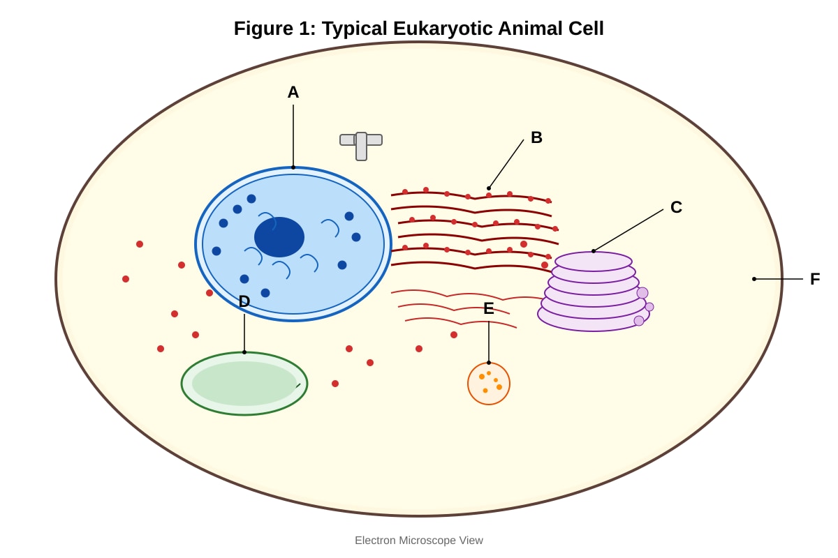
11. Figure 1 shows the structure of a typical eukaryotic animal cell as seen under an electron microscope.
Generated diagram for Q11.
(a) Identify the organelles labelled A and C in Figure 1. [2]
(b) State one function of the organelle labelled B. [1]
(c) Explain why the organelle labelled D is described as the "powerhouse of the cell". Your answer should refer to the specific structure visible in Figure 1. [3]
(d) Organelle E contains hydrolytic enzymes. Explain the consequence for the cell if the membrane of organelle E were to rupture. [2]
(e) Give one structural difference between a prokaryotic cell and the eukaryotic cell shown in Figure 1. [1]
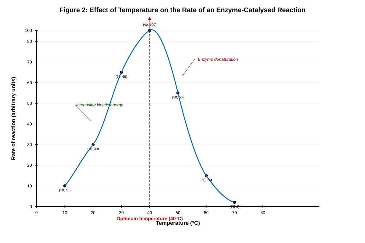
12. Figure 2 shows the effect of temperature on the rate of an enzyme-catalysed reaction.
Generated graph for Q12.
(a) With reference to Figure 2, describe the effect of temperature on the rate of the enzyme-catalysed reaction between 10 °C and 40 °C. [2]
(b) Explain the decrease in the rate of reaction above 40 °C. [3]
(c) A student repeated the experiment using an enzyme isolated from a thermophilic bacterium. Predict, with a reason, how the optimum temperature would differ from that shown in Figure 2. [2]
(d) Explain why the rate of reaction does not increase indefinitely with temperature, even before the enzyme begins to denature. [2]
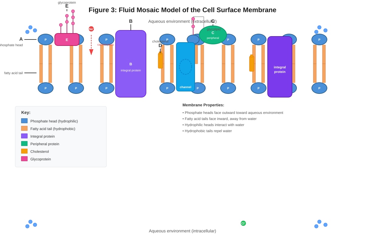
13. Figure 3 shows a section through a cell surface membrane, illustrating the fluid mosaic model.
Generated diagram for Q13.
(a) With reference to Figure 3, name the components labelled B, D, and E. [3]
(b) Explain how the structure of the phospholipid bilayer (component A) allows the membrane to act as a barrier to the movement of ions and large polar molecules. [3]
(c) Component D plays an important role in regulating membrane fluidity. Describe how cholesterol affects membrane fluidity at low temperatures and at high temperatures. [2]
(d) Explain the role of component E in cell signalling. [2]
14. A student investigated the effect of enzyme concentration on the rate of reaction using the enzyme catalase and hydrogen peroxide (H2O2). The volume of oxygen gas produced was measured over time. The results are shown in Table 1.
Table 1
| Catalase concentration (%) | Initial rate of reaction (cm³ O₂/min) |
|---|---|
| 0.0 | 0.0 |
| 2.0 | 1.8 |
| 4.0 | 3.5 |
| 6.0 | 5.2 |
| 8.0 | 6.8 |
| 10.0 | 8.0 |
| 12.0 | 8.0 |
| 14.0 | 8.0 |
(a) Plot a graph of catalase concentration against initial rate of reaction on the grid provided. [3]
(b) Describe the trend shown by the graph. [2]
(c) Explain why the rate of reaction remains constant at 8.0 cm³ O₂/min when the catalase concentration is increased from 10% to 14%. [2]
(d) State one variable that must be kept constant in this experiment to ensure valid results. [1]
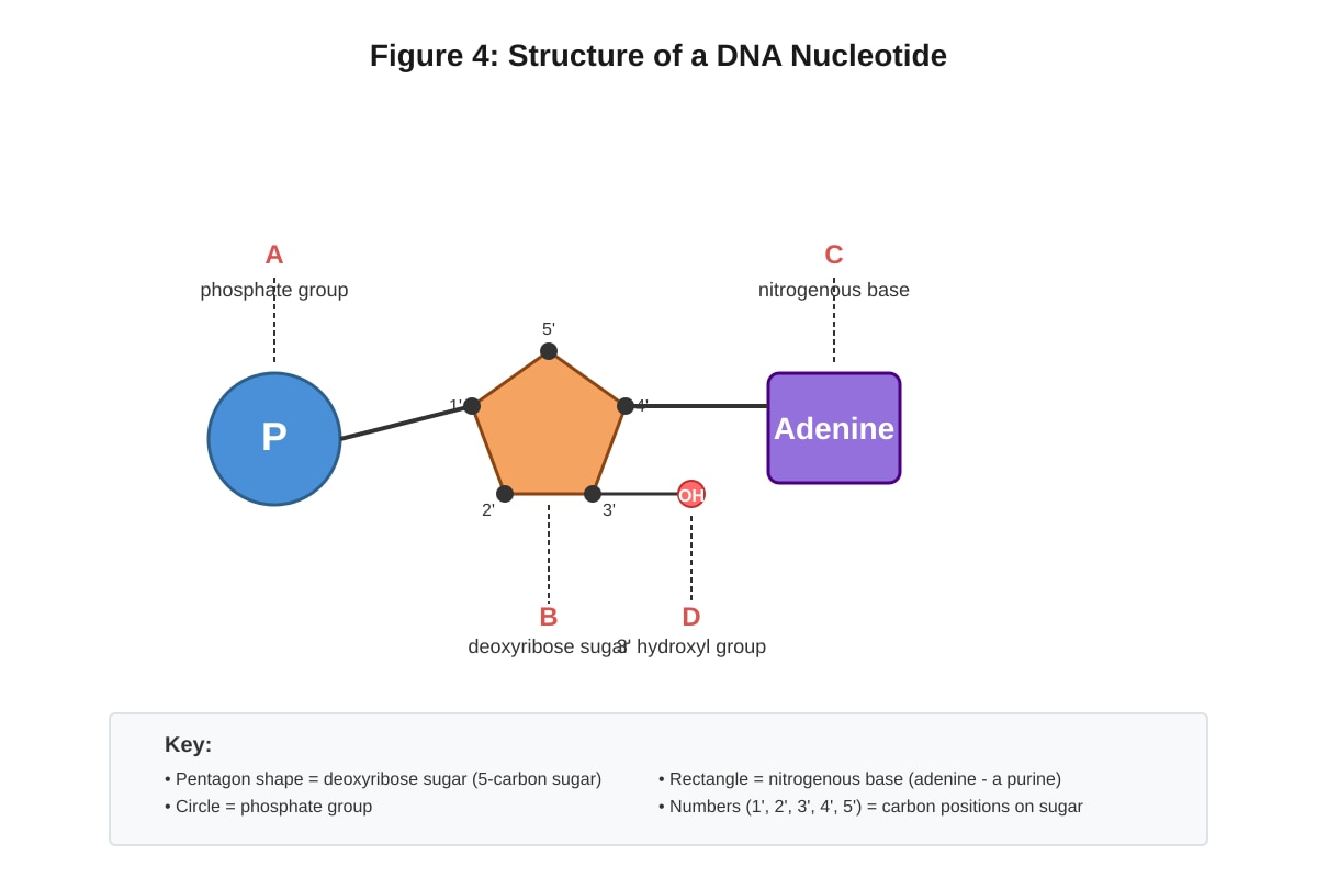
15. Figure 4 shows the structure of a nucleotide found in DNA.
Generated diagram for Q15.
(a) Name the component labelled B in Figure 4. [1]
(b) State two differences between the nucleotide in Figure 4 and a nucleotide found in RNA. [2]
(c) Explain how the structure of DNA allows it to carry genetic information. Your answer should refer to the sequence of bases and the double-helical structure. [3]
(d) During DNA replication, the enzyme DNA polymerase adds nucleotides to the growing strand. State the direction in which DNA polymerase synthesises the new strand and explain why. [2]
Section C: Data-Based and Extended Response Questions (15 marks)
Answer all questions.
16. Read the following passage and answer the questions that follow.
Prion diseases, such as Creutzfeldt-Jakob disease (CJD) in humans and bovine spongiform encephalopathy (BSE) in cattle, are caused by misfolded proteins called prions. The normal cellular prion protein, PrP^C, is found on the surface of neurons and has a predominantly alpha-helical secondary structure. The misfolded form, PrP^Sc, has a higher proportion of beta-sheet structure. PrP^Sc is able to induce the misfolding of normal PrP^C molecules upon contact, leading to an accumulation of insoluble protein aggregates in the brain. These aggregates cause neuronal death, resulting in progressive brain damage characterised by sponge-like holes in brain tissue.
Unlike viruses and bacteria, prions do not contain nucleic acids. They are also remarkably resistant to treatments that destroy nucleic acids, such as UV radiation and nucleases. However, PrP^Sc can be denatured by strong alkali treatment (e.g., 2M NaOH) or prolonged autoclaving at 134 °C.
(a) Explain why PrP^C and PrP^Sc have different secondary structures despite being composed of the same sequence of amino acids. [2]
(b) Suggest how the change from alpha-helical to beta-sheet structure could cause PrP^Sc to form insoluble aggregates. [2]
(c) Explain why prions are resistant to UV radiation and nuclease treatment. [2]
(d) A researcher treated a sample of PrP^Sc with 2M NaOH and found that it no longer caused misfolding of PrP^C. Explain this observation. [2]
(e) Explain why prion diseases are not considered infectious in the same way as viral diseases. [2]
17. Figure 5 shows a comparison of the structure of a typical prokaryotic cell and a typical eukaryotic plant cell.
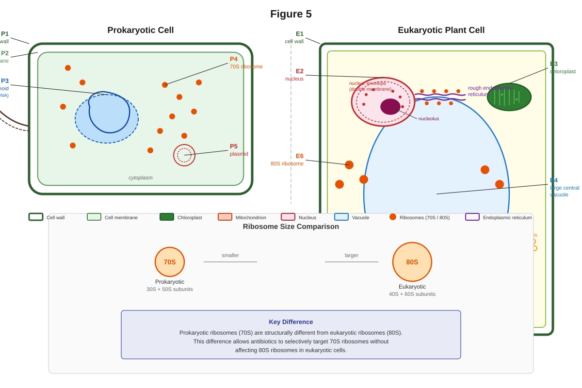
Generated diagram for Q17.
(a) With reference to Figure 5, state three structural differences between the prokaryotic cell and the eukaryotic plant cell. [3]
(b) The ribosomes in the prokaryotic cell (P4) are 70S, while those in the eukaryotic cell (E6) are 80S. Explain why antibiotics that target 70S ribosomes can kill prokaryotic cells without harming eukaryotic cells. [2]
(c) Explain the advantage to the eukaryotic plant cell of having membrane-bound organelles such as the chloroplast (E3) and mitochondrion (E5). [2]
(d) A student claims that the cell wall is a feature unique to plant cells. Evaluate this claim using evidence from Figure 5. [2]
18. Figure 6 shows the process of transcription and translation in a eukaryotic cell.
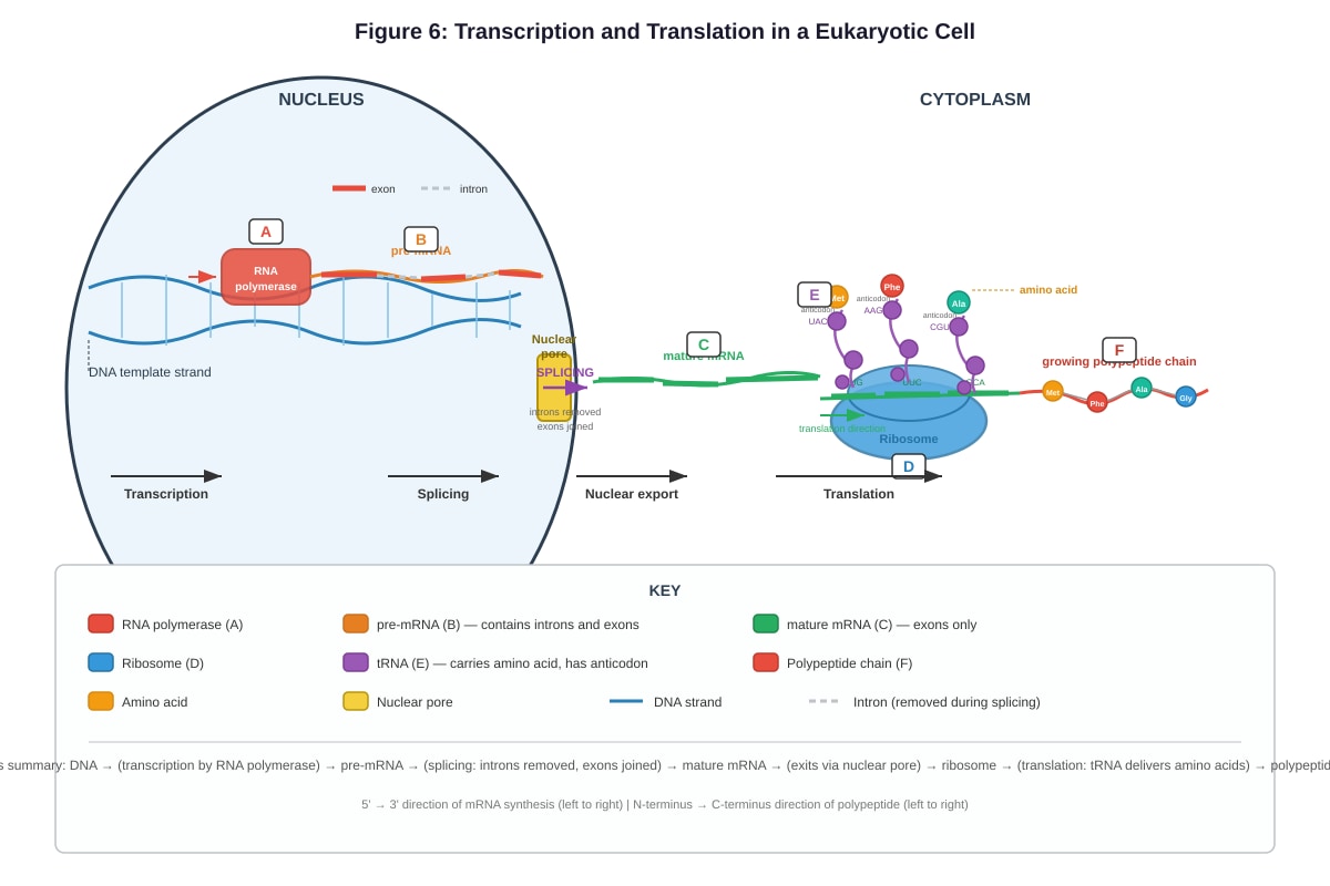
Generated diagram for Q18.
(a) With reference to Figure 6, describe the role of RNA polymerase (component A) in transcription. [2]
(b) Explain the significance of the conversion of pre-mRNA (component B) to mature mRNA (component C) before translation. [2]
(c) During translation, component E delivers amino acids to the ribosome. Explain how the correct amino acid is selected for addition to the growing polypeptide chain. [3]
(d) A mutation occurs in the DNA that changes a single base in an exon. Explain how this could result in a non-functional protein. [3]
End of Paper
Mark Summary
| Section | Marks |
|---|---|
| Section A: Questions 1–10 | 10 |
| Section B: Questions 11–15 | 35 |
| Section C: Questions 16–18 | 15 |
| Total | 60 |
Answers
TuitionGoWhere Practice Paper — Biology H2 A-Level
Answer Key — Cells & Biomolecules (Version 4 of 5)
Section A: Multiple Choice Questions (10 marks)
1. Answer: C (Ribosome)
Teaching notes: Ribosomes are the only organelle listed that are found in both prokaryotic and eukaryotic cells. They are the site of protein synthesis and are essential for all living cells. The nucleus (A), mitochondrion (B), and endoplasmic reticulum (D) are all membrane-bound organelles found only in eukaryotic cells. Prokaryotic cells lack membrane-bound organelles entirely. This is a fundamental distinction in cell biology.
[1 mark]
2. Answer: C (The fatty acid tails are hydrophobic and orient away from water.)
Teaching notes: Phospholipids are amphipathic molecules — they have a hydrophilic (water-loving) phosphate head and two hydrophobic (water-fearing) fatty acid tails. In an aqueous environment, the hydrophobic tails orient themselves away from water, forming the interior of the bilayer, while the hydrophilic heads face the aqueous environment on both sides. Option A is incorrect because the tails are hydrophobic, not hydrophilic. Option B is incorrect because the phosphate heads are hydrophilic, not hydrophobic. Option D is incorrect because the phosphate heads are polar, not non-polar.
[1 mark]
3. Answer: C (Hydrogen bonds)
Teaching notes: The two strands of the DNA double helix are held together by hydrogen bonds between complementary base pairs: adenine forms two hydrogen bonds with thymine, and guanine forms three hydrogen bonds with cytosine. Covalent bonds (A) form the sugar-phosphate backbone within each strand. Ionic bonds (B) are not involved in holding the strands together. Phosphodiester bonds (D) are the covalent bonds linking adjacent nucleotides within a single strand.
[1 mark]
4. Answer: B (The active site of the enzyme is altered due to disruption of ionic and hydrogen bonds in the tertiary structure.)
Teaching notes: Enzymes have an optimal pH at which their tertiary structure — and therefore their active site shape — is maintained. At extreme pH values (such as pH 3), excess H+ ions disrupt the ionic bonds and hydrogen bonds that maintain the enzyme's three-dimensional tertiary structure. This alters the shape of the active site so that the substrate can no longer bind effectively (the "lock and key" or "induced fit" model no longer applies). Option A is incorrect because it is the enzyme, not the substrate, that is denatured. Option C is too extreme — the enzyme is denatured, not fully hydrolysed. Option D is vague and does not explain the specific mechanism.
[1 mark]
5. Answer: C (A phospholipid bilayer with proteins floating within it, and the components can move laterally.)
Teaching notes: The fluid mosaic model, proposed by Singer and Nicolson in 1972, describes the cell membrane as a fluid phospholipid bilayer with proteins embedded within it or attached to its surface. The term "fluid" refers to the ability of phospholipids and some proteins to move laterally within the plane of the membrane. "Mosaic" refers to the scattered arrangement of proteins within the lipid bilayer. Option A describes an outdated model. Option B incorrectly describes a single layer. Option D is incorrect because cholesterol is found within the bilayer, not as the outermost layer.
[1 mark]
6. Answer: B (To hydrolyse the non-reducing sugar into its constituent reducing monosaccharides.)
Teaching notes: Non-reducing sugars (such as sucrose) do not react with Benedict's reagent because they lack a free aldehyde or ketone group. Adding dilute hydrochloric acid and heating hydrolyses the glycosidic bond in the non-reducing sugar, breaking it down into its constituent monosaccharides (e.g., glucose and fructose from sucrose). These monosaccharides are reducing sugars and will give a positive Benedict's test (brick-red precipitate) after neutralisation. Option A is incorrect because hydrolysis, not oxidation, occurs. Option C is incorrect because the acid provides an acidic, not alkaline, environment. Option D is not the primary purpose.
[1 mark]
7. Answer: C (Anaphase)
Teaching notes: During anaphase of mitosis, the centromeres joining sister chromatids divide, and the spindle fibres shorten, pulling the sister chromatids apart to opposite poles of the cell. In prophase (A), chromosomes condense and the spindle forms. In metaphase (B), chromosomes align at the cell's equator. In telophase (D), the nuclear envelope reforms around the separated chromosomes. Remember the sequence: PMAT — Prophase, Metaphase, Anaphase, Telophase.
[1 mark]
8. Answer: B (ATP)
Teaching notes: ATP (adenosine triphosphate) is the universal energy currency of the cell. When the terminal phosphate bond is hydrolysed (ATP → ADP + Pᵢ), energy is released that can be used to drive cellular processes. Glucose (A) is a fuel molecule that must be respired to produce ATP. NADH (C) is an electron carrier used in respiration. Starch (D) is a storage polysaccharide in plants and is not a direct energy source for cellular reactions.
[1 mark]
9. Answer: B (Translation will terminate prematurely, producing a truncated polypeptide of three amino acids.)
Teaching notes: A stop codon (UAA, UAG, or UGA) signals the termination of translation. If the codon for Val (which is the third amino acid: Met–Leu–Val–Arg–Ser) is replaced by a stop codon, the ribosome will stop translation after incorporating the second amino acid (Leu). The resulting polypeptide will be Met–Leu, which is only two amino acids long (the stop codon itself does not code for an amino acid). However, the question asks for the "most likely consequence" — the key point is premature termination producing a truncated polypeptide. Option B is the best answer because it correctly identifies premature termination and a truncated product. Option A is incorrect because the polypeptide is not simply "one amino acid shorter" — it is drastically shortened. Option C is incorrect because stop codons are not skipped. Option D is incorrect because the mutation affects translation, not transcription.
[1 mark]
10. Answer: C (Water is produced during condensation reactions and consumed during hydrolysis reactions.)
Teaching notes: In a condensation reaction, two molecules are joined together with the removal of a water molecule (e.g., amino acids joining to form a dipeptide, or monosaccharides joining to form a disaccharide). In a hydrolysis reaction, a molecule is broken down by the addition of a water molecule (e.g., proteins being digested into amino acids). This is a fundamental concept in biochemistry. Option A and B are incorrect because water is involved in both types of reactions, not just one. Option D reverses the correct relationship.
[1 mark]
Section B: Structured Questions (35 marks)
11.
(a) Identify the organelles labelled A and C. [2]
- A = Nucleus [1]
- C = Golgi apparatus (or Golgi body) [1]
Teaching notes: The nucleus is the largest organelle in a eukaryotic cell and contains the cell's genetic material. It is surrounded by a double membrane (nuclear envelope) with nuclear pores. The Golgi apparatus consists of a stack of flattened membrane-bound cisternae and is involved in modifying, sorting, and packaging proteins for secretion or delivery to other organelles.
(b) State one function of the organelle labelled B (rough endoplasmic reticulum). [1]
- Synthesis of proteins (for secretion / for transport to the Golgi apparatus) [1]
Teaching notes: The rough endoplasmic reticulum (RER) is studded with ribosomes on its cytoplasmic surface. These ribosomes synthesise proteins, particularly those destined for secretion, incorporation into membranes, or transport to other organelles. The RER also provides a pathway for the transport of these proteins to the Golgi apparatus.
(c) Explain why the organelle labelled D (mitochondrion) is described as the "powerhouse of the cell". [3]
- The mitochondrion is the site of aerobic respiration [1]
- Specifically, the Krebs cycle occurs in the matrix and the electron transport chain occurs on the inner membrane (cristae) [1]
- These processes produce large amounts of ATP, which is the energy currency used by the cell for metabolic processes [1]
Teaching notes: The mitochondrion is called the "powerhouse" because it generates most of the cell's ATP through aerobic respiration. The cristae (visible in Figure 1 as folds of the inner membrane) provide a large surface area for the electron transport chain and ATP synthase. The matrix contains enzymes for the Krebs cycle. The folded inner membrane (cristae) is a key structural feature that increases the efficiency of ATP production.
(d) Explain the consequence for the cell if the membrane of organelle E (lysosome) were to rupture. [2]
- The hydrolytic enzymes contained within the lysosome would be released into the cytoplasm [1]
- These enzymes would digest/hydrolyse the cell's own organelles and macromolecules, leading to cell death (autolysis) [1]
Teaching notes: Lysosomes contain powerful hydrolytic enzymes (e.g., proteases, lipases, nucleases) that function optimally at acidic pH (~pH 5). The lysosomal membrane keeps these enzymes contained and separated from the neutral pH of the cytoplasm. If the membrane ruptures, the enzymes are released and can digest the cell's own components, a process called autolysis. This is why lysosomes are sometimes called "suicide bags."
(e) Give one structural difference between a prokaryotic cell and the eukaryotic cell shown in Figure 1. [1]
- Prokaryotic cells lack a membrane-bound nucleus (or: prokaryotic cells lack membrane-bound organelles / prokaryotic cells have 70S ribosomes instead of 80S / prokaryotic cells have a single circular chromosome) [1]
Teaching notes: The most fundamental difference is that prokaryotic cells do not have a membrane-bound nucleus — their DNA is found in a region called the nucleoid. They also lack other membrane-bound organelles such as mitochondria, ER, and Golgi apparatus.
12.
(a) With reference to Figure 2, describe the effect of temperature on the rate of the enzyme-catalysed reaction between 10 °C and 40 °C. [2]
- As temperature increases from 10 °C to 40 °C, the rate of reaction increases [1]
- The rate increases from approximately 10 to 100 arbitrary units (or: the rate increases steadily/rapidly, reaching a maximum/optimum at 40 °C) [1]
Teaching notes: When describing trends from a graph, always include the direction of change and reference specific values from the graph. The increase in rate is due to greater kinetic energy of both enzyme and substrate molecules, leading to more frequent and more energetic collisions.
(b) Explain the decrease in the rate of reaction above 40 °C. [3]
- Above 40 °C, the enzyme molecules gain excess kinetic energy [1]
- This disrupts the hydrogen bonds and ionic bonds that maintain the enzyme's tertiary structure [1]
- The shape of the active site is altered (denaturation), so the substrate can no longer bind effectively, and the rate of reaction decreases [1]
Teaching notes: Denaturation is the loss of an enzyme's three-dimensional shape due to disruption of the weak bonds (hydrogen bonds, ionic bonds, hydrophobic interactions, and disulphide bridges) that maintain the tertiary structure. The active site is no longer complementary to the substrate, so the enzyme-substrate complex cannot form. This is an irreversible process for most enzymes at high temperatures.
(c) Predict, with a reason, how the optimum temperature would differ for an enzyme from a thermophilic bacterium. [2]
- The optimum temperature would be higher than 40 °C [1]
- Because thermophilic bacteria live in high-temperature environments (e.g., hot springs), so their enzymes have evolved to have more stable tertiary structures (e.g., more disulphide bonds / stronger hydrophobic interactions) that resist denaturation at higher temperatures [1]
Teaching notes: Thermophilic organisms have adapted to survive at high temperatures. Their enzymes have structural adaptations (such as increased numbers of disulphide bonds, more compact structures, and stronger hydrophobic cores) that maintain their tertiary structure at temperatures that would denature enzymes from mesophilic organisms.
(d) Explain why the rate of reaction does not increase indefinitely with temperature, even before the enzyme begins to denature. [2]
- The rate increases with temperature due to increased kinetic energy of enzyme and substrate molecules, leading to more frequent successful collisions [1]
- However, the rate is also limited by the number of active sites available — at some point, all active sites are occupied at any given time (the enzyme becomes saturated), so further increases in temperature cannot increase the rate beyond this maximum [1]
Teaching notes: This question tests understanding that temperature is not the only factor limiting reaction rate. Even before denaturation, the rate is constrained by enzyme concentration and the availability of active sites. This connects to the concept of enzyme saturation and Vmax.
13.
(a) Name the components labelled B, D, and E. [3]
- B = Integral protein (or transmembrane protein) [1]
- D = Cholesterol [1]
- E = Glycoprotein [1]
Teaching notes: Integral proteins are embedded within the phospholipid bilayer and may span the entire membrane (transmembrane proteins). Cholesterol is a lipid molecule found between phospholipid tails in animal cell membranes. Glycoproteins are proteins with carbohydrate chains attached, found on the outer surface of the cell membrane.
(b) Explain how the structure of the phospholipid bilayer allows the membrane to act as a barrier to the movement of ions and large polar molecules. [3]
- The interior of the bilayer consists of hydrophobic fatty acid tails [1]
- Ions and large polar molecules are hydrophilic/charged and cannot dissolve in or pass through this hydrophobic core [1]
- This makes the membrane selectively permeable, allowing only small non-polar molecules (e.g., O2, CO2) and water to pass freely [1]
Teaching notes: The hydrophobic core of the phospholipid bilayer is the key barrier. Charged ions (e.g., Na+, K+, Ca2+, Cl−) and large polar molecules (e.g., glucose, amino acids) cannot pass through this non-polar region without the assistance of transport proteins (channel proteins or carrier proteins). This is the basis of selective permeability.
(c) Describe how cholesterol affects membrane fluidity at low temperatures and at high temperatures. [2]
- At low temperatures: cholesterol prevents the phospholipids from packing too closely together, maintaining membrane fluidity [1]
- At high temperatures: cholesterol restricts the movement of phospholipids, reducing membrane fluidity and preventing the membrane from becoming too fluid/permeable [1]
Teaching notes: Cholesterol acts as a "fluidity buffer." Its rigid ring structure interferes with the movement of phospholipid fatty acid tails. At low temperatures, it prevents the membrane from becoming too rigid; at high temperatures, it prevents it from becoming too fluid. This dual role is essential for maintaining membrane integrity across a range of temperatures.
(d) Explain the role of component E (glycoprotein) in cell signalling. [2]
- The carbohydrate chains on glycoproteins act as cell surface markers/antigens [1]
- These markers allow cells to recognise each other and are involved in cell-cell communication/signalling (e.g., receptor binding of hormones or neurotransmitters, immune cell recognition) [1]
Teaching notes: Glycoproteins play crucial roles in cell recognition and signalling. The carbohydrate portion (glycocalyx) on the cell surface is involved in cell-cell recognition (e.g., immune cells recognising self vs. non-self), receptor-mediated signalling (e.g., hormone receptors), and cell adhesion. Examples include MHC glycoproteins in immune recognition and insulin receptors.
14.
(a) Plot a graph of catalase concentration against initial rate of reaction. [3]
Marking scheme:
- Correctly labelled axes (x-axis: Catalase concentration (%); y-axis: Initial rate of reaction (cm³ O₂/min)) [1]
- Appropriate scale on both axes [1]
- All points correctly plotted and a smooth curve/line drawn (curve rises steeply then plateaus) [1]
Expected graph description: The graph should show a curve that rises steeply from (0, 0) through approximately (2, 1.8), (4, 3.5), (6, 5.2), (8, 6.8), (10, 8.0) and then plateaus horizontally from 10% onwards at a rate of 8.0 cm³ O₂/min.
(b) Describe the trend shown by the graph. [2]
- As catalase concentration increases from 0% to 10%, the initial rate of reaction increases [1]
- Above 10% catalase concentration, the rate of reaction remains constant / plateaus at 8.0 cm³ O₂/min [1]
Teaching notes: The initial increase is because more enzyme molecules mean more active sites available to bind substrate, leading to more enzyme-subductrate complexes per unit time. The plateau occurs because the substrate becomes the limiting factor.
(c) Explain why the rate of reaction remains constant at 8.0 cm³ O₂/min when the catalase concentration is increased from 10% to 14%. [2]
- The substrate (H2O2) concentration is limited / becomes the limiting factor [1]
- All substrate molecules are already bound to enzyme active sites (the enzyme is saturated), so adding more enzyme does not increase the rate [1]
Teaching notes: This is the concept of enzyme saturation. When all substrate molecules are being converted to product as fast as possible, the reaction has reached its maximum velocity (Vmax). Adding more enzyme cannot increase the rate because there is no additional substrate to bind to the extra active sites.
(d) State one variable that must be kept constant in this experiment. [1]
- Temperature (or: pH / volume of H2O2 / concentration of H2O2) [1]
Teaching notes: To ensure a valid experiment, all variables other than the independent variable (catalase concentration) must be controlled. Temperature and pH both affect enzyme activity, and the volume/concentration of substrate must be the same in each trial.
15.
(a) Name the component labelled B (deoxyribose sugar). [1]
- Deoxyribose [1]
Teaching notes: The sugar in DNA is deoxyribose, a pentose (5-carbon) sugar. It is called "deoxyribose" because it lacks an oxygen atom on the 2' carbon compared to ribose (the sugar in RNA).
(b) State two differences between the nucleotide in Figure 4 and a nucleotide found in RNA. [2]
- RNA nucleotides contain ribose instead of deoxyribose [1]
- RNA nucleotides contain the base uracil instead of thymine [1]
(Alternative acceptable answer: RNA is single-stranded while DNA is double-stranded — but this refers to the overall molecule, not the individual nucleotide.)
Teaching notes: The key differences between DNA and RNA nucleotides are: (1) the sugar — deoxyribose in DNA vs. ribose in RNA; (2) the bases — thymine in DNA vs. uracil in RNA. RNA also typically exists as a single strand, while DNA is double-stranded.
(c) Explain how the structure of DNA allows it to carry genetic information. [3]
- DNA consists of a sequence of four different nucleotide bases (A, T, G, C) along each strand [1]
- The sequence of these bases codes for the sequence of amino acids in proteins (genetic code) [1]
- The double-helical structure with complementary base pairing (A-T, G-C) allows the genetic information to be accurately copied/replicated during cell division [1]
Teaching notes: The genetic information is stored in the linear sequence of bases along the DNA strand. Each triplet of bases (codon) specifies a particular amino acid. The complementary base pairing ensures that during DNA replication, each strand can serve as a template for the synthesis of a new complementary strand, allowing faithful transmission of genetic information.
(d) State the direction in which DNA polymerase synthesises the new strand and explain why. [2]
- DNA polymerase synthesises the new strand in the 5' to 3' direction [1]
- Because DNA polymerase can only add nucleotides to the 3' hydroxyl (-OH) group of the growing strand (nucleotides are added to the 3' end) [1]
Teaching notes: DNA polymerase catalyses the formation of a phosphodiester bond between the 3'-OH group of the growing strand and the 5'-phosphate group of the incoming nucleotide. This means synthesis always proceeds in the 5'→3' direction. This is a fundamental constraint of all DNA polymerases and has important consequences for the mechanism of replication (leading and lagging strands).
Section C: Data-Based and Extended Response Questions (15 marks)
16.
(a) Explain why PrP^C and PrP^Sc have different secondary structures despite being composed of the same sequence of amino acids. [2]
- The amino acid sequence (primary structure) determines how the polypeptide folds [1]
- PrP^Sc has undergone a change in folding/conformation, resulting in a different arrangement of secondary structural elements (more beta-sheets instead of alpha-helices), even though the primary structure is the same [1]
Teaching notes: The primary structure (sequence of amino acids) determines the tertiary structure through interactions between R-groups. However, proteins can sometimes misfold into alternative conformations. In prion diseases, the same polypeptide chain can adopt two different folds — the normal PrP^C (alpha-helical) and the misfolded PrP^Sc (beta-sheet-rich). This is unusual because most proteins have one stable native conformation.
(b) Suggest how the change from alpha-helical to beta-sheet structure could cause PrP^Sc to form insoluble aggregates. [2]
- Beta-sheets can form intermolecular hydrogen bonds between adjacent polypeptide chains [1]
- This causes PrP^Sc molecules to stack together / form fibrils, creating large insoluble aggregates that precipitate out of solution [1]
Teaching notes: Alpha-helices are stabilised by intramolecular hydrogen bonds (within the same polypeptide chain), while beta-sheets can form both intra- and intermolecular hydrogen bonds. The increased beta-sheet content in PrP^Sc allows the protein molecules to form extensive intermolecular hydrogen bonding networks, leading to the formation of amyloid fibrils — rigid, insoluble protein aggregates that accumulate in brain tissue.
(c) Explain why prions are resistant to UV radiation and nuclease treatment. [2]
- UV radiation and nucleases target/destroy nucleic acids (DNA and RNA) [1]
- Prions are composed only of protein and do not contain any nucleic acids, so these treatments have no effect on them [1]
Teaching notes: UV radiation causes damage to nucleic acids (e.g., thymine dimers in DNA), and nucleases are enzymes that hydrolyse phosphodiester bonds in nucleic acids. Since prions are purely proteinaceous infectious particles with no DNA or RNA, these treatments — which are effective against viruses and bacteria — are ineffective against prions.
(d) Explain why treatment with 2M NaOH eliminates the ability of PrP^Sc to cause misfolding of PrP^C. [2]
- 2M NaOH is a strong alkali that denatures the PrP^Sc protein [1]
- Denaturation alters the tertiary structure / shape of PrP^Sc, so it can no longer act as a template to induce misfolding of normal PrP^C [1]
Teaching notes: The misfolding activity of PrP^Sc depends on its specific three-dimensional shape. Strong alkali disrupts the hydrogen bonds, ionic bonds, and other interactions that maintain the protein's tertiary structure. Once denatured, PrP^Sc loses its ability to act as a template for converting PrP^C to the misfolded form.
(e) Explain why prion diseases are not considered infectious in the same way as viral diseases. [2]
- Viruses contain nucleic acids (DNA or RNA) that carry genetic information and can be replicated inside host cells [1]
- Prions contain no nucleic acids and do not replicate in the traditional sense — they propagate by inducing conformational changes in existing normal proteins, rather than by directing the synthesis of new genetic material [1]
Teaching notes: Viruses are infectious because they contain genetic material that hijacks the host cell's machinery to produce new viral particles. Prions, in contrast, are misfolded proteins that cause disease by converting normal proteins into the misfolded form through direct contact. There is no replication of genetic material involved. This makes prions unique among infectious agents.
17.
(a) State three structural differences between the prokaryotic cell and the eukaryotic plant cell. [3]
- Prokaryotic cells have no membrane-bound nucleus (DNA is in the nucleoid region), while eukaryotic cells have a membrane-bound nucleus [1]
- Prokaryotic cells have 70S ribosomes, while eukaryotic cells have 80S ribosomes [1]
- Prokaryotic cells lack membrane-bound organelles (e.g., no chloroplasts, no mitochondria, no ER, no Golgi), while eukaryotic cells have these organelles [1]
(Alternative acceptable differences: Prokaryotic cells have a single circular chromosome; eukaryotic cells have multiple linear chromosomes. Prokaryotic cells may have a plasmid; eukaryotic plant cells do not.)
(b) Explain why antibiotics that target 70S ribosomes can kill prokaryotic cells without harming eukaryotic cells. [2]
- Prokaryotic cells have 70S ribosomes, which are structurally different from the 80S ribosomes found in eukaryotic cells [1]
- The antibiotic binds specifically to the 70S ribosome and inhibits protein synthesis in prokaryotic cells, but cannot bind to the structurally different 80S ribosomes in eukaryotic cells [1]
Teaching notes: This is the basis of selective toxicity in antibiotic therapy. Examples include tetracycline and chloramphenicol, which target the 70S ribosome. The structural differences between 70S and 80S ribosomes (in both RNA and protein composition) mean that antibiotics can selectively inhibit bacterial protein synthesis without affecting the host's protein synthesis.
(c) Explain the advantage to the eukaryotic plant cell of having membrane-bound organelles such as the chloroplast and mitochondrion. [2]
- Membrane-bound organelles compartmentalise specific biochemical reactions, allowing incompatible reactions to occur simultaneously in different parts of the cell [1]
- The membranes of these organelles provide a large surface area for the attachment of enzymes and electron transport chain components, increasing the efficiency of processes like photosynthesis (chloroplast) and aerobic respiration (mitochondrion) [1]
Teaching notes: Compartmentalisation is a key advantage of eukaryotic cells. It allows the cell to maintain different conditions (e.g., pH, enzyme concentrations) in different organelles, optimising the efficiency of each metabolic pathway. The internal membranes of chloroplasts (thylakoids) and mitochondria (cristae) greatly increase the surface area available for the reactions of photosynthesis and respiration.
(d) Evaluate the claim that the cell wall is a feature unique to plant cells. [2]
- The claim is incorrect [1]
- Figure 5 shows that the prokaryotic cell also has a cell wall (labelled P1), so the cell wall is not unique to plant cells. Additionally, fungi also have cell walls (made of chitin) [1]
Teaching notes: Cell walls are found in plants (cellulose), bacteria (peptidoglycan), fungi (chitin), and some protists. The composition of the cell wall differs between these groups, but the presence of a cell wall is not unique to plant cells. This is a common misconception that should be addressed.
18.
(a) Describe the role of RNA polymerase (component A) in transcription. [2]
- RNA polymerase binds to the promoter region of a gene on the DNA template strand [1]
- It unwinds the DNA double helix and catalyses the formation of phosphodiester bonds between RNA nucleotides, synthesising a complementary mRNA strand in the 5' to 3' direction [1]
Teaching notes: RNA polymerase is the key enzyme in transcription. It reads the DNA template strand in the 3'→5' direction and synthesises mRNA in the 5'→3' direction. It also has helicase activity to unwind the DNA. No primer is required (unlike DNA polymerase in replication).
(b) Explain the significance of the conversion of pre-mRNA (component B) to mature mRNA (component C) before translation. [2]
- Pre-mRNA contains both exons (coding sequences) and introns (non-coding sequences) [1]
- During splicing, introns are removed and exons are joined together, ensuring that only the coding sequences are translated into the correct polypeptide [1]
Teaching notes: RNA splicing is essential in eukaryotic cells because the initial transcript (pre-mRNA) includes non-coding introns that would disrupt the reading frame if translated. The removal of introns and joining of exons produces mature mRNA that contains only the coding sequence. Additionally, alternative splicing allows a single gene to produce multiple different proteins by including or excluding different exons.
(c) Explain how the correct amino acid is selected for addition to the growing polypeptide chain. [3]
- Each tRNA molecule has a specific anticodon that is complementary to a specific codon on the mRNA [1]
- The anticodon on the tRNA base-pairs with the codon on the mRNA in the ribosome [1]
- Each tRNA is attached to a specific amino acid (determined by aminoacyl-tRNA synthetase enzymes), so the correct amino acid is delivered according to the mRNA codon sequence [1]
Teaching notes: The genetic code is read in triplets (codons) on the mRNA. Each codon specifies a particular amino acid. The tRNA acts as an adapter molecule, matching the codon to the correct amino acid through complementary base pairing between the anticodon and codon. The enzyme aminoacyl-tRNA synthetase ensures that each tRNA is charged with the correct amino acid.
(d) Explain how a single base mutation in an exon could result in a non-functional protein. [3]
- A single base change (point mutation) could change a codon so that it codes for a different amino acid (missense mutation) [1]
- If this amino acid is in a critical region of the protein (e.g., the active site of an enzyme), it could alter the protein's tertiary structure [1]
- The altered tertiary structure could change the shape of the active site (or other functional region), preventing the protein from carrying out its function, rendering it non-functional [1]
(Alternative: The point mutation could create a stop codon (nonsense mutation), causing premature termination of translation and producing a truncated, non-functional polypeptide.)
Teaching notes: Not all point mutations have severe effects — some are silent (due to the degeneracy of the genetic code) or have minimal impact. However, a single amino acid substitution in a critical region (e.g., the active site of an enzyme or a structural region) can dramatically alter protein function. Sickle cell anaemia is a classic example where a single base change (GAG → GTG) results in glutamic acid being replaced by valine in the beta-globin chain, causing the protein to malfunction.
Mark Summary
| Section | Marks |
|---|---|
| Section A: Questions 1–10 | 10 |
| Section B: Questions 11–15 | 35 |
| Section C: Questions 16–18 | 15 |
| Total | 60 |
Free quiz and exam paper access
Enter your details to view this paper
Your access is remembered on this device.