From Real Exams Exam Paper
A Level H2 Biology Practice Paper 3
Free A Level H2 Biology Practice Paper 3, LongCat Exam version, with questions, answers, and A Level-style practice for Singapore students.
These static practice materials are generated from the site's syllabus and paper-generation workflow, with source and model context shown so students and parents can evaluate the material before use.
Questions
TuitionGoWhere Practice Paper - Biology H2 A-Level
TuitionGoWhere Secondary School (AI)
Subject: Biology Level: A-Level H2 (9477) Paper: Practice Paper — Cells & Biomolecules Version: 3 of 5 Duration: 60 minutes Total Marks: 50
Name: ___________________________ Class: ___________________________ Date: ___________________________
Instructions
- Answer all questions in the spaces provided.
- Write your answers in dark blue or black pen.
- You may use a pencil for any diagrams or graphs.
- The number of marks for each question is shown in brackets [ ].
- The total marks for this paper is 50.
- You are advised to spend no more than 60 minutes on this paper.
Section A: Structured Questions (30 marks)
Answer all questions 1–10.
1. Fig. 1.1 shows the structure of a typical eukaryotic animal cell as seen under an electron microscope.
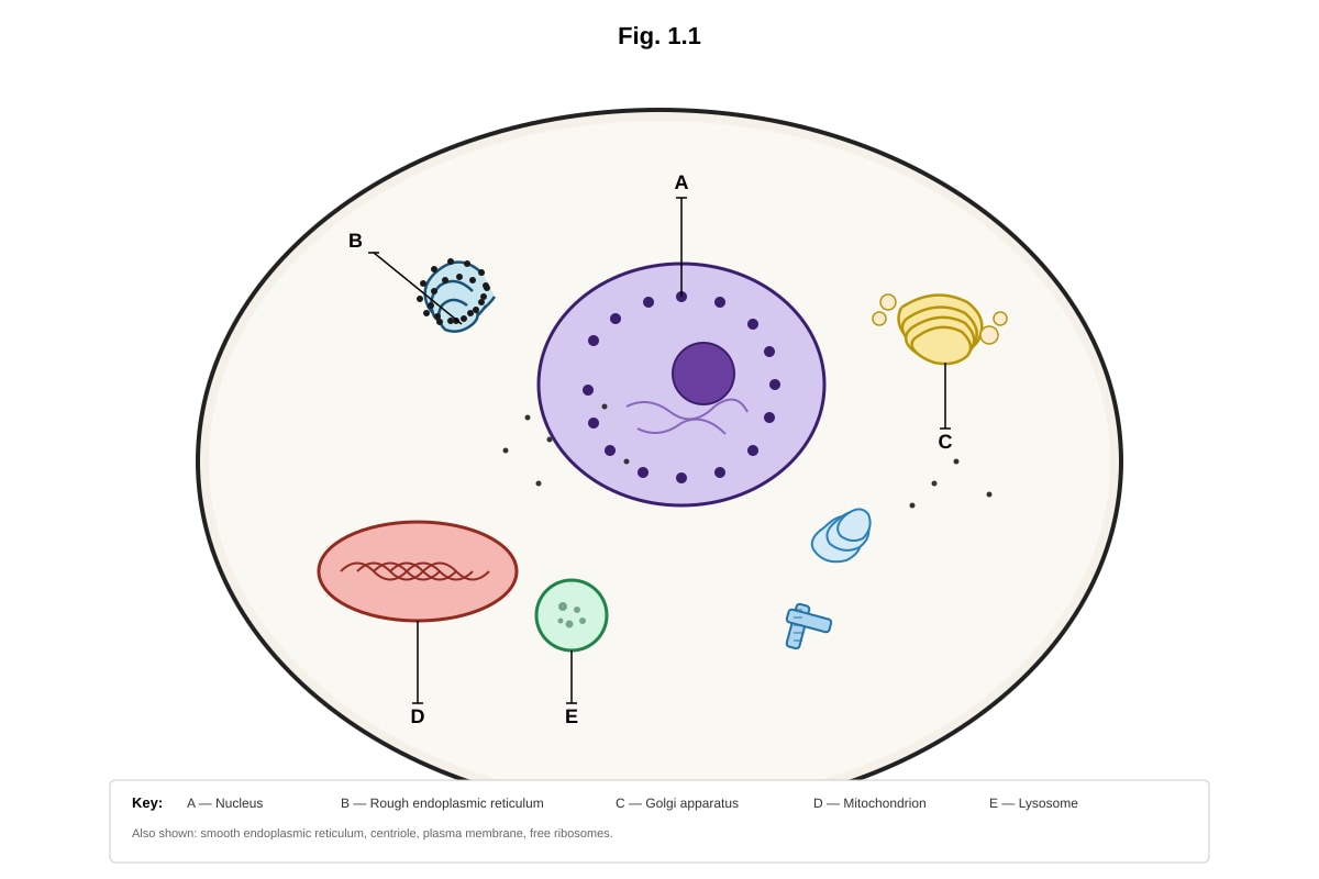
Generated diagram for Q1.
(a) Identify the organelles labelled A and D in Fig. 1.1. State one function of each. [4]
(b) Explain why the organelle labelled B appears different from the smooth endoplasmic reticulum when viewed under an electron microscope. [2]
(c) Describe two structural features of the organelle labelled D that are adaptations to its function. [4]
2. Table 2.1 shows the approximate percentage composition of four biological molecules in a typical animal cell and a plant cell.
| Biological molecule | Animal cell (% dry mass) | Plant cell (% dry mass) |
|---|---|---|
| Protein | 50 | 30 |
| Lipid | 15 | 5 |
| Carbohydrate | 3 | 40 |
| Nucleic acid | 15 | 10 |
| Other | 17 | 15 |
Table 2.1
(a) Calculate the difference in percentage dry mass of carbohydrate between the animal cell and the plant cell. Show your working. [2]
(b) Suggest one reason for the higher percentage of protein in the animal cell compared to the plant cell. [1]
(c) Explain why the percentage of nucleic acid is relatively low in both cell types despite its critical importance. [2]
3. Fig. 3.1 shows the structure of a phospholipid molecule.
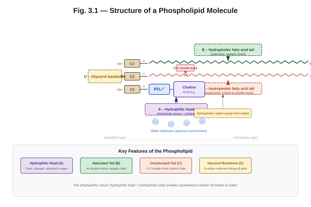
Generated diagram for Q3.
(a) Label the parts A, B, C, and D in Fig. 3.1 using the terms provided in the description above. [2]
(b) Explain how the property of the phospholipid molecule shown in Fig. 3.1 enables the spontaneous formation of a phospholipid bilayer in aqueous environments. [3]
(c) Explain the significance of having one saturated and one unsaturated fatty acid tail in a phospholipid molecule for membrane fluidity. [2]
4. Fig. 4.1 shows a graph of the effect of temperature on the rate of an enzyme-catalysed reaction.
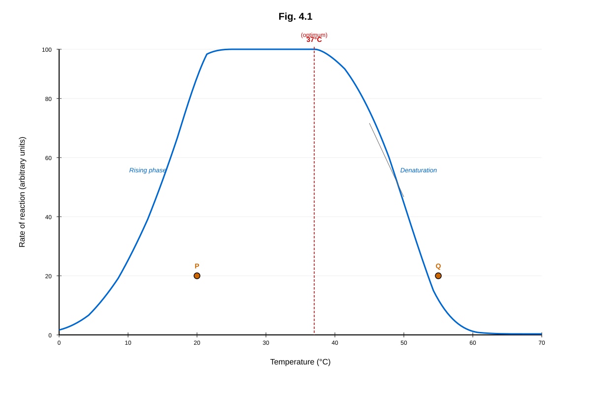
Generated graph for Q4.
(a) With reference to Fig. 4.1, describe the effect of temperature on the rate of the enzyme-catalysed reaction from 0 °C to 37 °C. Explain this effect in terms of molecular kinetic energy. [3]
(b) Explain why the rate of reaction decreases sharply above 37 °C. [2]
(c) A student claims that the enzyme is completely inactive at 0 °C. Evaluate this claim using evidence from Fig. 4.1. [2]
5. Describe the process of osmosis and explain how it differs from diffusion. Include in your answer the role of a partially permeable membrane. [4]
6. Fig. 6.1 shows the structure of a molecule of glucose.
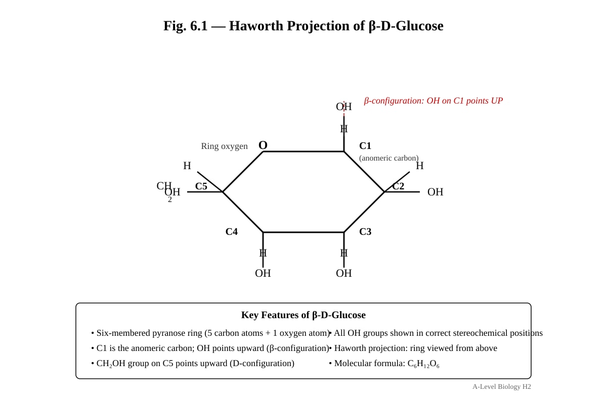
Generated diagram for Q6.
(a) State the molecular formula of glucose. [1]
(b) Glucose molecules can join together by condensation reactions to form polysaccharides. Name two polysaccharides formed from glucose and state where each is found in living organisms. [4]
(c) Explain how the structure of one of the polysaccharides you named in (b) is related to its function. [3]
7. Fig. 7.1 shows a diagram of the fluid mosaic model of the cell surface membrane.
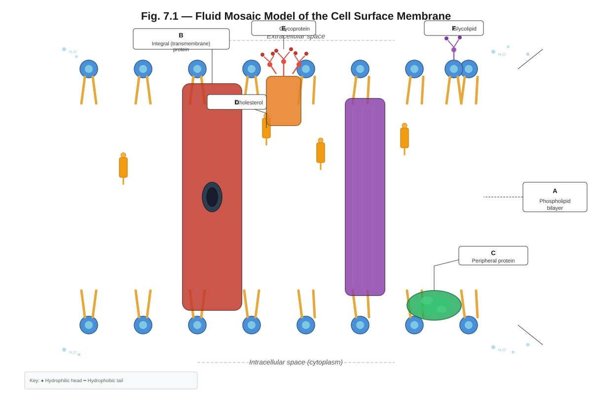
Generated diagram for Q7.
(a) With reference to Fig. 7.1, describe the roles of cholesterol (D) in the cell surface membrane. [2]
(b) Explain how the structure of integral protein B enables it to remain embedded in the phospholipid bilayer. [2]
(c) Glycoproteins (E) and glycolipids (F) are found only on the extracellular surface of the membrane. Suggest one biological function of glycoproteins and explain how their position on the extracellular surface is important for this function. [2]
8. A student carried out an experiment to investigate the effect of pH on the activity of the enzyme catalase. The results are shown in Table 8.1.
| pH | Volume of oxygen gas collected in 1 minute / cm³ |
|---|---|
| 3 | 2.1 |
| 5 | 5.8 |
| 7 | 12.4 |
| 9 | 8.3 |
| 11 | 1.6 |
Table 8.1
(a) Plot a graph of the results shown in Table 8.1. [3]
(b) Describe the trend shown by the graph. [2]
(c) Explain the effect of pH on catalase activity at pH 3 and pH 11. [3]
9. Compare and contrast prokaryotic and eukaryotic cells. Include in your answer reference to at least four structural differences. [6]
10. Fig. 10.1 shows the structure of an amino acid.
Image pending generation: diagram for Q10.
(a) Name the type of reaction that joins two amino acids together. Identify the bond that is formed. [2]
(b) Explain how the R group of an amino acid influences the three-dimensional structure and function of a protein. [4]
(c) Describe the secondary and tertiary levels of protein structure. For each level, name the type(s) of bond that stabilises the structure. [4]
Section B: Data-Based Question (10 marks)
Answer all parts of Question 11.
11. Read the following passage and answer the questions that follow.
Aquaporins and Membrane Transport
Aquaporins are integral membrane proteins that form channels specifically for the transport of water molecules across the cell surface membrane. They are found in many cell types, including red blood cells and cells of the kidney nephron. Each aquaporin protein is composed of six transmembrane alpha-helices that form a central pore. The pore contains a conserved Asn-Pro-Ala (NPA) motif that acts as a selectivity filter, allowing only water molecules to pass through while excluding protons (H⁺ ions) and other solutes.
In the collecting duct of the kidney, the hormone antidiuretic hormone (ADH, also known as vasopressin) triggers the insertion of aquaporin-2 (AQP2) channels into the apical membrane of the duct cells. This increases the permeability of the membrane to water, allowing more water to be reabsorbed from the filtrate back into the blood. When ADH levels are low, AQP2 channels are removed from the membrane by endocytosis and stored in cytoplasmic vesicles.
A rare genetic condition called nephrogenic diabetes insipidus (NDI) can result from mutations in the AQP2 gene. Individuals with this condition produce large volumes of dilute urine because their kidney cells cannot respond properly to ADH.
(a) With reference to the passage, explain why aquaporins are described as having a "selectivity filter". [2]
(b) Using information from the passage, explain how the action of ADH on the collecting duct cells helps to maintain water balance in the body. [3]
(c) Suggest how a mutation in the AQP2 gene could lead to the symptoms described in nephrogenic diabetes insipidus. [3]
(d) Explain why aquaporins are classified as integral membrane proteins rather than peripheral membrane proteins. [2]
Section C: Extended Response (10 marks)
Answer Question 12.
12. A student investigated the effect of different sugar solutions on the mass of potato cylinders. Identical cylinders of potato tissue were placed in sucrose solutions of different concentrations for 30 minutes. The percentage change in mass of each cylinder was recorded.
The results are shown in Table 12.1.
| Sucrose concentration / mol dm⁻³ | Mean percentage change in mass |
|---|---|
| 0.0 | +8.2 |
| 0.2 | +3.1 |
| 0.4 | −1.5 |
| 0.6 | −5.8 |
| 0.8 | −9.4 |
Table 12.1
(a) Explain why the potato cylinders gained mass in 0.0 mol dm⁻³ sucrose solution. In your answer, refer to water potential. [3]
(b) At approximately what sucrose concentration would the potato cells be at incipient plasmolysis? Explain how you determined this from the data. [3]
(c) Describe how the structure of the cell wall and the cell surface membrane differ, and explain how these structural differences are important in the osmotic behaviour of plant cells. [4]
End of Paper
Answers
TuitionGoWhere Practice Paper - Biology H2 A-Level
Answer Key — Cells & Biomolecules (Version 3 of 5)
Section A: Structured Questions
Question 1
(a) Identify the organelles labelled A and D in Fig. 1.1. State one function of each. [4]
- A – Nucleus [1]
- Function: Contains DNA / genetic material / controls cell activities / site of transcription / stores genetic information [1]
- D – Mitochondrion [1]
- Function: Site of aerobic respiration / produces ATP / site of the Krebs cycle and oxidative phosphorylation [1]
Marking notes: Award 1 mark for correct identification of each organelle and 1 mark for a correct function. Accept any valid function. Do not accept "site of respiration" without specifying aerobic for mitochondrion.
(b) Explain why the organelle labelled B (rough endoplasmic reticulum) appears different from the smooth endoplasmic reticulum when viewed under an electron microscope. [2]
- The rough endoplasmic reticulum has ribosomes attached to its cytoplasmic surface [1]
- These ribosomes appear as dark/electron-dense granules/studded structures on the membrane surface, giving it a "rough" appearance, whereas the smooth ER lacks ribosomes and appears smooth [1]
Teaching note: The key distinction is the presence of ribosomes. Under an electron microscope, ribosomes are visible as small dark dots bound to the cytoplasmic face of the RER membrane. The SER lacks these and therefore has a smooth, tubular appearance.
(c) Describe two structural features of the organelle labelled D (mitochondrion) that are adaptations to its function. [4]
- Double membrane / inner membrane folded into cristae – increases the surface area for the attachment of electron transport chain proteins and ATP synthase, maximising ATP production [2]
- Matrix contains enzymes – the fluid-filled matrix contains enzymes for the Krebs cycle (e.g., enzymes that catalyse the oxidation of acetyl-CoA), enabling the second stage of aerobic respiration [2]
Alternative acceptable answers (any two):
- Inner membrane is folded into cristae – large surface area for oxidative phosphorylation / electron transport chain
- Matrix contains circular DNA and 70S ribosomes – allows the mitochondrion to synthesise some of its own proteins, enabling rapid production of respiratory enzymes
- Small size (0.5–1.0 μm diameter) – short diffusion distances for substrates and products
Marking notes: Award 2 marks per feature: 1 mark for identifying the structural feature and 1 mark for linking it to its functional significance. Answers must link structure to function to gain full marks.
Question 2
(a) Calculate the difference in percentage dry mass of carbohydrate between the animal cell and the plant cell. Show your working. [2]
- Working: 40% − 3% = 37% [1 mark for correct working, 1 mark for correct answer]
Teaching note: The question asks for the difference, so subtract the animal cell value from the plant cell value. The plant cell has 40% carbohydrate (mainly cellulose and starch) while the animal cell has only 3% (mainly glycogen).
(b) Suggest one reason for the higher percentage of protein in the animal cell compared to the plant cell. [1]
- Animal cells have more enzymes / structural proteins / membrane proteins due to greater metabolic diversity / more cellular activities / more organelles [1]
- OR: Plant cells have a large central vacuole and cell wall (cellulose), which dilute the relative proportion of protein [1]
Accept any valid reason.
(c) Explain why the percentage of nucleic acid is relatively low in both cell types despite its critical importance. [2]
- Nucleic acids (DNA and RNA) are required in very small quantities relative to other molecules because each DNA molecule can be transcribed many times to produce multiple mRNA molecules, and each mRNA can be translated many times to produce many protein molecules [1]
- DNA is a stable, long-lived molecule that does not need to be present in large amounts — a single copy of each gene is sufficient to direct the synthesis of many protein molecules [1]
Teaching note: This question tests understanding that the functional importance of a molecule is not directly proportional to its abundance. DNA and RNA act as templates/information carriers, so small amounts can direct the production of large quantities of protein.
Question 3
(a) Label the parts A, B, C, and D in Fig. 3.1. [2]
- A = Hydrophilic head (phosphate group + choline) [½]
- B = Saturated fatty acid tail (hydrophobic, straight) [½]
- C = Unsaturated fatty acid tail (hydrophobic, kinked) [½]
- D = Glycerol backbone [½]
Marking notes: Award ½ mark per correct label.
(b) Explain how the property of the phospholipid molecule enables the spontaneous formation of a phospholipid bilayer in aqueous environments. [3]
- The phospholipid molecule is amphipathic — it has a hydrophilic (water-loving) head and hydrophobic (water-fearing) tails [1]
- In water, the hydrophilic heads orientate towards the aqueous environment (interacting with water via electrostatic forces / hydrogen bonding), while the hydrophobic tails orientate away from water (towards each other) to minimise contact with water [1]
- This results in the spontaneous formation of a bilayer with heads facing the aqueous medium on both sides and tails facing each other in the interior, which is the most thermodynamically stable arrangement [1]
Teaching note: The key concept is amphipathicity. Students should use the terms "hydrophilic" and "hydrophobic" and explain the orientation of each part relative to water. The term "spontaneous" indicates this is a thermodynamically favourable process driven by the hydrophobic effect.
(c) Explain the significance of having one saturated and one unsaturated fatty acid tail in a phospholipid molecule for membrane fluidity. [2]
- Saturated fatty acid tails are straight and can pack tightly together, which would reduce membrane fluidity [1]
- Unsaturated fatty acid tails have kinks (caused by double bonds) that prevent close packing of phospholipid molecules, increasing the space between them and thereby increasing membrane fluidity [1]
Teaching note: This is a common exam question. The kink introduced by the C=C double bond in unsaturated fatty acids disrupts the regular packing of phospholipid tails, maintaining fluidity at lower temperatures. This is why membranes with more unsaturated phospholipids remain fluid at cooler temperatures.
Question 4
(a) With reference to Fig. 4.1, describe the effect of temperature on the rate of the enzyme-catalysed reaction from 0 °C to 37 °C. Explain this effect in terms of molecular kinetic energy. [3]
- As temperature increases from 0 °C to 37 °C, the rate of reaction increases [1]
- This is because molecules have greater kinetic energy at higher temperatures, leading to more frequent successful collisions between enzyme active sites and substrate molecules [1]
- The increased kinetic energy also means that a greater proportion of molecules possess energy equal to or greater than the activation energy of the reaction [1]
Marking notes: Award 1 mark for describing the trend (increase), 1 mark for linking to collision frequency, and 1 mark for mentioning activation energy or proportion of molecules exceeding activation energy.
(b) Explain why the rate of reaction decreases sharply above 37 °C. [2]
- Above 37 °C, the enzyme denatures — the high temperature disrupts the hydrogen bonds, ionic bonds, and other weak interactions that maintain the enzyme's tertiary structure [1]
- This changes the shape of the active site so that the substrate can no longer bind effectively (the enzyme-substrate complex cannot form), and the rate of reaction decreases sharply [1]
Teaching note: Students must specify that it is the tertiary structure (3D shape) that is disrupted, and that this alters the active site shape. Simply saying "the enzyme is destroyed" is insufficient — the correct term is "denatured."
(c) A student claims that the enzyme is completely inactive at 0 °C. Evaluate this claim using evidence from Fig. 4.1. [2]
- The claim is incorrect [1]
- Fig. 4.1 shows that there is still a measurable rate of reaction at 0 °C (the curve does not touch the x-axis at 0 °C), which means the enzyme is still catalysing the reaction, albeit at a low rate [1]
Teaching note: At low temperatures, enzyme activity is reduced because molecules have less kinetic energy and collide less frequently, but the enzyme is not denatured and retains some activity. This is a key distinction: low temperature reduces activity reversibly, while high temperature denatures the enzyme irreversibly.
Question 5
**Describe the process of osmosis and explain how it differs from diffusion. Include in your answer the role of a partially permeable membrane. [4]
- Osmosis is the net movement of water molecules from a region of higher water potential (dilute solution) to a region of lower water potential (concentrated solution), across a partially permeable membrane [1]
- A partially permeable membrane allows the passage of water molecules but not (or restricts) the passage of solute molecules [1]
- Diffusion is the net movement of molecules or ions from a region of higher concentration to a region of lower concentration (down a concentration gradient) [1]
- The key difference is that osmosis specifically involves the movement of water across a partially permeable membrane and is driven by a water potential gradient, whereas diffusion involves any molecules/ions and does not require a membrane [1]
Marking notes: Award 1 mark for each valid point. Students must clearly distinguish between the two processes. Common error: stating that osmosis is "movement of solute" — this is incorrect; osmosis involves water movement.
Question 6
(a) State the molecular formula of glucose. [1]
- C₆H₁₂O₆ [1]
(b) Name two polysaccharides formed from glucose and state where each is found in living organisms. [4]
- Starch — found in plant cells (as energy storage in chloroplasts and amyloplasts) [2]
- Glycogen — found in animal cells (as energy storage in liver and muscle cells) [2]
Alternative acceptable answer:
- Cellulose — found in plant cell walls (as a structural polysaccharide) [2]
Marking notes: Award 1 mark for the name and 1 mark for the location/organism for each polysaccharide.
(c) Explain how the structure of one of the polysaccharides you named in (b) is related to its function. [3]
If starch:
- Starch is composed of amylose (helical/unbranched chains) and amylopectin (branched chains with α-1,6 glycosidic bonds) [1]
- The helical/compact shape of amylose makes it insoluble in water, so it does not affect water potential within the cell [1]
- The branched structure of amylopectin provides many terminal glucose residues that can be rapidly hydrolysed by enzymes, allowing quick release of glucose for respiration [1]
If glycogen:
- Glycogen is highly branched (more branched than amylopectin, with α-1,6 glycosidic bonds every 8–12 glucose residues) [1]
- The highly branched structure provides many terminal/non-reducing ends from which glucose can be rapidly released by glycogen phosphorylase [1]
- This is important for animals that need rapid mobilisation of glucose for metabolic demands (e.g., during exercise or fight-or-flight response) [1]
If cellulose:
- Cellulose consists of long, straight chains of β-glucose linked by β-1,4 glycosidic bonds [1]
- Adjacent cellulose chains are linked by hydrogen bonds to form strong microfibrils [1]
- This gives cellulose high tensile strength, making it suitable as a structural component of plant cell walls that can withstand the turgor pressure of the cell [1]
Marking notes: Award up to 3 marks. The answer must clearly link structural features to function. Generic statements without specific structural detail will not gain full marks.
Question 7
(a) With reference to Fig. 7.1, describe the roles of cholesterol (D) in the cell surface membrane. [2]
- Cholesterol regulates membrane fluidity — at high temperatures, it restricts the movement of phospholipids, reducing fluidity; at low temperatures, it prevents the phospholipid tails from packing too closely, maintaining fluidity [1]
- Cholesterol also increases the mechanical stability of the membrane and reduces its permeability to small polar molecules/ions [1]
Teaching note: Cholesterol acts as a "fluidity buffer." Its rigid steroid ring structure interacts with the fatty acid tails of phospholipids. This is a common exam question — students should mention both the fluidity regulation and the stability/permeability aspects.
(b) Explain how the structure of integral protein B enables it to remain embedded in the phospholipid bilayer. [2]
- Integral proteins have hydrophobic amino acid residues (non-polar R groups) on their surface regions that span the membrane [1]
- These hydrophobic regions interact with the hydrophobic fatty acid tails of the phospholipids in the interior of the bilayer, anchoring the protein in place [1]
Teaching note: The transmembrane domains of integral proteins are typically α-helices composed of hydrophobic amino acids. This hydrophobic interaction is what keeps the protein embedded. Peripheral proteins, by contrast, lack these transmembrane domains and are held by weaker interactions on the membrane surface.
(c) Glycoproteins (E) are found only on the extracellular surface of the membrane. Suggest one biological function of glycoproteins and explain how their position on the extracellular surface is important for this function. [2]
- Function: Cell recognition / cell signalling / receptor for hormones or neurotransmitters [1]
- Explanation: The carbohydrate chains on glycoproteins act as antigens / recognition sites on the cell surface. Being on the extracellular surface allows them to interact with molecules in the extracellular environment (e.g., hormones, antibodies, or other cells) to trigger cellular responses [1]
Acceptable functions: cell-cell recognition, immune response (antigens), receptor binding, cell adhesion.
Marking notes: Award 1 mark for a valid function and 1 mark for explaining why the extracellular position is important.
Question 8
(a) Plot a graph of the results shown in Table 8.1. [3]
Expected graph:
- X-axis: pH (labelled, with scale from 2 to 12)
- Y-axis: Volume of oxygen gas collected in 1 minute / cm³ (labelled, with scale from 0 to 14)
- Points plotted correctly:
- pH 3 → 2.1 cm³
- pH 5 → 5.8 cm³
- pH 7 → 12.4 cm³
- pH 9 → 8.3 cm³
- pH 11 → 1.6 cm³
- Smooth curve drawn through the points, showing a peak at pH 7
Marking scheme:
- [1] Correctly labelled axes with units
- [1] Appropriate scales used and points plotted correctly
- [1] Smooth curve drawn through points
(b) Describe the trend shown by the graph. [2]
- As pH increases from 3 to 7, the volume of oxygen produced increases, reaching a maximum at pH 7 [1]
- As pH increases from 7 to 11, the volume of oxygen produced decreases [1]
Marking notes: Award 1 mark for describing the increase to the optimum and 1 mark for describing the decrease after the optimum. Students must reference the data (pH 7 as optimum) rather than giving a vague description.
(c) Explain the effect of pH on catalase activity at pH 3 and pH 11. [3]
- At pH 3 (acidic) and pH 11 (alkaline), the enzyme catalase is denatured [1]
- The extreme pH disrupts ionic bonds and hydrogen bonds that maintain the enzyme's tertiary structure / 3D shape [1]
- This changes the shape of the active site, so the substrate (hydrogen peroxide) can no longer bind effectively, and the rate of reaction decreases [1]
Teaching note: Students should understand that both acidic and alkaline conditions can denature enzymes. The optimum pH for catalase is around pH 7 (neutral). At extreme pH values, the charges on amino acid R groups are altered, breaking ionic bonds that hold the protein in its specific 3D shape.
Question 9
**Compare and contrast prokaryotic and eukaryotic cells. Include in your answer reference to at least four structural differences. [6]
| Feature | Prokaryotic cell | Eukaryotic cell |
|---|---|---|
| Nucleus | No true nucleus; DNA is in the nucleoid region (not enclosed by a membrane) | DNA enclosed within a membrane-bound nucleus with a double nuclear envelope |
| DNA structure | Circular DNA; no histones (or histone-like proteins) | Linear DNA associated with histone proteins to form chromosomes |
| Membrane-bound organelles | Absent — no mitochondria, ER, Golgi, lysosomes | Present — mitochondria, ER, Golgi apparatus, lysosomes, etc. |
| Ribosomes | 70S ribosomes (smaller) | 80S ribosomes (larger) in cytoplasm; 70S in mitochondria and chloroplasts |
| Cell wall | Present, made of murein (peptidoglycan) | Present in plants (made of cellulose), fungi (chitin); absent in animal cells |
| Size | Generally smaller (0.1–5.0 μm) | Generally larger (10–100 μm) |
Marking notes: Award 1 mark per valid comparison point, up to a maximum of 6 marks. Students need at least 4 differences for full marks. Each difference must compare both cell types (not just describe one). Award a maximum of 3 marks if only one cell type is described for each feature.
Question 10
(a) Name the type of reaction that joins two amino acids together. Identify the bond that is formed. [2]
- Condensation reaction (or dehydration synthesis) [1]
- Peptide bond [1]
Teaching note: A condensation reaction involves the removal of a water molecule (–OH from the carboxyl group of one amino acid and –H from the amino group of another) to form a peptide bond (–CO–NH–). The reverse reaction is hydrolysis.
(b) Explain how the R group of an amino acid influences the three-dimensional structure and function of a protein. [4]
- The R group determines the chemical properties of each amino acid (e.g., polar, non-polar, charged, hydrophilic, hydrophobic) [1]
- During protein folding, interactions between R groups (hydrophobic interactions, hydrogen bonds, ionic bonds, disulphide bridges, and van der Waals forces) determine the tertiary structure of the protein [1]
- The specific 3D shape of the protein determines the shape of the active site (in enzymes) or binding sites (in receptors, antibodies, etc.) [1]
- If the R groups are altered (e.g., by mutation), the interactions change, which can alter the protein's 3D shape and therefore its function (e.g., sickle cell anaemia caused by a single amino acid substitution: glutamic acid → valine in the β-globin chain) [1]
Marking notes: Award 1 mark for each valid point up to 4 marks. The answer should cover: R group properties → folding interactions → 3D structure → function. Award a maximum of 2 marks if the answer only describes R group types without linking to structure and function.
(c) Describe the secondary and tertiary levels of protein structure. For each level, name the type(s) of bond that stabilises the structure. [4]
Secondary structure:
- The polypeptide chain is folded into regular shapes, most commonly the α-helix or β-pleated sheet [1]
- Stabilised by hydrogen bonds between the C=O group of one peptide bond and the N–H group of another peptide bond along the backbone [1]
Tertiary structure:
- The secondary structure is further folded into a complex 3D shape [1]
- Stabilised by interactions between R groups: hydrophobic interactions (between non-polar R groups), hydrogen bonds (between polar R groups), ionic bonds (between positively and negatively charged R groups), disulphide bridges (covalent bonds between cysteine residues), and van der Waals forces [1]
Marking notes: Award 1 mark for describing each level and 1 mark for naming the correct bond type(s) for each. For tertiary structure, students need to name at least two types of R-group interactions to gain the mark.
Section B: Data-Based Question
Question 11
(a) With reference to the passage, explain why aquaporins are described as having a "selectivity filter". [2]
- The NPA (Asn-Pro-Ala) motif in the pore acts as a selectivity filter that allows only water molecules to pass through [1]
- It excludes protons (H⁺ ions) and other solutes, ensuring that only water is transported and that the proton gradient across the membrane is maintained [1]
Teaching note: The selectivity filter is a narrow region of the channel that is sized and charged to permit water molecules (small, polar) while blocking ions and larger solutes. The NPA motif creates an electrostatic barrier that prevents proton conduction via the Grotthuss mechanism.
(b) Using information from the passage, explain how the action of ADH on the collecting duct cells helps to maintain water balance in the body. [3]
- ADH triggers the insertion of AQP2 channels into the apical membrane of collecting duct cells [1]
- This increases the permeability of the apical membrane to water [1]
- More water is reabsorbed from the filtrate (urine) back into the blood by osmosis, producing a smaller volume of more concentrated urine and conserving water in the body [1]
Marking notes: Award 1 mark for each logical step in the chain of reasoning. Students must link ADH → AQP2 insertion → increased permeability → water reabsorption → water balance.
(c) Suggest how a mutation in the AQP2 gene could lead to the symptoms described in nephrogenic diabetes insipidus. [3]
- A mutation in the AQP2 gene could result in a change in the amino acid sequence of the AQP2 protein [1]
- This could alter the 3D shape / tertiary structure of the AQP2 protein, meaning it may not fold correctly or may not be inserted properly into the apical membrane [1]
- As a result, the collecting duct cells would have fewer functional AQP2 channels in the membrane, so water cannot be reabsorbed efficiently from the filtrate, leading to the production of large volumes of dilute urine [1]
Teaching note: This question tests the gene → protein → function chain. Students should demonstrate understanding that a gene mutation can alter protein structure, which in turn affects function at the cellular and organism level.
(d) Explain why aquaporins are classified as integral membrane proteins rather than peripheral membrane proteins. [2]
- Aquaporins have transmembrane domains (six transmembrane alpha-helices) that span the entire phospholipid bilayer [1]
- These transmembrane regions contain hydrophobic amino acid residues that interact with the hydrophobic fatty acid tails of the phospholipids, anchoring the protein permanently within the bilayer. Peripheral proteins, by contrast, are only loosely attached to the membrane surface and do not span the bilayer [1]
Teaching note: Integral proteins are embedded within the bilayer and can only be removed by disrupting the membrane (e.g., with detergents). Peripheral proteins are attached to the surface via electrostatic interactions and can be removed more easily.
Section C: Extended Response
Question 12
(a) Explain why the potato cylinders gained mass in 0.0 mol dm⁻³ sucrose solution. In your answer, refer to water potential. [3]
- The 0.0 mol dm⁻³ sucrose solution is pure water, which has the highest water potential (0 kPa) [1]
- The potato cell sap has a lower (more negative) water potential due to the presence of dissolved solutes [1]
- Therefore, there is a water potential gradient from the solution into the cells, and water moves into the potato cells by osmosis, causing them to gain mass [1]
Marking notes: Award 1 mark for each valid point. Students must use the term "water potential" correctly. The direction of water movement is from high water potential (pure water) to low water potential (cell sap).
(b) At approximately what sucrose concentration would the potato cells be at incipient plasmolysis? Explain how you determined this from the data. [3]
- Incipient plasmolysis occurs when there is no net movement of water into or out of the cells, meaning the percentage change in mass is 0% [1]
- From the table, the percentage change in mass is positive at 0.2 mol dm⁻³ (+3.1%) and negative at 0.4 mol dm⁻³ (−1.5%) [1]
- Therefore, the concentration at which the change is 0% lies between 0.2 and 0.4 mol dm⁻³, approximately 0.3 mol dm⁻³ (by interpolation) [1]
Teaching note: Incipient plasmolysis is the point at which the cell just begins to lose water and the plasma membrane starts to pull away from the cell wall. At this point, the water potential of the external solution equals the water potential of the cell sap. Students should interpolate between the two data points that bracket 0%.
(c) Describe how the structure of the cell wall and the cell surface membrane differ, and explain how these structural differences are important in the osmotic behaviour of plant cells. [4]
Structural differences:
- The cell wall is made of cellulose microfibrils embedded in a matrix of hemicellulose and pectin; it is fully permeable to water and solutes and is rigid/strong [1]
- The cell surface membrane is made of a phospholipid bilayer with embedded proteins (fluid mosaic model); it is partially/selectively permeable and is flexible [1]
Importance in osmotic behaviour:
- Because the cell wall is fully permeable, it does not act as a barrier to osmosis — water and solutes can pass through freely. The cell surface membrane is the partially permeable membrane across which osmosis occurs [1]
- The rigid cell wall resists expansion and provides structural support, preventing the cell from bursting when water enters by osmosis (in hypotonic solutions). This allows the cell to become turgid, which is important for maintaining the structural support of plant tissues. In hypertonic solutions, the cell wall maintains the cell's shape even as the cytoplasm shrinks (plasmolysis) [1]
Marking notes: Award 1 mark for each valid point up to 4 marks. Students must describe both structures and explain their roles in osmotic behaviour. Common error: stating that the cell wall is partially permeable — it is fully permeable; the cell surface membrane is the selectively permeable barrier.
Mark Summary
| Section | Question | Marks |
|---|---|---|
| A | 1(a) | 4 |
| A | 1(b) | 2 |
| A | 1(c) | 4 |
| A | 2(a) | 2 |
| A | 2(b) | 1 |
| A | 2(c) | 2 |
| A | 3(a) | 2 |
| A | 3(b) | 3 |
| A | 3(c) | 2 |
| A | 4(a) | 3 |
| A | 4(b) | 2 |
| A | 4(c) | 2 |
| A | 5 | 4 |
| A | 6(a) | 1 |
| A | 6(b) | 4 |
| A | 6(c) | 3 |
| A | 7(a) | 2 |
| A | 7(b) | 2 |
| A | 7(c) | 2 |
| A | 8(a) | 3 |
| A | 8(b) | 2 |
| A | 8(c) | 3 |
| A | 9 | 6 |
| A | 10(a) | 2 |
| A | 10(b) | 4 |
| A | 10(c) | 4 |
| B | 11(a) | 2 |
| B | 11(b) | 3 |
| B | 11(c) | 3 |
| B | 11(d) | 2 |
| C | 12(a) | 3 |
| C | 12(b) | 3 |
| C | 12(c) | 4 |
| Total | 50 |
Total Marks: 50
Free quiz and exam paper access
Enter your details to view this paper
Your access is remembered on this device.