From Real Exams Exam Paper
A Level H2 Biology Practice Paper 3
Free A Level H2 Biology Practice Paper 3, DeepSeek Exam version, with questions, answers, and A Level-style practice for Singapore students.
These static practice materials are generated from the site's syllabus and paper-generation workflow, with source and model context shown so students and parents can evaluate the material before use.
Questions
TuitionGoWhere Practice Paper – Biology H2 A-Level
PRACTICE PAPER 3 (Version 3 of 5)
Subject: Biology H2 (9477)
Level: A-Level
Paper: Structured Questions – Cells & Biomolecules
Duration: 1 hour 30 minutes
Total Marks: 60
Name: _________________________
Class: _________________________
Date: _________________________
Instructions to Candidates
- This paper consists of four sections: A, B, C, and D.
- Answer all questions in the spaces provided.
- The number of marks is given in brackets [ ] at the end of each question or part question.
- You are advised to spend no more than 45 minutes on Section A, and 45 minutes on Sections B, C, and D combined.
- Where appropriate, show your working and reasoning clearly.
- Use diagrams and labelled sketches where they help your explanation.
SECTION A: Structured Response (20 marks)
Answer all questions in this section.
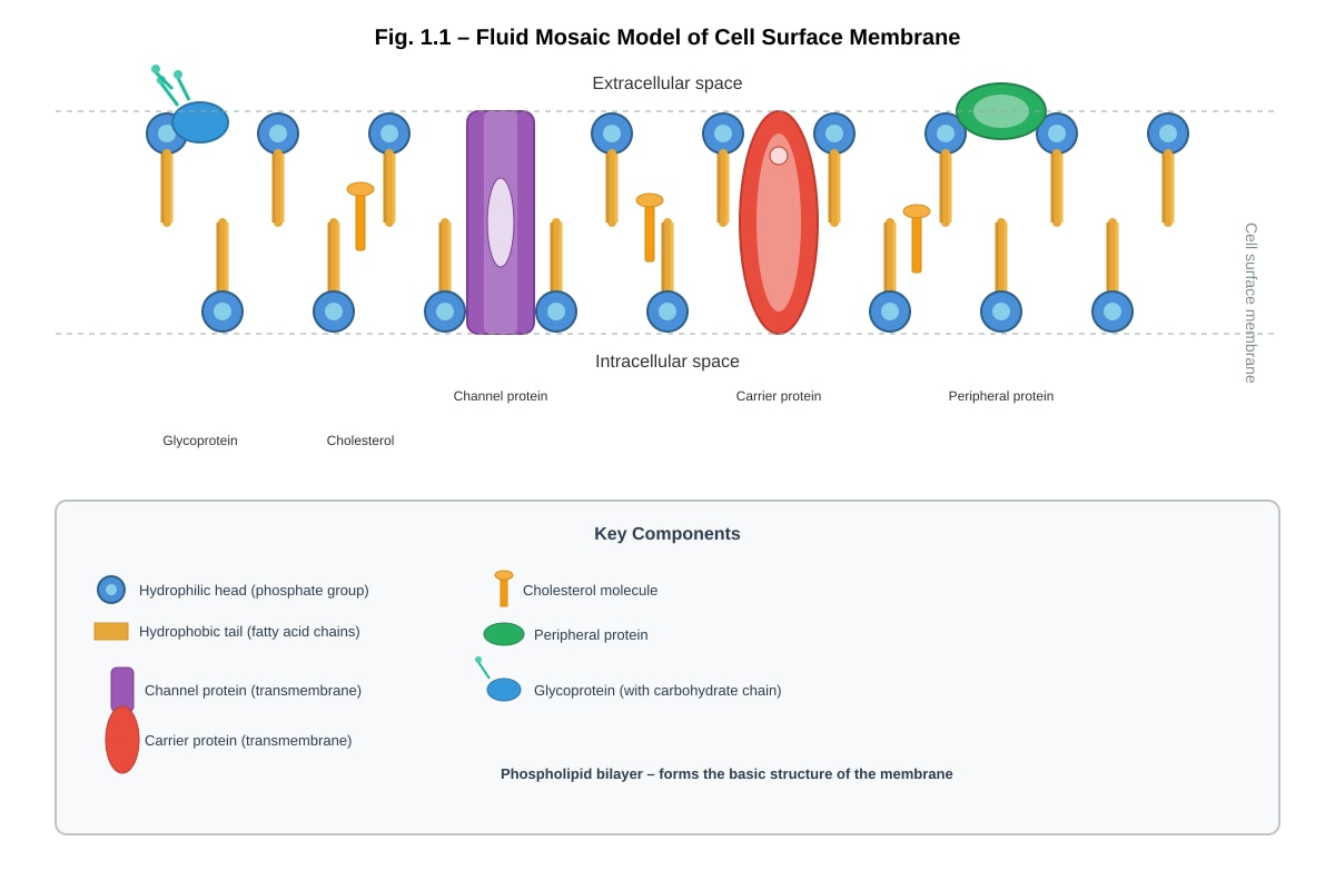
1. With reference to Fig. 1.1, which shows the fluid mosaic model of a cell surface membrane, explain how the structure of phospholipids contributes to the selective permeability of the membrane.
Generated diagram for this question.
[3 marks]
2. A suspension of mitochondria was prepared in a buffer containing ADP and inorganic phosphate (Pi). The oxygen concentration in the buffer was monitored over time. At point X, sodium azide (an inhibitor of cytochrome c oxidase) was added to the suspension.
With reference to the expected changes in oxygen concentration, explain the role of oxygen in aerobic respiration and predict the effect of sodium azide addition.
[4 marks]
3. Describe and explain how gel electrophoresis can be used to diagnose sickle cell anaemia. In your answer, explain why individuals who are heterozygous for the sickle cell allele (Hb^A/Hb^S) show two bands on the gel, while homozygous individuals show only one band.
[4 marks]
4. The lac operon in E. coli codes for inducible enzymes involved in lactose metabolism. In contrast, the trp operon codes for repressible enzymes involved in tryptophan synthesis.
Suggest and explain why it is advantageous for a prokaryote to have an inducible operon such as the lac operon, rather than expressing the genes constitutively.
[3 marks]
5. With reference to Fig. 5.1, which shows the molecular structure of a misfolded prion protein (PrP^Sc) alongside the normal cellular prion protein (PrP^C), suggest why misfolded prion proteins tend to aggregate in the cell and explain how this aggregation leads to cellular dysfunction.
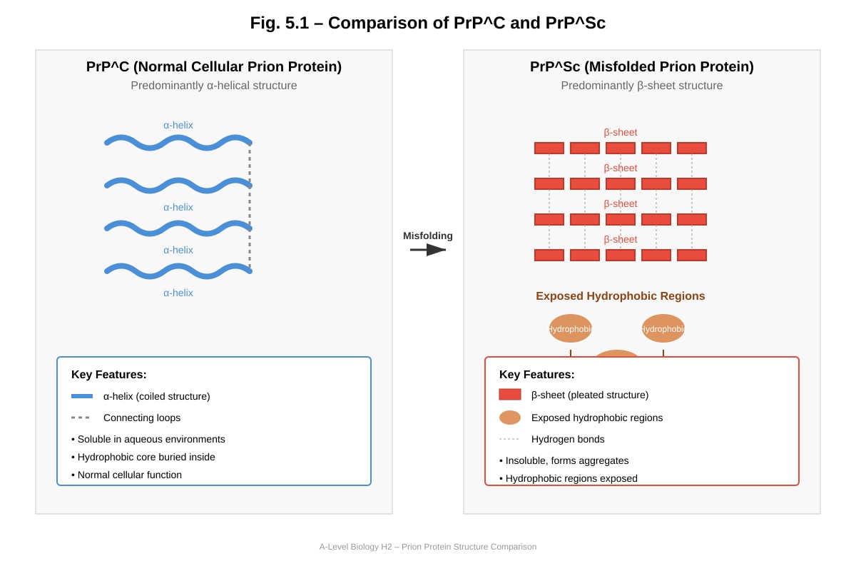
Generated diagram for this question.
[3 marks]
6. A student prepared a serial dilution of a protein solution and used the biuret test to estimate protein concentration. The results are shown in Table 6.1.
Table 6.1: Biuret test results for protein standards and unknown sample
| Protein concentration (mg cm⁻³) | Absorbance at 540 nm |
|---|---|
| 0.0 | 0.00 |
| 0.2 | 0.12 |
| 0.4 | 0.24 |
| 0.6 | 0.36 |
| 0.8 | 0.48 |
| 1.0 | 0.60 |
| Unknown sample | 0.42 |
(a) Plot a calibration curve of absorbance against protein concentration on the grid provided. [2 marks]
(b) Use your calibration curve to determine the protein concentration of the unknown sample. [1 mark]
[Grid space for graph plotting]
SECTION B: Diagram and Data Interpretation (15 marks)
Answer all questions in this section.
7. Fig. 7.1 shows the oxygen dissociation curves for adult haemoglobin (HbA) and fetal haemoglobin (HbF).
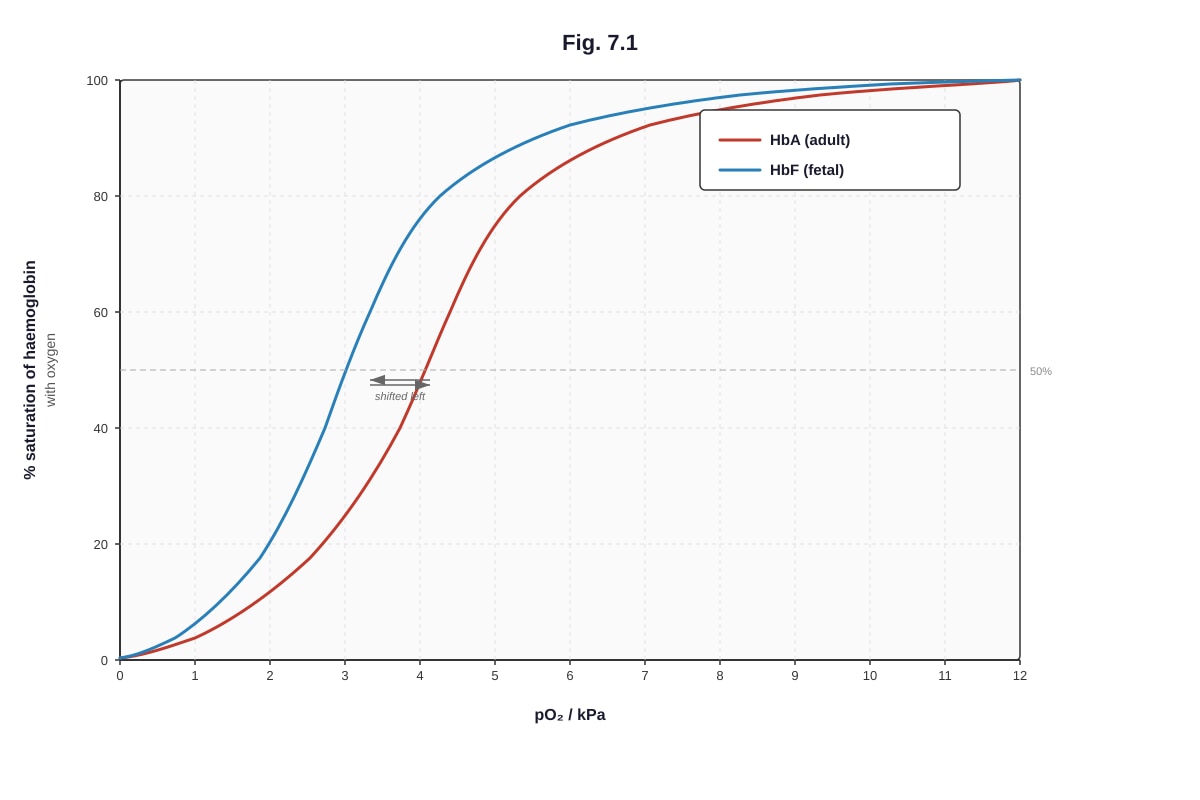
Generated graph for this question.
(a) State the pO₂ at which HbA is 50% saturated with oxygen. [1 mark]
(b) Explain why the oxygen dissociation curve of HbF is shifted to the left relative to that of HbA. [2 marks]
(c) Explain the physiological significance of the difference in oxygen affinity between HbF and HbA for the developing fetus. [2 marks]
8. Fig. 8.1 shows the molecular structure of a nucleotide triphosphate.
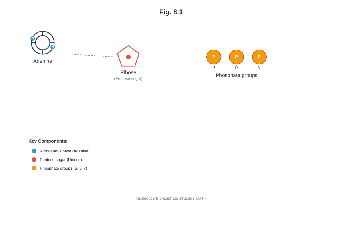
Generated diagram for this question.
(a) Name the molecule shown in Fig. 8.1. [1 mark]
(b) Explain why the hydrolysis of the bond between the β and γ phosphate groups releases a large amount of free energy. [2 marks]
(c) State two uses of the molecule named in (a) in cellular metabolism, other than as an energy source. [2 marks]
9. Fig. 9.1 shows the effect of temperature on the rate of an enzyme-catalysed reaction.
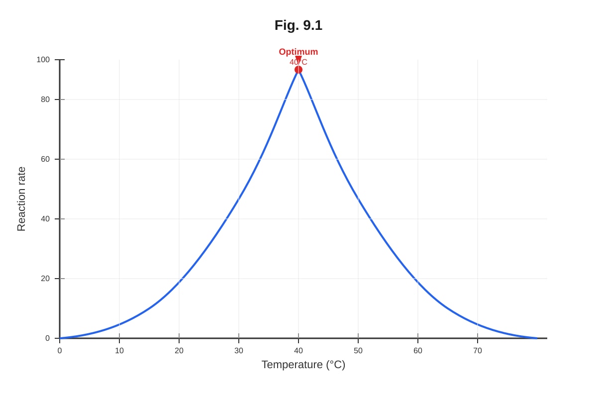
Generated graph for this question.
(a) Explain the increase in reaction rate between 10°C and 40°C. [2 marks]
(b) Explain the decrease in reaction rate above 45°C. [3 marks]
SECTION C: Source/Data-Based Questions (15 marks)
Answer all questions in this section.
10. Read the following passage and answer the questions that follow.
Cystic fibrosis (CF) is caused by mutations in the CFTR gene, which codes for a chloride ion channel protein. The most common mutation, ΔF508, results in the deletion of a phenylalanine residue at position 508 of the CFTR protein. This mutation causes the protein to misfold in the endoplasmic reticulum (ER). The misfolded protein is recognised by the ER quality control system and targeted for degradation via the ubiquitin-proteasome pathway, preventing it from reaching the cell surface membrane.
In some individuals, treatment with a combination of drugs—a 'corrector' (e.g., lumacaftor) and a 'potentiator' (e.g., ivacaftor)—has been shown to improve CFTR function. Correctors help the ΔF508-CFTR protein fold correctly and traffic to the cell surface, while potentiators increase the open probability of the channel once it reaches the membrane.
(a) With reference to the passage, explain why the ΔF508 mutation results in a loss of CFTR function at the cell surface. [3 marks]
(b) Suggest how the combination therapy of a corrector and a potentiator restores CFTR function. [3 marks]
(c) Discuss the limitations of using the combination therapy described in the passage for all CF patients. [2 marks]
11. A research study investigated the effect of cholesterol content on membrane fluidity in mammalian cells. Cells were treated to alter their membrane cholesterol content, and membrane fluidity was measured using fluorescence recovery after photobleaching (FRAP). The results are shown in Table 11.1.
Table 11.1: Effect of cholesterol content on membrane fluidity
| Cholesterol content (% of total membrane lipid) | Fluidity (arbitrary units) |
|---|---|
| 0 | 8.2 |
| 10 | 6.5 |
| 20 | 5.1 |
| 30 | 4.0 |
| 40 | 3.8 |
| 50 | 3.7 |
(a) Describe the relationship between cholesterol content and membrane fluidity shown in Table 11.1. [2 marks]
(b) Explain how cholesterol modulates membrane fluidity at the molecular level. [3 marks]
(c) Suggest why membrane fluidity is important for the function of integral membrane proteins such as ion channels and receptors. [2 marks]
SECTION D: Extended Response (10 marks)
Answer all questions in this section.
12. (a) Compare and contrast the structure and function of DNA and RNA. [5 marks]
(b) Discuss the importance of complementary base pairing in DNA replication and protein synthesis. [5 marks]
END OF PAPER
This paper was prepared by TuitionGoWhere Exam Practice (AI). Version 3 of 5.
Answers
TuitionGoWhere Practice Paper – Biology H2 A-Level
PRACTICE PAPER 3 (Version 3 of 5) – ANSWER KEY AND MARKING SCHEME
Subject: Biology H2 (9477)
Total Marks: 60
SECTION A: Structured Response (20 marks)
1. With reference to Fig. 1.1, explain how the structure of phospholipids contributes to the selective permeability of the membrane. [3 marks]
Answer:
- Phospholipids are amphipathic molecules, with hydrophilic (polar) phosphate heads and hydrophobic (non-polar) fatty acid tails; [1 mark]
- The phospholipids arrange into a bilayer, with hydrophilic heads facing the aqueous environments on both sides of the membrane and hydrophobic tails facing inwards, forming a hydrophobic core; [1 mark]
- This hydrophobic core acts as a barrier to the free passage of polar molecules, ions, and large molecules, allowing only small, non-polar molecules (e.g., O₂, CO₂) and water (to a limited extent) to pass through freely, thus contributing to selective permeability. [1 mark]
Marking notes:
- Award [1] for amphipathic nature/structure description.
- Award [1] for bilayer arrangement with hydrophobic core.
- Award [1] for linking hydrophobic core to selective permeability (barrier to polar/charged molecules).
- Reference to Fig. 1.1 must be explicit or clearly implied.
2. Explain the role of oxygen in aerobic respiration and predict the effect of sodium azide addition. [4 marks]
Answer:
- Oxygen acts as the final electron acceptor in the electron transport chain (ETC); [1 mark]
- It accepts electrons and combines with protons (H⁺) to form water, catalysed by cytochrome c oxidase (Complex IV); [1 mark]
- This maintains the flow of electrons through the ETC, which is necessary for the continued pumping of protons across the inner mitochondrial membrane to maintain the proton gradient for ATP synthesis via chemiosmosis; [1 mark]
- Sodium azide inhibits cytochrome c oxidase, preventing electron transfer to oxygen. This would cause oxygen consumption to cease (or decrease sharply), as oxygen can no longer be reduced to water. The ETC would back up, proton pumping would stop, the proton gradient would dissipate, and ATP synthesis would halt. [1 mark]
Marking notes:
- Award [1] for final electron acceptor.
- Award [1] for formation of water (accepts electrons and protons).
- Award [1] for linking oxygen consumption to maintenance of proton gradient/ATP synthesis.
- Award [1] for prediction: oxygen consumption stops/decreases, with explanation linking to cytochrome c oxidase inhibition and consequences for ETC/ATP synthesis.
3. Describe and explain how gel electrophoresis can be used to diagnose sickle cell anaemia. Explain why heterozygous individuals show two bands. [4 marks]
Answer:
- A sample of haemoglobin is extracted from the patient's red blood cells and loaded into a well in a gel (e.g., polyacrylamide or agarose gel); [1 mark]
- An electric potential difference (electric field) is applied across the gel. Haemoglobin proteins, being charged, migrate through the gel matrix. The rate of migration depends on the net charge and molecular mass/shape of the protein; [1 mark]
- Normal haemoglobin (HbA) and sickle cell haemoglobin (HbS) differ by a single amino acid substitution (glutamic acid → valine), which alters the net charge of the protein. HbS has a less negative charge than HbA and therefore migrates more slowly towards the positive electrode, resulting in different band positions; [1 mark]
- Heterozygous individuals (Hb^A/Hb^S) produce both normal and sickle cell haemoglobin. Since each allele codes for a protein with a different charge, two distinct bands appear on the gel—one corresponding to HbA and one to HbS. Homozygous individuals produce only one type of haemoglobin and thus show a single band. [1 mark]
Marking notes:
- Award [1] for sample loading and application of electric field.
- Award [1] for explanation of separation based on charge/mass.
- Award [1] for linking amino acid difference to charge difference and different migration.
- Award [1] for explanation of two bands in heterozygotes (two different proteins from two different alleles).
4. Suggest and explain why it is advantageous for a prokaryote to have an inducible operon such as the lac operon. [3 marks]
Answer:
- An inducible operon is only transcribed (switched on) when its substrate (e.g., lactose) is present in the environment; [1 mark]
- This prevents the wasteful synthesis of enzymes (e.g., β-galactosidase, lactose permease) when their substrate is absent, conserving energy and resources (amino acids, ATP, ribosomes); [1 mark]
- This metabolic efficiency provides a selective advantage in competitive environments where nutrients are scarce or variable, allowing the prokaryote to allocate resources to the synthesis of other essential proteins. [1 mark]
Marking notes:
- Award [1] for stating that transcription occurs only when substrate is present.
- Award [1] for explaining energy/resource conservation.
- Award [1] for linking to selective advantage/metabolic efficiency.
- Accept reference to the lac operon specifically (lactose → allolactose → inducer, inactivates repressor).
5. Suggest why misfolded prion proteins tend to aggregate and explain how this aggregation leads to cellular dysfunction. [3 marks]
Answer:
- Misfolded prion proteins (PrP^Sc) have a different conformation from normal PrP^C, with a higher proportion of β-sheet structure. This conformational change exposes hydrophobic amino acid residues that are normally buried in the protein core; [1 mark]
- The exposed hydrophobic regions on different PrP^Sc molecules interact with each other via hydrophobic interactions, causing the proteins to clump together and form insoluble aggregates; [1 mark]
- These aggregates accumulate in cells (particularly neurons), disrupting normal cellular functions. They are resistant to protease degradation and cannot be cleared effectively. The accumulation interferes with intracellular transport, organelle function, and ultimately leads to cell death, causing the spongiform degeneration characteristic of prion diseases. [1 mark]
Marking notes:
- Award [1] for exposure of hydrophobic residues due to conformational change.
- Award [1] for hydrophobic interactions driving aggregation.
- Award [1] for linking aggregation to cellular dysfunction (insoluble, resistant to degradation, disrupts cell function, leads to cell death).
6. (a) Plot a calibration curve of absorbance against protein concentration. [2 marks]
Answer:
- Axes correctly labelled: x-axis = Protein concentration (mg cm⁻³), y-axis = Absorbance at 540 nm; [1 mark]
- All six data points plotted accurately (0.0, 0.00), (0.2, 0.12), (0.4, 0.24), (0.6, 0.36), (0.8, 0.48), (1.0, 0.60); straight line of best fit drawn through origin. [1 mark]
Marking notes:
- Award [1] for correct axes labels with units.
- Award [1] for accurate plotting and appropriate line of best fit (should be linear and pass through origin).
- Deduct [1] if line is not straight or does not pass through origin.
(b) Use your calibration curve to determine the protein concentration of the unknown sample. [1 mark]
Answer:
- 0.70 mg cm⁻³ (accept 0.68–0.72 mg cm⁻³). [1 mark]
Marking notes:
- Award [1] for correct reading from graph within acceptable range.
- Method: absorbance 0.42 corresponds to 0.42/0.60 = 0.70 mg cm⁻³ on linear calibration.
SECTION B: Diagram and Data Interpretation (15 marks)
7. (a) State the pO₂ at which HbA is 50% saturated with oxygen. [1 mark]
Answer:
- Approximately 3.5–4.0 kPa (accept 26–30 mm Hg if units are in mm Hg on the graph). [1 mark]
Marking notes:
- Award [1] for correct reading from the graph. Accept values within a reasonable range depending on graph scale.
(b) Explain why the oxygen dissociation curve of HbF is shifted to the left relative to that of HbA. [2 marks]
Answer:
- Fetal haemoglobin (HbF) has a higher affinity for oxygen than adult haemoglobin (HbA); [1 mark]
- This is because HbF has a different subunit composition (α₂γ₂) compared to HbA (α₂β₂). The γ-subunits bind 2,3-bisphosphoglycerate (2,3-BPG) less strongly than β-subunits. Since 2,3-BPG reduces haemoglobin's affinity for oxygen, the reduced binding of 2,3-BPG to HbF results in a higher oxygen affinity, shifting the curve to the left. [1 mark]
Marking notes:
- Award [1] for stating HbF has higher oxygen affinity.
- Award [1] for explanation involving reduced 2,3-BPG binding (or different subunit composition).
(c) Explain the physiological significance of the difference in oxygen affinity between HbF and HbA for the developing fetus. [2 marks]
Answer:
- In the placenta, fetal blood is in close proximity to maternal blood. The higher oxygen affinity of HbF allows it to load oxygen at the relatively low pO₂ of the placental blood supply, where maternal HbA is unloading oxygen; [1 mark]
- This ensures efficient transfer of oxygen from maternal blood to fetal blood across the placenta, providing the fetus with sufficient oxygen for aerobic respiration and growth. [1 mark]
Marking notes:
- Award [1] for explaining oxygen transfer from mother to fetus at placental pO₂.
- Award [1] for linking to fetal oxygen supply/requirement.
8. (a) Name the molecule shown in Fig. 8.1. [1 mark]
Answer:
- Adenosine triphosphate (ATP). [1 mark]
Marking notes:
- Award [1] for ATP. Accept adenosine triphosphate.
(b) Explain why the hydrolysis of the bond between the β and γ phosphate groups releases a large amount of free energy. [2 marks]
Answer:
- The three phosphate groups are negatively charged and repel each other strongly. The repulsion between the closely spaced negative charges creates an unstable, high-energy bond; [1 mark]
- Hydrolysis of the terminal (γ) phosphate group relieves this electrostatic repulsion, and the products (ADP and inorganic phosphate, Pi) are more stable than ATP. The difference in free energy between ATP and the products is released as useful energy. [1 mark]
Marking notes:
- Award [1] for electrostatic repulsion between negatively charged phosphate groups.
- Award [1] for products being more stable / relief of repulsion releasing energy.
(c) State two uses of ATP in cellular metabolism, other than as an energy source. [2 marks]
Answer:
- Any two from:
- As a substrate for RNA synthesis (incorporated as a nucleotide during transcription). [1 mark]
- As a phosphate donor in phosphorylation reactions (e.g., by kinases to activate or deactivate enzymes). [1 mark]
- As a signalling molecule (e.g., acting as an extracellular signalling molecule in purinergic signalling). [1 mark]
- As a coenzyme in certain enzymatic reactions. [1 mark]
Marking notes:
- Award [1] each for any two valid uses, up to [2 marks].
- Do not accept "energy source" or "energy currency" as this is excluded by the question.
9. (a) Explain the increase in reaction rate between 10°C and 40°C. [2 marks]
Answer:
- As temperature increases, the kinetic energy of both enzyme and substrate molecules increases; [1 mark]
- This results in more frequent collisions between enzyme and substrate molecules, and a greater proportion of collisions have sufficient energy to overcome the activation energy barrier, leading to more enzyme-substrate complexes formed per unit time and an increased rate of reaction. [1 mark]
Marking notes:
- Award [1] for increased kinetic energy / more frequent collisions.
- Award [1] for more successful collisions / more ES complexes formed.
(b) Explain the decrease in reaction rate above 45°C. [3 marks]
Answer:
- Above the optimum temperature (45°C), the increased thermal energy disrupts the weak bonds (hydrogen bonds, ionic bonds, hydrophobic interactions) that maintain the enzyme's specific three-dimensional tertiary structure; [1 mark]
- This causes the enzyme to denature—the active site loses its specific complementary shape; [1 mark]
- As a result, the substrate can no longer bind to the active site, fewer enzyme-substrate complexes form, and the rate of reaction decreases sharply. The denaturation is usually irreversible. [1 mark]
Marking notes:
- Award [1] for disruption of bonds maintaining tertiary structure.
- Award [1] for denaturation / loss of active site shape.
- Award [1] for consequence: substrate cannot bind / fewer ES complexes / rate decreases.
SECTION C: Source/Data-Based Questions (15 marks)
10. (a) With reference to the passage, explain why the ΔF508 mutation results in a loss of CFTR function at the cell surface. [3 marks]
Answer:
- The ΔF508 mutation causes the deletion of a phenylalanine residue at position 508 of the CFTR protein; [1 mark]
- This deletion causes the CFTR protein to misfold in the endoplasmic reticulum (ER); [1 mark]
- The misfolded protein is recognised by the ER quality control system and is targeted for degradation via the ubiquitin-proteasome pathway, preventing it from being transported to the cell surface membrane. Without CFTR at the cell surface, chloride ion transport cannot occur. [1 mark]
Marking notes:
- Award [1] for identifying the mutation (deletion of Phe508).
- Award [1] for protein misfolding in the ER.
- Award [1] for degradation preventing trafficking to cell surface (must reference the passage).
(b) Suggest how the combination therapy of a corrector and a potentiator restores CFTR function. [3 marks]
Answer:
- The corrector (e.g., lumacaftor) acts as a pharmacological chaperone, helping the ΔF508-CFTR protein to fold correctly in the ER, thereby allowing it to pass the ER quality control system; [1 mark]
- This enables the correctly folded CFTR protein to be trafficked to the cell surface membrane; [1 mark]
- The potentiator (e.g., ivacaftor) increases the open probability of the CFTR chloride channel once it is at the cell surface, enhancing chloride ion transport even if the channel has reduced function. [1 mark]
Marking notes:
- Award [1] for corrector helping protein folding/trafficking.
- Award [1] for CFTR reaching cell surface.
- Award [1] for potentiator increasing channel open probability/activity.
(c) Discuss the limitations of using the combination therapy described in the passage for all CF patients. [2 marks]
Answer:
- The therapy is specific to the ΔF508 mutation. CF can be caused by over 2000 different mutations in the CFTR gene, including nonsense mutations, splice-site mutations, and deletions other than ΔF508. Patients with mutations that produce no CFTR protein at all (e.g., nonsense mutations) would not benefit from a corrector/potentiator; [1 mark]
- The therapy is expensive and may not be accessible to all patients. Additionally, individual responses vary; some patients may not respond adequately, and there may be side effects or drug interactions. [1 mark]
Marking notes:
- Award [1] for mutation specificity (only works for ΔF508 or certain misfolding mutations).
- Award [1] for any other valid limitation: cost, accessibility, variable efficacy, side effects, does not cure the disease (symptomatic treatment).
- Accept other reasonable limitations.
11. (a) Describe the relationship between cholesterol content and membrane fluidity shown in Table 11.1. [2 marks]
Answer:
- As cholesterol content increases from 0% to 30%, membrane fluidity decreases sharply (from 8.2 to 4.0 arbitrary units); [1 mark]
- Between 30% and 50% cholesterol, the decrease in fluidity is much smaller, with fluidity levelling off at approximately 3.7–3.8 arbitrary units. [1 mark]
Marking notes:
- Award [1] for describing the initial sharp decrease.
- Award [1] for describing the plateau/levelling off at higher cholesterol concentrations.
- Award only [1] if the description is vague (e.g., "fluidity decreases as cholesterol increases").
(b) Explain how cholesterol modulates membrane fluidity at the molecular level. [3 marks]
Answer:
- Cholesterol is an amphipathic molecule that intercalates between phospholipid molecules in the membrane; [1 mark]
- At moderate to high temperatures, the rigid steroid ring structure of cholesterol restricts the movement of phospholipid fatty acid tails, reducing membrane fluidity (making the membrane less fluid); [1 mark]
- At low temperatures, cholesterol prevents the tight packing of phospholipid fatty acid tails, inhibiting crystallisation and maintaining membrane fluidity (preventing the membrane from becoming too rigid). Thus, cholesterol acts as a fluidity buffer. [1 mark]
Marking notes:
- Award [1] for cholesterol intercalating between phospholipids.
- Award [1] for reducing fluidity at higher temperatures (restricts movement).
- Award [1] for maintaining fluidity at low temperatures (prevents tight packing) / fluidity buffer concept.
(c) Suggest why membrane fluidity is important for the function of integral membrane proteins such as ion channels and receptors. [2 marks]
Answer:
- Membrane fluidity allows integral membrane proteins to diffuse laterally within the plane of the membrane, enabling them to interact with other proteins (e.g., receptor clustering for signal transduction); [1 mark]
- Fluidity also allows conformational changes in membrane proteins (e.g., opening and closing of ion channels, binding of ligands to receptors) that are necessary for their function. A membrane that is too rigid would restrict these conformational changes and impair protein function. [1 mark]
Marking notes:
- Award [1] for lateral diffusion / protein movement / interaction.
- Award [1] for conformational changes / flexibility required for function.
- Accept other valid points: membrane fusion, endocytosis/exocytosis, cell signalling.
SECTION D: Extended Response (10 marks)
12. (a) Compare and contrast the structure and function of DNA and RNA. [5 marks]
Answer: Similarities:
- Both are nucleic acids composed of nucleotide monomers, each consisting of a nitrogenous base, a pentose sugar, and a phosphate group; [1 mark]
- Both contain the purine bases adenine (A) and guanine (G), and the pyrimidine base cytosine (C); [1 mark]
- Both have a sugar-phosphate backbone formed by phosphodiester bonds between the 3' carbon of one sugar and the 5' carbon of the next; [1 mark]
Differences:
- DNA contains the sugar deoxyribose, while RNA contains ribose (which has an –OH group on the 2' carbon); [1 mark]
- DNA contains the pyrimidine base thymine (T), while RNA contains uracil (U) instead; [1 mark]
- DNA is typically double-stranded, forming a double helix with antiparallel strands held together by hydrogen bonds between complementary base pairs (A-T, G-C). RNA is typically single-stranded, although it can form secondary structures (e.g., hairpin loops) through intra-strand base pairing; [1 mark]
- Functionally, DNA stores genetic information long-term and serves as the template for replication and transcription. RNA has multiple functions: mRNA carries genetic information from DNA to ribosomes for translation; tRNA transfers amino acids to ribosomes during translation; rRNA is a structural and catalytic component of ribosomes. [1 mark]
Marking notes:
- Award marks for valid similarities and differences. Maximum [5 marks].
- At least one similarity and at least one difference must be included for full marks.
- Award [1] each for up to 5 distinct, correct points covering both structure and function.
- Points can be awarded in any order.
(b) Discuss the importance of complementary base pairing in DNA replication and protein synthesis. [5 marks]
Answer: DNA replication:
- Complementary base pairing (A with T, G with C) ensures that each strand of the parental DNA molecule acts as a template for the synthesis of a new complementary strand; [1 mark]
- This ensures accurate, semi-conservative replication, where each new DNA molecule consists of one parental strand and one newly synthesised strand, preserving the genetic information; [1 mark]
- The specificity of base pairing (purine with pyrimidine) also maintains the uniform width of the DNA double helix. [1 mark]
Protein synthesis (transcription and translation):
- During transcription, complementary base pairing (A with U, T with A, G with C, C with G) allows RNA polymerase to synthesise a pre-mRNA strand that is complementary to the template strand of DNA, accurately copying the genetic code; [1 mark]
- During translation, complementary base pairing between the codons on mRNA and the anticodons on tRNA molecules ensures that the correct amino acid is added to the growing polypeptide chain according to the genetic code; [1 mark]
- This specificity is fundamental to the fidelity of gene expression, ensuring that the correct sequence of amino acids is assembled to produce a functional protein. Errors in base pairing (mutations) can lead to incorrect amino acid incorporation and potentially non-functional proteins. [1 mark]
Marking notes:
- Award [1] for each valid point, up to [5 marks].
- Points must cover both DNA replication and protein synthesis for full marks.
- Award marks for: template function, semi-conservative replication, accuracy/fidelity, transcription (mRNA synthesis), translation (codon-anticodon pairing), consequences of errors.
- Accept well-explained points in any logical order.
END OF ANSWER KEY
This answer key was prepared by TuitionGoWhere Exam Practice (AI). Version 3 of 5.
Free quiz and exam paper access
Enter your details to view this paper
Your access is remembered on this device.