From Real Exams Exam Paper
A Level H2 Biology Practice Paper 2
Free A Level H2 Biology Practice Paper 2, LongCat Exam version, with questions, answers, and A Level-style practice for Singapore students.
These static practice materials are generated from the site's syllabus and paper-generation workflow, with source and model context shown so students and parents can evaluate the material before use.
Questions
TuitionGoWhere Practice Paper - Biology H2 A-Level
TuitionGoWhere Secondary School (AI)
| Field | Details |
|---|---|
| Subject: | Biology |
| Level: | A-Level H2 (9477) |
| Paper: | Practice Paper — Cells & Biomolecules |
| Version: | 2 of 5 |
| Duration: | 60 minutes |
| Total Marks: | 50 |
| Name: | |
| Class: | |
| Date: |
Instructions
- Answer all questions in the spaces provided.
- Write your answers in dark blue or black pen.
- You may use a pencil for any diagrams or graphs.
- No calculators are allowed.
- The total mark for this paper is 50.
- The number of marks for each question or part question is shown in brackets [ ].
Section A: Structured Questions (30 marks)
Questions 1–6
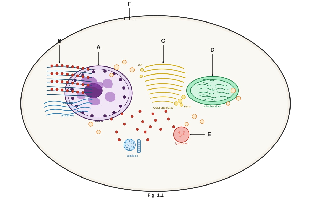
1. Fig. 1.1 shows the structure of a typical eukaryotic animal cell as seen under an electron microscope.
Generated diagram for Q1.
(a) Identify the organelles labelled A, B, C, and D in Fig. 1.1.
| Label | Organelle |
|---|---|
| A | _________________________________ |
| B | _________________________________ |
| C | _________________________________ |
| D | _________________________________ |
[4]
(b) State two structural features of the organelle labelled D that are adapted for its function.
[2]
(c) Describe one function of the organelle labelled E.
[1]
2. Table 2.1 shows the approximate composition of four different cell types in terms of the percentage of water and protein by mass.
Table 2.1
| Cell type | Water (%) | Protein (%) |
|---|---|---|
| Red blood cell | 65 | 30 |
| Liver cell | 70 | 18 |
| Bacterial cell | 75 | 15 |
| Skin cell | 68 | 22 |
(a) Calculate the ratio of water to protein in a red blood cell. Show your working.
Answer: _________________________________ [2]
(b) Suggest one reason why the protein content of a red blood cell is higher than that of a bacterial cell.
[1]
(c) Water has several important properties as a biological molecule. Explain two properties of water that make it important for living organisms.
[2]
3. Fig. 3.1 shows the structure of a phospholipid molecule.
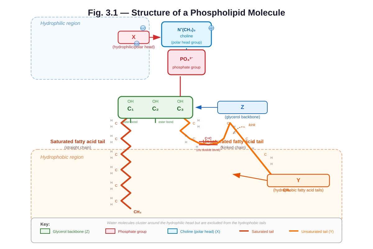
Generated diagram for Q3.
(a) Identify the parts labelled X and Y in Fig. 3.1.
X: _________________________________
Y: _________________________________
[2]
(b) With reference to Fig. 3.1, explain how phospholipids are arranged in the cell membrane and why this arrangement occurs.
[3]
(c) Explain how the presence of an unsaturated fatty acid tail affects the fluidity of the cell membrane.
[2]
4. Fig. 4.1 shows the effect of temperature on the rate of an enzyme-catalysed reaction.
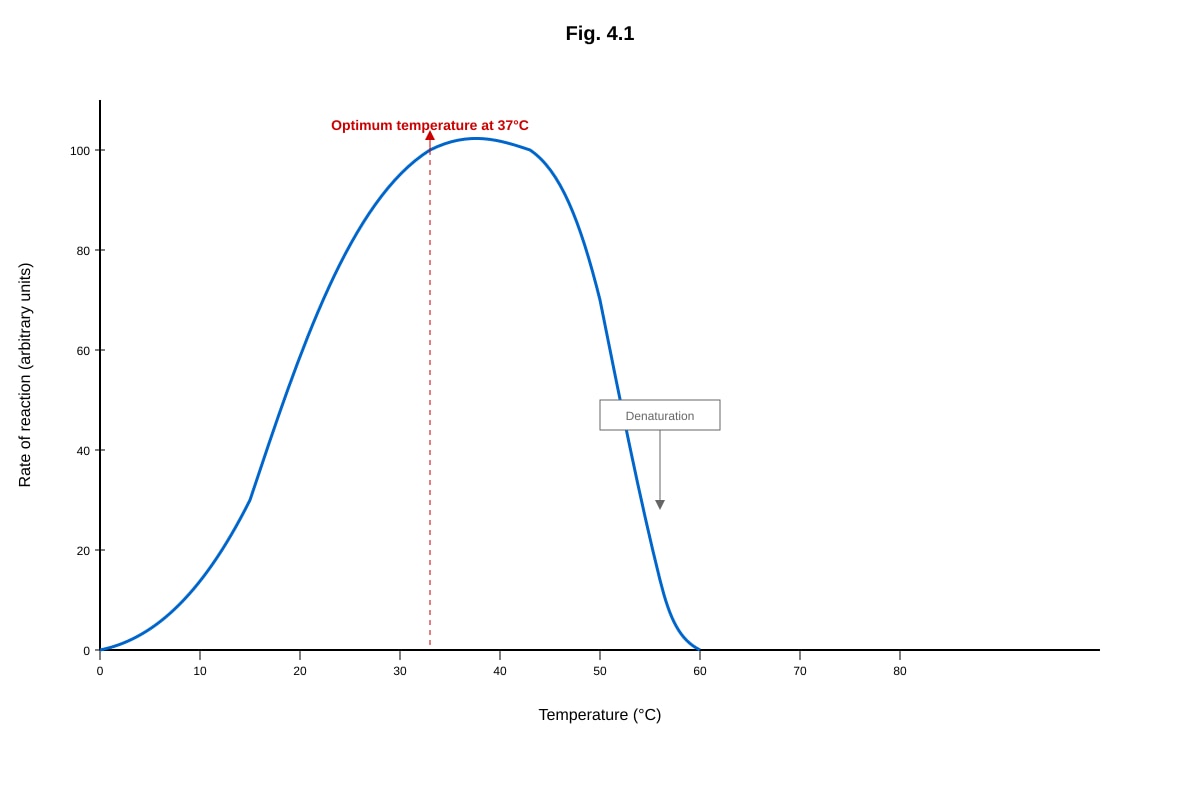
Generated graph for Q4.
(a) With reference to Fig. 4.1, describe the effect of temperature on the rate of the enzyme-catalysed reaction between 0 °C and 37 °C.
[2]
(b) Explain why the rate of reaction decreases sharply above 37 °C.
[2]
(c) A student repeated the experiment using an enzyme isolated from a thermophilic bacterium. Predict and explain how the graph would differ.
[2]
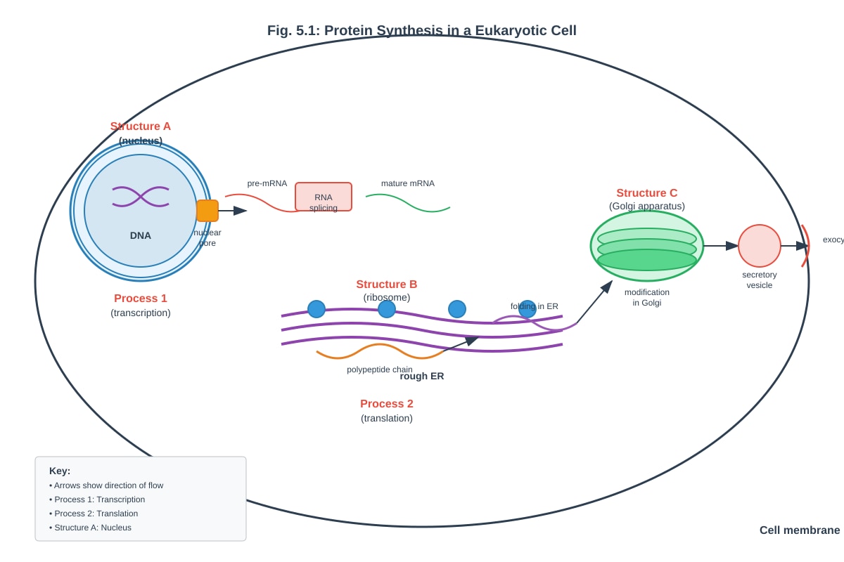
5. Fig. 5.1 shows a simplified diagram of the process of protein synthesis in a eukaryotic cell.
Generated diagram for Q5.
(a) Name the processes labelled Process 1 and Process 2 in Fig. 5.1.
Process 1: _________________________________
Process 2: _________________________________
[2]
(b) With reference to Fig. 5.1, describe what happens during Process 1.
[2]
(c) Explain why the protein produced at the ribosome in Fig. 5.1 is transported to the Golgi apparatus rather than being released directly into the cytoplasm.
[2]
6. A student carried out an experiment to test for the presence of biological molecules in three unknown solutions, P, Q, and R. The results are shown in Table 6.1.
Table 6.1
| Test | Solution P | Solution Q | Solution R |
|---|---|---|---|
| Benedict's test (heated) | Blue | Brick-red precipitate | Blue |
| Biuret test | Purple | Blue | Purple |
| Ethanol emulsion test | Clear | Clear | Milky-white |
(a) Identify the biological molecule(s) present in each solution.
Solution P: _________________________________
Solution Q: _________________________________
Solution R: _________________________________
[3]
(b) State one precaution the student should take when carrying out the Benedict's test.
[1]
(c) Describe how the Biuret test is carried out.
[2]
Section B: Data Interpretation (12 marks)
Questions 7–8
7. Fig. 7.1 shows the results of an experiment investigating the effect of different concentrations of sucrose solution on the mass of potato cylinders after 30 minutes of immersion.
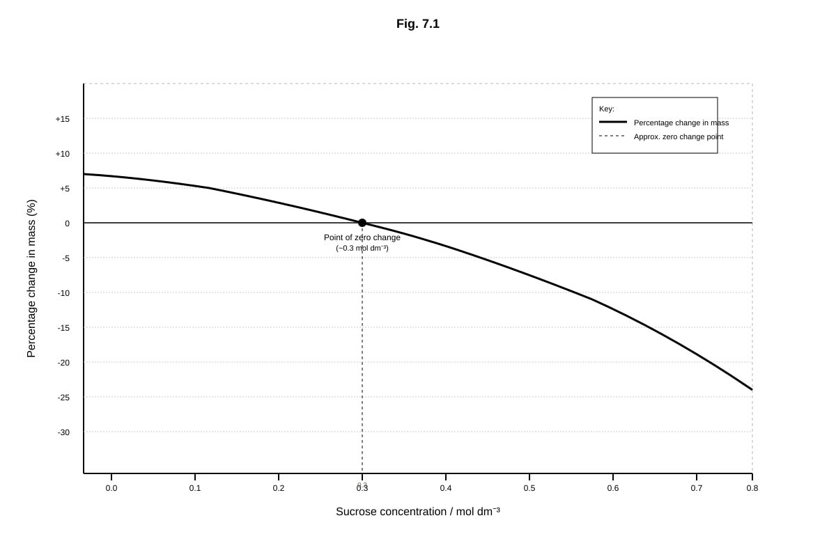
Generated graph for Q7.
(a) With reference to Fig. 7.1, state the sucrose concentration at which there is no net movement of water into or out of the potato cells. Explain your answer.
[2]
(b) Explain why the potato cylinders in 0.1 mol dm⁻³ sucrose solution gained mass.
[2]
(c) A student suggested that the cell sap concentration of the potato cells is exactly 0.3 mol dm⁻³. Evaluate this suggestion.
[2]
8. Fig. 8.1 shows the structure of a molecule of ATP (adenosine triphosphate).

Generated diagram for Q8.
(a) Identify the components labelled A, B, and C in Fig. 8.1.
A: _________________________________
B: _________________________________
C: _________________________________
[3]
(b) ATP is described as the universal energy currency of the cell. Explain why ATP is a suitable molecule for this role.
[3]
Section C: Extended Response (8 marks)
Question 9
9. Collagen is the most abundant protein in the human body. It is a fibrous structural protein found in connective tissues such as tendons, ligaments, and skin.
Describe the structure of collagen and explain how its structure is related to its function as a structural protein.
In your answer, you should refer to the following levels of protein structure:
- primary structure
- secondary structure
- tertiary structure
- quaternary structure
[8]
End of Paper
Total: 50 marks
Answers
TuitionGoWhere Practice Paper - Biology H2 A-Level
Answer Key — Cells & Biomolecules (Version 2 of 5)
Section A: Structured Questions
Question 1
(a) Identify the organelles labelled A, B, C, and D. [4]
| Label | Organelle |
|---|---|
| A | Nucleus |
| B | Rough endoplasmic reticulum (rough ER) |
| C | Golgi apparatus (Golgi body) |
| D | Mitochondrion |
[1 mark each, total 4]
Teaching notes: These are the four most commonly identified organelles in electron micrograph questions. The nucleus is identified by its double membrane (nuclear envelope) with nuclear pores and the presence of chromatin/nucleolus. Rough ER is identified by the presence of ribosomes on its cytoplasmic surface. The Golgi apparatus appears as a stack of flattened cisternae (membrane-bound sacs). The mitochondrion is identified by its double membrane and internal folds called cristae.
(b) State two structural features of the organelle labelled D (mitochondrion) that are adapted for its function. [2]
- Cristae (inner membrane folds) — increase the surface area for the attachment of electron transport chain proteins and ATP synthase, maximising ATP production during oxidative phosphorylation.
- Matrix — contains enzymes required for the Krebs cycle (link reaction and citric acid cycle), providing the necessary environment for these reactions to occur.
Acceptable alternatives:
- Double membrane — compartmentalises the mitochondrion, creating distinct environments for different stages of aerobic respiration.
- Small size / circular DNA — allows rapid replication and localised protein synthesis.
- Inner membrane is selectively permeable — essential for establishing the proton gradient.
[1 mark each, total 2]
Teaching notes: The key exam skill here is linking structure to function. Students must not merely describe a structural feature but must explain how it aids the mitochondrion's role in aerobic respiration. Common mistake: stating "has cristae" without explaining the increased surface area benefit.
(c) Describe one function of the organelle labelled E (lysosome). [1]
Lysosomes contain hydrolytic (digestive) enzymes that break down worn-out organelles, engulfed pathogens, or cellular debris through intracellular digestion.
Acceptable alternatives:
- Autolysis — self-digestion of the cell after the lysosome membrane ruptures (important in programmed cell death / apoptosis).
- Digestion of material taken in by endocytosis/phagocytosis.
[1 mark]
Teaching notes: Lysosomes are membrane-bound organelles containing hydrolytic enzymes (e.g., proteases, lipases, nucleases) that function optimally at acidic pH (~pH 5). The membrane prevents these enzymes from digesting the cell's own contents.
Question 2
(a) Calculate the ratio of water to protein in a red blood cell. Show your working. [2]
Working:
- Water content = 65%
- Protein content = 30%
- Ratio of water : protein = 65 : 30
- Simplify by dividing both by 5: 13 : 6 (or approximately 2.2 : 1)
Answer: 13 : 6 (or 2.2 : 1)
[1 mark for correct working, 1 mark for correct simplified ratio]
Teaching notes: Students must show their working to gain full marks. Accept any correctly simplified ratio. Common mistake: writing 65:30 without simplifying, or inverting the ratio.
(b) Suggest one reason why the protein content of a red blood cell is higher than that of a bacterial cell. [1]
Red blood cells contain a large amount of haemoglobin (a protein), which is essential for oxygen transport. Bacterial cells have a lower proportion of protein relative to water because they have different metabolic requirements and do not require haemoglobin.
Acceptable alternatives:
- Red blood cells are packed with haemoglobin to maximise oxygen-carrying capacity.
- Bacterial cells have a cell wall and different structural composition.
[1 mark]
(c) Explain two properties of water that make it important for living organisms. [2]
-
High specific heat capacity — Water can absorb or release a large amount of heat energy with only a small change in temperature. This helps organisms maintain a stable internal body temperature (thermoregulation) and provides a stable aquatic environment for aquatic organisms.
-
Cohesion and surface tension — Water molecules are polar and form hydrogen bonds with each other (cohesion). This allows water to be pulled up through xylem vessels in plants (transpiration stream) and creates surface tension that supports small organisms.
Acceptable alternatives:
- Universal solvent — Water's polarity allows it to dissolve many ionic and polar substances, making it an excellent medium for biochemical reactions in the cytoplasm.
- High latent heat of vaporisation — Evaporation of water (sweating, transpiration) is an effective cooling mechanism.
- Incompressible — Provides turgor pressure in plant cells and acts as a hydrostatic skeleton in some animals.
- Transparent — Allows light to penetrate for photosynthesis in aquatic environments.
[1 mark each, total 2]
Teaching notes: Students must explain the property AND its biological significance to gain the mark. Simply stating "water is a solvent" without explaining why this is biologically important is insufficient.
Question 3
(a) Identify the parts labelled X and Y in Fig. 3.1. [2]
X: Hydrophilic (polar) head (composed of phosphate group and glycerol)
Y: Hydrophobic fatty acid tails (composed of hydrocarbon chains)
[1 mark each, total 2]
(b) Explain how phospholipids are arranged in the cell membrane and why this arrangement occurs. [3]
Phospholipids are arranged as a bilayer in the cell membrane. The hydrophilic (polar) heads face outwards towards the aqueous environment on both sides of the membrane (the extracellular fluid and the cytoplasm), while the hydrophobic fatty acid tails face inwards, away from water, forming the interior of the membrane.
This arrangement occurs because:
- The hydrophilic heads are attracted to water (polar interactions, hydrogen bonding) on both the outside and inside of the cell.
- The hydrophobic tails are repelled by water and therefore orientate themselves away from the aqueous environment, towards the centre of the bilayer.
- This is the most thermodynamically stable arrangement — it minimises the interaction between hydrophobic tails and water while maximising the interaction between hydrophilic heads and water.
[1 mark for bilayer arrangement, 1 mark for orientation of heads/tails, 1 mark for explanation of why (hydrophilic/hydrophobic interactions)]
Teaching notes: This is a classic A-Level question testing understanding of the fluid mosaic model. Students must use the terms "hydrophilic" and "hydrophobic" correctly. Common mistake: confusing the orientation of heads and tails.
(c) Explain how the presence of an unsaturated fatty acid tail affects the fluidity of the cell membrane. [2]
Unsaturated fatty acid tails contain one or more double bonds between carbon atoms, which introduce kinks/bends in the hydrocarbon chain. These kinks prevent the phospholipid molecules from packing closely together, increasing the fluidity of the membrane. This is because the irregular shape of unsaturated tails creates more space between phospholipid molecules, reducing the strength of the van der Waals forces between adjacent tails.
[1 mark for kinks/double bonds, 1 mark for reduced packing/increased fluidity]
Teaching notes: Students should connect the molecular structure (double bond → kink) to the macroscopic property (increased fluidity). Common mistake: stating that unsaturated fats "make the membrane more permeable" without explaining the structural basis.
Question 4
(a) Describe the effect of temperature on the rate of the enzyme-catalysed reaction between 0 °C and 37 °C. [2]
As temperature increases from 0 °C to 37 °C, the rate of the enzyme-catalysed reaction increases. This is because:
- Both the enzyme and substrate molecules gain kinetic energy as temperature rises.
- This leads to more frequent successful collisions between enzyme active sites and substrate molecules.
- More enzyme-substrate complexes are formed per unit time, increasing the rate of reaction.
- The rate approximately doubles for every 10 °C rise (Q₁₀ effect).
[1 mark for stating rate increases, 1 mark for explanation involving kinetic energy/collisions]
(b) Explain why the rate of reaction decreases sharply above 37 °C. [2]
Above 37 °C, the rate decreases sharply because the enzyme denatures. The high temperature disrupts the hydrogen bonds, ionic bonds, and other weak interactions that maintain the enzyme's tertiary (3D) structure. This causes the active site to change shape so that the substrate can no longer fit (no longer complementary). The enzyme-substrate complex can no longer form, and the reaction rate drops sharply.
[1 mark for denaturation, 1 mark for explanation of bond disruption/active site shape change]
Teaching notes: Students must be precise: the enzyme is denatured, not "killed" (enzymes are not living). The active site shape changes — the primary structure (amino acid sequence) is usually not broken at moderate denaturing temperatures.
(c) Predict and explain how the graph would differ if an enzyme from a thermophilic bacterium were used. [2]
The optimum temperature would be higher (e.g., around 60–80 °C instead of 37 °C). The curve would peak at a higher temperature and would not drop off sharply until a much higher temperature is reached. This is because enzymes from thermophilic bacteria have stronger bonds (e.g., more disulphide bridges, more ionic bonds, more hydrophobic interactions) in their tertiary structure, making them more resistant to thermal denaturation.
[1 mark for higher optimum temperature, 1 mark for explanation involving stronger bonds/heat resistance]
Question 5
(a) Name the processes labelled Process 1 and Process 2. [2]
Process 1: Transcription
Process 2: Translation
[1 mark each, total 2]
(b) Describe what happens during Process 1 (transcription). [2]
During transcription:
- The DNA double helix is unwound and unzipped by the enzyme RNA polymerase, breaking the hydrogen bonds between complementary base pairs.
- RNA polymerase reads the template (antisense) strand of DNA in the 3' → 5' direction.
- Free ribonucleotides line up alongside the template strand by complementary base pairing (A-U, T-A, G-C, C-G).
- RNA polymerase catalyses the formation of phosphodiester bonds between adjacent ribonucleotides, synthesising a pre-mRNA strand in the 5' → 3' direction.
[1 mark for unwinding/unzipping/RNA polymerase, 1 mark for complementary base pairing/phosphodiester bonds/pre-mRNA synthesis]
Teaching notes: Students should specify that it is the template/antisense strand that is transcribed, and that uracil replaces thymine in RNA.
(c) Explain why the protein produced at the ribosome in Fig. 5.1 is transported to the Golgi apparatus rather than being released directly into the cytoplasm. [2]
The protein is likely a secretory protein (e.g., an enzyme or hormone) that needs to be modified, processed, and packaged before being exported from the cell. The Golgi apparatus:
- Modifies proteins (e.g., by adding carbohydrate groups — glycosylation).
- Folds proteins into their correct 3D conformation.
- Packages proteins into secretory vesicles for transport to the cell membrane for exocytosis.
If the protein were released directly into the cytoplasm, it would not be correctly processed and could not be secreted from the cell.
[1 mark for secretory protein/modification, 1 mark for specific Golgi function — glycosylation/packaging/vesicles]
Question 6
(a) Identify the biological molecule(s) present in each solution. [3]
- Solution P: Protein (Biuret test positive → purple; Benedict's negative → no reducing sugar; ethanol emulsion negative → no lipid)
- Solution Q: Reducing sugar (Benedict's test positive → brick-red precipitate; Biuret negative → no protein; ethanol emulsion negative → no lipid)
- Solution R: Lipid (Ethanol emulsion test positive → milky-white; Benedict's negative → no reducing sugar; Biuret positive → also contains protein)
[1 mark each, total 3]
Teaching notes: Solution R tests positive for both lipid and protein. Students must identify both to gain the mark. Common mistake: only identifying one molecule when two tests are positive.
(b) State one precaution the student should take when carrying out the Benedict's test. [1]
- Heat the mixture in a water bath (not directly over a Bunsen flame) to ensure even heating.
- Ensure the solution is heated to boiling for sufficient time (at least 2 minutes) to allow the reaction to occur.
- Use equal volumes of Benedict's reagent and test solution.
Accept any one valid precaution.
[1 mark]
(c) Describe how the Biuret test is carried out. [2]
- Add sodium hydroxide (NaOH) solution to the test solution to create an alkaline environment.
- Add a few drops of copper(II) sulfate (CuSO₄) solution (dilute) to the mixture.
- If protein is present, the solution turns from blue to purple/violet. If no protein is present, the solution remains blue.
[1 mark for NaOH, 1 mark for CuSO₄ and colour change]
Teaching notes: The Biuret test detects peptide bonds. The copper(II) ions form a violet-coloured complex with peptide bonds in alkaline solution. Students must mention both reagents and the colour change.
Section B: Data Interpretation
Question 7
(a) State the sucrose concentration at which there is no net movement of water. Explain your answer. [2]
The concentration is approximately 0.3 mol dm⁻³. At this point, the percentage change in mass is zero, meaning there is no net movement of water into or out of the potato cells. The water potential of the sucrose solution is equal to the water potential of the cell sap (equilibrium).
[1 mark for 0.3 mol dm⁻³, 1 mark for explanation involving zero net movement/equal water potential]
(b) Explain why the potato cylinders in 0.1 mol dm⁻³ sucrose solution gained mass. [2]
The 0.1 mol dm⁻³ sucrose solution has a higher water potential (less negative / more dilute) than the cell sap of the potato cells. Therefore, water molecules move by osmosis from the solution (higher water potential) into the potato cells (lower water potential) across the partially permeable cell membrane. This causes the potato cells to become turgid and the cylinders to gain mass.
[1 mark for higher water potential in solution, 1 mark for osmosis/water moving into cells]
Teaching notes: Students must use the term "osmosis" and refer to water potential. Common mistake: saying "water moves from high to low concentration" without mentioning water potential — at A-Level, water potential language is expected.
(c) Evaluate the suggestion that the cell sap concentration of the potato cells is exactly 0.3 mol dm⁻³. [2]
The suggestion is partially correct but not entirely accurate. The point where percentage change in mass is zero (0.3 mol dm⁻³) indicates that the water potential of the external solution equals the water potential of the cell sap. However, the cell sap contains various dissolved substances (ions, sugars, amino acids), not just sucrose. Therefore, 0.3 mol dm⁻³ is the equivalent sucrose concentration that has the same water potential as the cell sap, but the actual concentration of solutes in the cell sap may differ in composition.
[1 mark for agreeing it indicates equal water potential, 1 mark for explaining that cell sap composition differs from pure sucrose solution]
Question 8
(a) Identify the components labelled A, B, and C in Fig. 8.1. [3]
A: Adenine (nitrogenous base)
B: Ribose (pentose sugar)
C: Three phosphate groups
[1 mark each, total 3]
Teaching notes: Together, adenine + ribose form adenosine. Adenosine + three phosphate groups = adenosine triphosphate (ATP). Students should be able to distinguish the base, sugar, and phosphate components.
(b) Explain why ATP is a suitable molecule as the universal energy currency of the cell. [3]
-
Small, soluble, and easily transported — ATP is a small, water-soluble molecule that can be readily transported within the cell to wherever energy is needed.
-
Releases energy in small, manageable amounts — When ATP is hydrolysed to ADP + Pᵢ, a relatively small amount of energy is released (~30.5 kJ mol⁻¹), which is suitable for coupling to individual cellular reactions without wasting energy as excess heat.
-
Rapidly regenerated — ATP can be quickly resynthesised from ADP + Pᵢ during cellular respiration (oxidative phosphorylation) and photosynthesis (photophosphorylation), ensuring a continuous supply.
-
Universal — ATP is used by all living organisms, from bacteria to humans, as the immediate source of energy for cellular processes.
Accept any three valid points.
[1 mark each, maximum 3]
Teaching notes: This is a standard A-Level question. Students should avoid vague statements like "ATP stores energy" and instead explain the specific properties that make ATP suitable as an energy carrier.
Section C: Extended Response
Question 9
Describe the structure of collagen and explain how its structure is related to its function as a structural protein. [8]
Marking scheme:
| Level | Marks | Descriptors |
|---|---|---|
| Level 3 | 7–8 | All four levels of protein structure are described accurately with clear links to collagen's function. Answer is well-organised, uses appropriate terminology, and demonstrates comprehensive understanding. |
| Level 2 | 4–6 | At least three levels of structure are described with some links to function. Answer shows good understanding but may lack detail or precision in places. |
| Level 1 | 1–3 | One or two levels of structure are mentioned but descriptions may be vague or lack clear links to function. Limited use of terminology. |
| 0 | 0 | No relevant content. |
Expected content points (for 8 marks, award 1 mark per valid point, maximum 8):
Primary structure:
- Collagen has a primary structure consisting of a repeating sequence of three amino acids: glycine-proline-hydroxyproline (Gly-X-Y, where X is often proline and Y is often hydroxyproline). Glycine is the smallest amino acid, which is critical for the tight packing of the triple helix.
Secondary structure: 2. Each individual collagen polypeptide chain forms a left-handed helix (not an α-helix — it is a distinct polyproline II-type helix). This is the secondary structure of collagen.
Tertiary structure: 3. Three of these left-handed helical polypeptide chains are wound around each other to form a right-handed triple helix (also called tropocollagen). This is stabilised by hydrogen bonds between the chains (particularly involving the -OH group of hydroxyproline) and by the close packing of glycine residues in the interior of the triple helix.
Quaternary structure: 4. Multiple tropocollagen molecules (triple helices) are assembled into collagen fibrils, which are further bundled into collagen fibres. This represents the quaternary structure. The tropocollagen molecules are staggered and cross-linked by covalent bonds (aldol cross-links and Schiff bases) between lysine and hydroxylysine residues of adjacent molecules.
Links to function: 5. The triple helix structure provides great tensile strength, making collagen resistant to stretching and pulling forces — essential for its role in tendons, ligaments, and skin. 6. The cross-links between tropocollagen molecules within fibrils provide additional mechanical strength and stability to the connective tissue. 7. The staggered arrangement of tropocollagen molecules in fibrils, combined with cross-links, distributes mechanical stress evenly throughout the tissue, preventing tearing. 8. The high proportion of glycine (the smallest amino acid) allows the three chains to pack tightly together in the triple helix, contributing to the structural stability of the molecule.
Teaching notes: This question tests the ability to apply knowledge of protein structure levels to a specific, well-known protein. Students often lose marks by not being specific about collagen's unique features (e.g., the triple helix is NOT the same as an α-helix; the primary structure has a characteristic Gly-X-Y repeat). The key skill is linking each level of structure to the mechanical properties that make collagen an effective structural protein.
Mark Summary
| Question | Marks |
|---|---|
| 1(a) | 4 |
| 1(b) | 2 |
| 1(c) | 1 |
| 2(a) | 2 |
| 2(b) | 1 |
| 2(c) | 2 |
| 3(a) | 2 |
| 3(b) | 3 |
| 3(c) | 2 |
| 4(a) | 2 |
| 4(b) | 2 |
| 4(c) | 2 |
| 5(a) | 2 |
| 5(b) | 2 |
| 5(c) | 2 |
| 6(a) | 3 |
| 6(b) | 1 |
| 6(c) | 2 |
| 7(a) | 2 |
| 7(b) | 2 |
| 7(c) | 2 |
| 8(a) | 3 |
| 8(b) | 3 |
| 9 | 8 |
| Total | 50 |
Free quiz and exam paper access
Enter your details to view this paper
Your access is remembered on this device.