AI Generated Quiz
A Level H1 Biology Human Physiology Quiz
Free A Level H1 Biology Human Physiology quiz, AI version, with questions, answers, and A Level-style practice for Singapore students.
These static practice materials are generated from the site's syllabus and paper-generation workflow, with source and model context shown so students and parents can evaluate the material before use.
Questions
Free quiz and exam paper access
Enter your details to view this paper
Your access is remembered on this device.
Answers
A-Level Biology H1 Quiz - Human Physiology: Answer Key
Total Marks: 50
Section A: Multiple Choice (10 marks)
1. B. It initiates the electrical impulse that sets the heart rate.
- Marks: 2
- Explanation: The sinoatrial (SA) node, located in the wall of the right atrium, is the natural pacemaker of the heart. It spontaneously generates electrical impulses that spread across the atria, causing them to contract. This sets the basic heart rate. Option A describes the role of the atrioventricular (AV) node, which delays the impulse. Option C describes the AV node and the Purkinje fibres. Option D describes the AV node and the bundle of His.
- Common Mistake: Confusing the SA node with the AV node. Remember: SA node = pacemaker; AV node = delay.
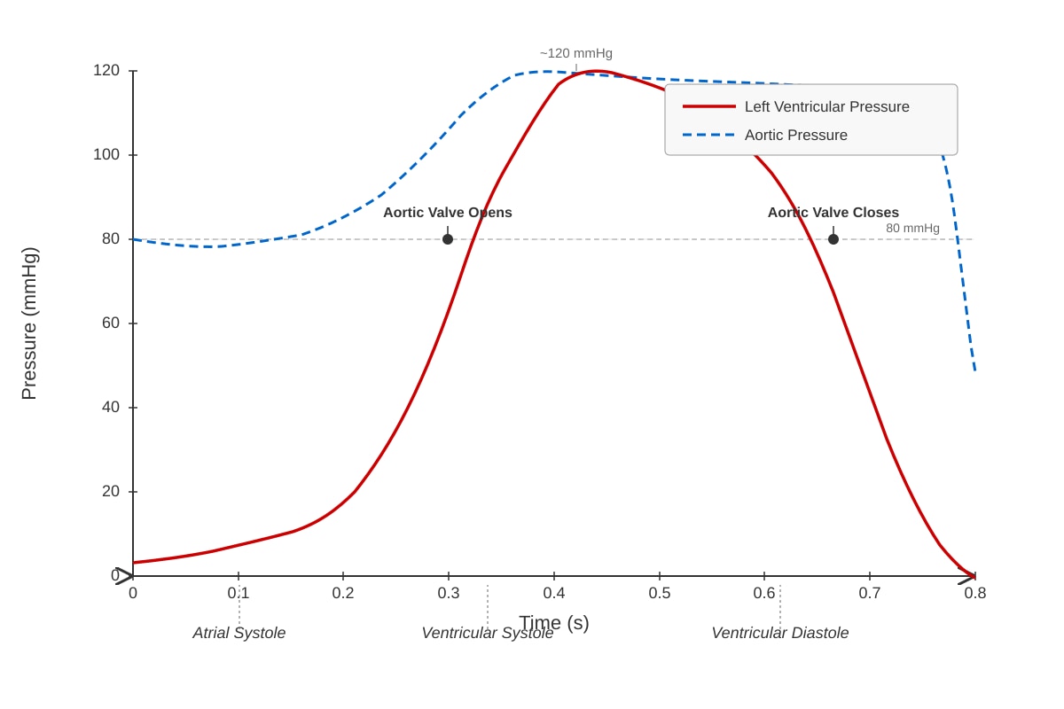
2. B. When ventricular pressure falls below aortic pressure.
- Marks: 2
- Explanation: The aortic valve closes to prevent backflow of blood from the aorta into the left ventricle. This occurs at the end of ventricular systole when the left ventricle relaxes and its pressure drops below the pressure in the aorta. The graph shows the dashed line (aortic pressure) and the solid line (ventricular pressure) crossing at this point. Option A describes when the aortic valve opens.
- Common Mistake: Confusing valve opening and closing. Opening occurs when ventricular pressure exceeds aortic pressure; closing occurs when ventricular pressure falls below aortic pressure.
3. B. Oxygen moves from the alveoli to the capillaries by simple diffusion down its concentration gradient.
- Marks: 2
- Explanation: Oxygen is a small, non-polar molecule that can diffuse directly through the phospholipid bilayer. It moves from an area of high partial pressure (alveoli, 100 mmHg) to an area of low partial pressure (capillaries, 40 mmHg) down its concentration gradient. This is a passive process requiring no energy. Options A and C are incorrect because oxygen does not require active transport or facilitated diffusion. Option D is incorrect because osmosis is the movement of water.
- Common Mistake: Forgetting that oxygen is a small, non-polar molecule that diffuses directly through the membrane.
4. A. ADH increases the permeability of the collecting duct to water, leading to increased water reabsorption.
- Marks: 2
- Explanation: Antidiuretic hormone (ADH) is released from the posterior pituitary gland in response to increased blood plasma osmolarity (dehydration). It acts on the collecting duct of the nephron by increasing the insertion of aquaporin water channels into the cell membrane. This increases the permeability of the collecting duct to water, allowing more water to be reabsorbed into the bloodstream, concentrating the urine and conserving water. Options C and D are incorrect because ADH primarily affects water, not sodium, permeability.
- Common Mistake: Confusing the effect of ADH with aldosterone. Aldosterone increases sodium reabsorption; ADH increases water reabsorption.
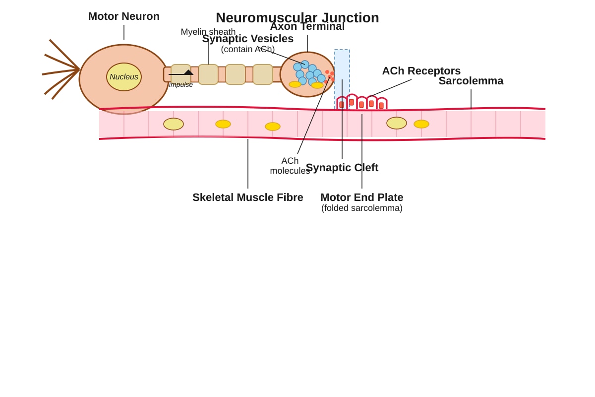
5. B. The depolarisation of the sarcolemma, generating an action potential.
- Marks: 2
- Explanation: Acetylcholine (ACh) is a neurotransmitter released from the axon terminal of a motor neuron. It diffuses across the synaptic cleft and binds to ACh receptors on the sarcolemma (muscle cell membrane). This binding opens ligand-gated ion channels, allowing sodium ions (Na⁺) to enter the muscle cell. The influx of positive charge depolarises the sarcolemma, generating an action potential that propagates along the muscle fibre. This action potential then triggers the release of calcium ions from the sarcoplasmic reticulum (option A), which is a subsequent step. Option C is incorrect because contraction is a later step. Option D is incorrect because reuptake occurs after the signal is transmitted.
- Common Mistake: Confusing the immediate effect (depolarisation) with the later effect (calcium release and contraction).
Section B: Structured Questions (25 marks)
6. (a) Two structural differences:
- Marks: 2 (1 mark for each correct difference)
- Acceptable answers:
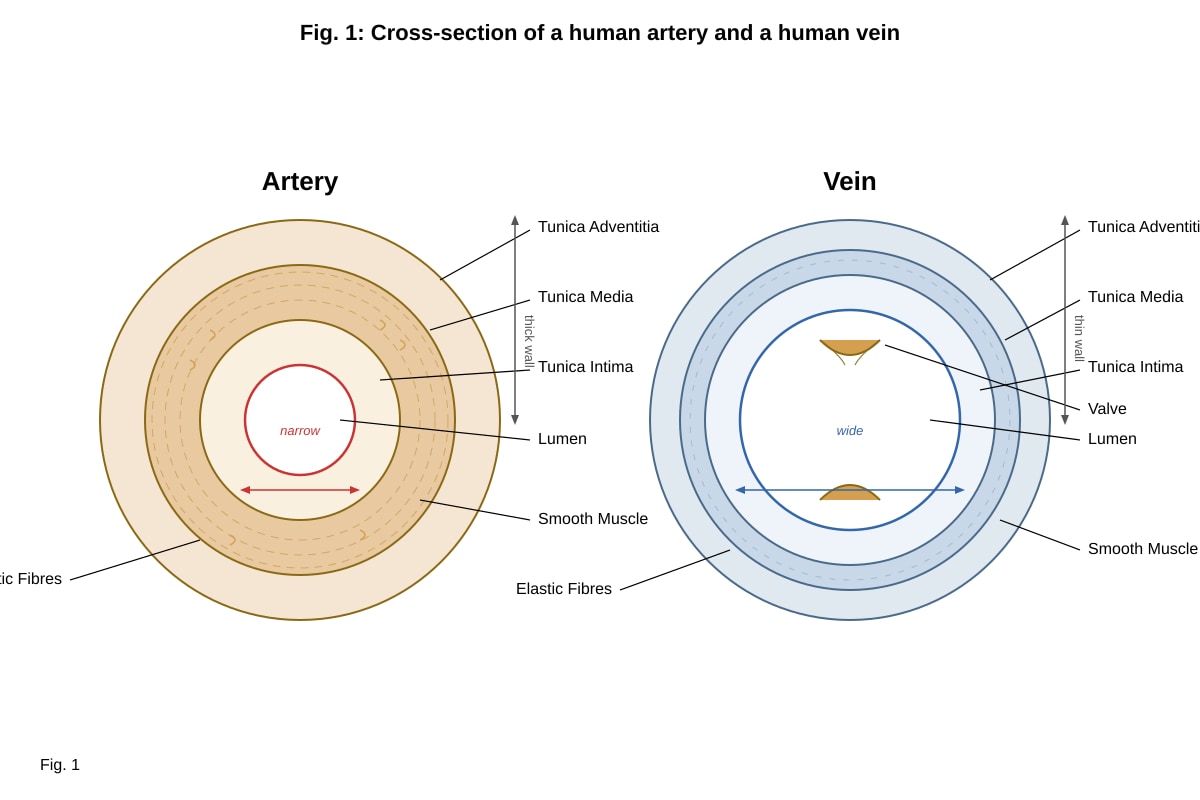
- The artery has a thicker wall (specifically a thicker tunica media) than the vein.
- The artery has a narrower lumen than the vein.
- The vein has valves, whereas the artery does not.
- The artery has more elastic fibres and smooth muscle in its wall than the vein.
- Teaching Note: These differences are directly related to their functions. Arteries carry blood away from the heart under high pressure, so they need thick, elastic walls to withstand and maintain that pressure. Veins carry blood back to the heart under low pressure, so they have thinner walls and valves to prevent backflow.
(b) Explanation of artery structure adaptation:
- Marks: 3
- Expected points:
- The thick wall (especially the tunica media with smooth muscle and elastic fibres) provides strength to withstand the high pressure of blood pumped from the heart. (1 mark)
- The elastic fibres allow the artery to stretch and recoil, helping to maintain blood pressure and smooth out the pulsatile flow. (1 mark)
- The smooth muscle allows the artery to constrict or dilate (vasoconstriction/vasodilation) to regulate blood flow and blood pressure. (1 mark)
- Teaching Note: Think of the artery as a strong, flexible tube. The elastic fibres are like rubber bands that stretch when blood is pumped in and recoil to push the blood along. The smooth muscle acts like a control valve to adjust the diameter.
7. (a) Explanation for absence of plasma proteins in filtrate:
- Marks: 2
- Expected points:
- Plasma proteins are large molecules (e.g., albumin, globulins) with a high molecular weight. (1 mark)
- They are too large to pass through the filtration slits in the glomerular capillaries and the basement membrane during ultrafiltration. (1 mark)
- Teaching Note: Ultrafiltration in the glomerulus acts like a sieve. Small molecules like water, glucose, urea, and ions can pass through, but large molecules like plasma proteins and blood cells are too big and remain in the blood.
(b) Process of glucose reabsorption:
- Marks: 1
- Answer: Active transport (specifically co-transport with sodium ions).
- Teaching Note: Glucose is reabsorbed in the proximal convoluted tubule. It is transported against its concentration gradient using energy from ATP. This is achieved by a sodium-glucose co-transporter (SGLT) on the apical membrane.
(c) Prediction and explanation for urea concentration in urine:
- Marks: 2
- Expected points:
- The concentration of urea in urine is much higher than in plasma. (1 mark)
- Explanation: Water is reabsorbed along the nephron (especially in the collecting duct under the influence of ADH), but urea is not reabsorbed to the same extent. This means the same amount of urea is dissolved in a much smaller volume of water, concentrating it. (1 mark)
- Teaching Note: The nephron reabsorbs most of the water from the filtrate, but it does not reabsorb urea as efficiently. This makes the urine more concentrated in urea than the original plasma.
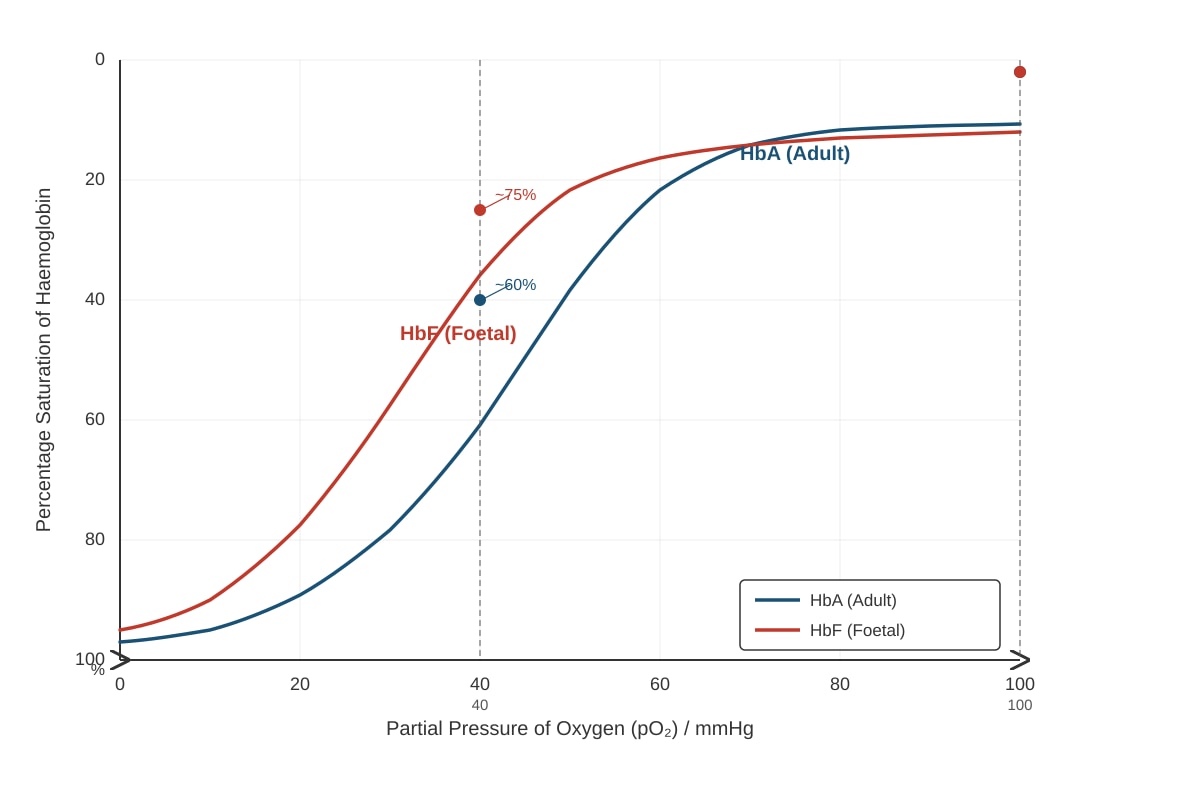
8. (a) Percentage saturation of HbA at pO₂ of 40 mmHg:
- Marks: 1
- Answer: Approximately 60% (accept 55–65%).
- Teaching Note: Read the value directly from the graph by finding 40 mmHg on the x-axis, drawing a vertical line to the HbA curve, and then reading the corresponding value on the y-axis.
(b) Significance of difference in oxygen affinity:
- Marks: 3
- Expected points:
- Foetal haemoglobin (HbF) has a higher affinity for oxygen than adult haemoglobin (HbA), as shown by its dissociation curve being to the left. (1 mark)
- At the placenta, the pO₂ is relatively low (around 30–40 mmHg). At this pO₂, HbF is more saturated with oxygen than HbA would be. (1 mark)
- This allows efficient transfer of oxygen from the mother's blood (where it is released from HbA) to the foetus's blood (where it is picked up by HbF). (1 mark)
- Teaching Note: The left-shifted curve of HbF means it can "hold on" to oxygen more tightly. This is crucial in the placenta, where the oxygen concentration is not as high as in the lungs. It allows the foetus to extract oxygen from the mother's blood.
9. Role of hypothalamus in thermoregulation (high external temperature):
- Marks: 4
- Expected points:
- The hypothalamus contains thermoreceptors that detect the temperature of the blood. It also receives nerve impulses from peripheral thermoreceptors in the skin. (1 mark)
- When the external temperature is high, the hypothalamus initiates cooling mechanisms. (1 mark)
- It sends nerve impulses to the skin to cause vasodilation of arterioles, increasing blood flow to the surface and increasing heat loss by radiation and convection. (1 mark)
- It also stimulates sweat glands to produce more sweat. Evaporation of sweat from the skin surface removes latent heat, cooling the body. (1 mark)
- Teaching Note: The hypothalamus acts as the body's thermostat. It compares the body's temperature to a set point (around 37°C) and triggers responses to correct any deviation. For high temperatures, the goal is to increase heat loss.
10. (a) Region that shortens during contraction:
- Marks: 1
- Answer: The I-band (and the H-zone).
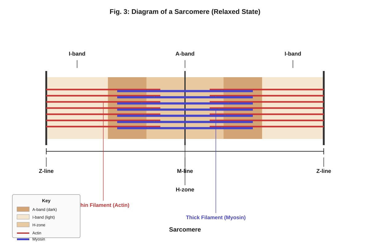
- Teaching Note: During the sliding filament theory, the thin filaments (actin) slide inwards over the thick filaments (myosin). This pulls the Z-lines closer together, shortening the sarcomere. The I-band (the region of only thin filaments) and the H-zone (the region of only thick filaments) both decrease in width. The A-band (the length of the thick filaments) remains constant.
(b) Role of calcium ions in muscle contraction:
- Marks: 3
- Expected points:
- An action potential arrives at the neuromuscular junction, causing the release of calcium ions from the sarcoplasmic reticulum into the sarcoplasm. (1 mark)
- Calcium ions bind to troponin, a protein on the thin filament. This causes a conformational change in troponin, which moves tropomyosin away from the myosin-binding sites on the actin filament. (1 mark)
- This exposes the myosin-binding sites, allowing myosin heads to bind to actin and form cross-bridges, initiating the power stroke and muscle contraction. (1 mark)
- Teaching Note: Calcium ions are the key that unlocks the interaction between actin and myosin. Without calcium, tropomyosin blocks the binding sites, and contraction cannot occur.
Section C: Free-Response Questions (15 marks)
11. Adaptations of the human lung for efficient gas exchange:
- Marks: 4
- Expected points:
- Large surface area: The alveoli provide a vast surface area for gas exchange due to their large number and spherical shape. (1 mark)
- Thin diffusion distance: The alveolar walls are only one cell thick (squamous epithelium), and the capillary walls are also one cell thick, creating a very short diffusion pathway for gases. (1 mark)
- Rich blood supply: The alveoli are surrounded by a dense network of capillaries, maintaining a steep concentration gradient for oxygen and carbon dioxide. (1 mark)
- Ventilation: The lungs are constantly ventilated (breathing in and out), which renews the air in the alveoli and maintains the concentration gradient. (1 mark)
- Teaching Note: Think of the "SAID" principle: Surface area, thinness (diffusion distance), blood supply, and ventilation. All four are essential for efficient gas exchange.
12. Role of the nephron in urine formation:
- Marks: 5
- Expected points:
- Ultrafiltration: Blood enters the glomerulus under high pressure. Small molecules (water, glucose, ions, urea) are forced through the capillary walls, the basement membrane, and the podocyte filtration slits into the Bowman's capsule. Large molecules (proteins, blood cells) remain in the blood. (2 marks)
- Selective reabsorption: The filtrate passes through the proximal convoluted tubule (PCT). Useful substances like glucose, amino acids, and ions are actively reabsorbed back into the blood. Most of the water is also reabsorbed by osmosis. (2 marks)
- Further processing: The loop of Henle creates a concentration gradient in the medulla. The distal convoluted tubule and collecting duct fine-tune the reabsorption of water and ions under hormonal control (ADH and aldosterone), producing urine of variable concentration. (1 mark)
- Teaching Note: The nephron's job is to filter the blood, take back what the body needs, and excrete the waste. Ultrafiltration is the initial filter, and selective reabsorption is the process of reclaiming useful substances.
13. (a) Explanation for increase in lactic acid during exercise:
- Marks: 2
- Expected points:
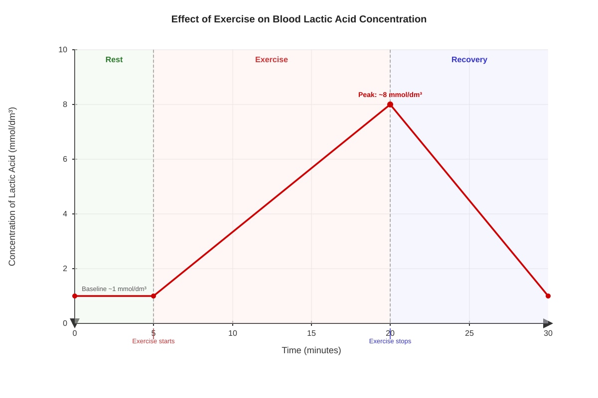
- During intense exercise, the demand for ATP in muscles exceeds the supply of oxygen. (1 mark)
- The muscles switch to anaerobic respiration, where pyruvate is converted to lactate (lactic acid) to regenerate NAD⁺, allowing glycolysis to continue producing a small amount of ATP. (1 mark)
- Teaching Note: Anaerobic respiration is a less efficient way to produce ATP, but it is faster and does not require oxygen. The build-up of lactic acid is a by-product of this process.
(b) Process of lactic acid removal after exercise:
- Marks: 4
- Expected points:
- After exercise, the body continues to breathe heavily (oxygen debt) to provide the oxygen needed to remove the lactic acid. (1 mark)
- Lactic acid is transported from the muscles to the liver via the bloodstream. (1 mark)
- In the liver, lactic acid is converted back to pyruvate. (1 mark)
- The pyruvate is then used in the Krebs cycle (aerobic respiration) to produce ATP, or it is converted back to glucose via gluconeogenesis (the Cori cycle). (1 mark)
- Teaching Note: The "oxygen debt" is the extra oxygen needed after exercise to metabolise the lactic acid that built up during anaerobic respiration. The liver plays a central role in this process.
14. Role of the nervous system in controlling heart rate:
- Marks: 4
- Expected points:
- The heart rate is initiated by the sinoatrial (SA) node, but it is modified by the autonomic nervous system. (1 mark)
- The cardiovascular centre in the medulla oblongata receives input from chemoreceptors (detecting blood pH, CO₂, and O₂ levels) and baroreceptors (detecting blood pressure). (1 mark)
- The sympathetic nervous system releases noradrenaline, which binds to β-adrenergic receptors on the SA node, increasing heart rate (e.g., during exercise or stress). (1 mark)
- The parasympathetic nervous system (via the vagus nerve) releases acetylcholine, which binds to muscarinic receptors on the SA node, decreasing heart rate (e.g., during rest). (1 mark)
- Teaching Note: The SA node is the natural pacemaker, but its rate is constantly adjusted by the brain to meet the body's needs. The sympathetic system is the "accelerator," and the parasympathetic system is the "brake."
15. Adaptations of red blood cells for oxygen transport:
- Marks: 3
- Expected points:
- Biconcave disc shape: This increases the surface area to volume ratio, allowing for faster diffusion of oxygen into and out of the cell. (1 mark)
- No nucleus or organelles: This maximises the space available for haemoglobin, the oxygen-carrying protein. (1 mark)
- Contains haemoglobin: Haemoglobin binds reversibly to oxygen, allowing the cell to transport a large amount of oxygen. (1 mark)
- Teaching Note: Every feature of a red blood cell is optimised for its single purpose: carrying oxygen. The shape, the lack of a nucleus, and the high concentration of haemoglobin all contribute to this.
16. Process of blood clotting:
- Marks: 4
- Expected points:
- Vasoconstriction: Damaged blood vessels constrict to reduce blood flow. (1 mark)
- Platelet plug formation: Platelets adhere to the exposed collagen at the wound site and become activated, releasing chemicals that attract more platelets, forming a temporary plug. (1 mark)
- Coagulation cascade: A series of reactions involving clotting factors (e.g., thromboplastin) converts prothrombin (an inactive plasma protein) into thrombin (an active enzyme). (1 mark)
- Fibrin formation: Thrombin converts soluble fibrinogen into insoluble fibrin. Fibrin threads form a mesh that traps red blood cells and platelets, forming a stable clot. (1 mark)
- Teaching Note: Blood clotting is a complex cascade that must be carefully regulated. The final step is the formation of a fibrin mesh that stabilises the platelet plug and prevents further blood loss.
17. Role of the pancreas in regulating blood glucose concentration:
- Marks: 5
- Expected points:
- The pancreas contains clusters of cells called islets of Langerhans, which contain alpha (α) cells and beta (β) cells. (1 mark)
- When blood glucose is high (e.g., after a meal): β-cells detect the rise and secrete insulin into the blood. Insulin stimulates the uptake of glucose by body cells (especially muscle and liver cells), promotes the conversion of glucose to glycogen (glycogenesis) in the liver, and inhibits gluconeogenesis. This lowers blood glucose. (2 marks)
- When blood glucose is low (e.g., between meals): α-cells detect the fall and secrete glucagon into the blood. Glucagon stimulates the breakdown of glycogen to glucose (glycogenolysis) in the liver and promotes gluconeogenesis. This raises blood glucose. (2 marks)
- Teaching Note: Insulin and glucagon are antagonistic hormones that work together to maintain blood glucose homeostasis. The pancreas acts as the sensor and the effector, detecting changes and releasing the appropriate hormone.
18. Structure of a synapse and impulse transmission:
- Marks: 5
- Expected points:
- Structure: A synapse is a junction between two neurones (or a neurone and an effector). It consists of a presynaptic knob (containing synaptic vesicles with neurotransmitter), a synaptic cleft (a narrow gap), and a postsynaptic membrane (with receptors). (2 marks)
- Transmission:
- An action potential arrives at the presynaptic knob, causing voltage-gated calcium channels to open. (1 mark)
- Calcium ions enter the knob, causing synaptic vesicles to fuse with the presynaptic membrane and release neurotransmitter (e.g., acetylcholine) into the synaptic cleft by exocytosis. (1 mark)
- The neurotransmitter diffuses across the cleft and binds to specific receptors on the postsynaptic membrane, causing ion channels to open and generating a new action potential (or an excitatory/inhibitory postsynaptic potential). (1 mark)
- Note: The neurotransmitter is then broken down by enzymes (e.g., acetylcholinesterase) to prevent continuous stimulation.
- Teaching Note: The synapse is a one-way junction. The electrical signal (action potential) is converted into a chemical signal (neurotransmitter) and then back into an electrical signal. This ensures the impulse travels in only one direction.
19. (a) Chamber with the thickest wall:
- Marks: 1
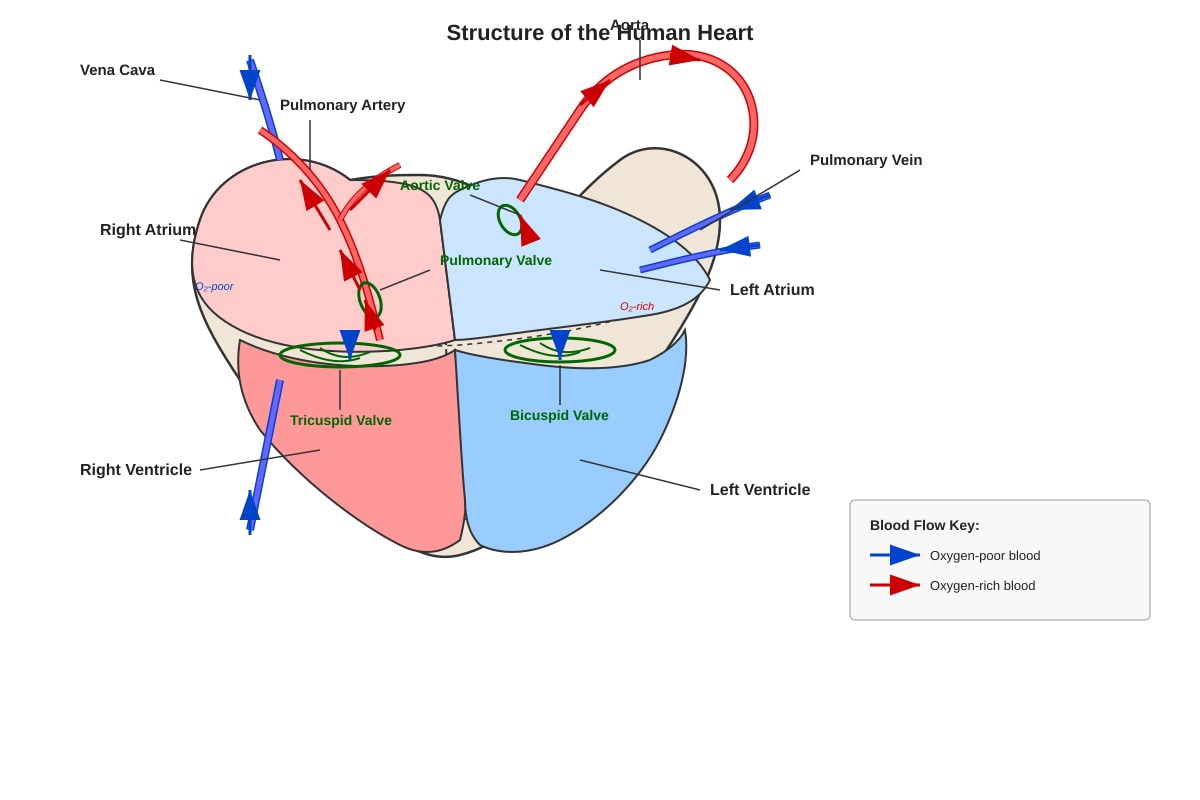
- Answer: The left ventricle.
(b) Explanation:
- Marks: 2
- Expected points:
- The left ventricle pumps blood to the entire body (systemic circulation), which requires it to generate a very high pressure to overcome the resistance of the systemic arteries. (1 mark)
- The thick muscular wall allows it to contract forcefully and generate this high pressure. (1 mark)
- Teaching Note: The right ventricle only pumps blood to the lungs (pulmonary circulation), which is a much shorter, lower-pressure circuit. Therefore, its wall is thinner.
20. Role of the liver in amino acid metabolism:
- Marks: 3
- Expected points:
- Deamination: The liver removes the amino group (-NH₂) from excess amino acids, producing ammonia (NH₃), which is toxic. (1 mark)
- Urea formation: The liver converts the toxic ammonia into urea (via the ornithine cycle), which is less toxic and can be safely transported in the blood to the kidneys for excretion. (1 mark)
- Transamination: The liver can also transfer amino groups from one amino acid to a keto acid to form a different amino acid, allowing the synthesis of non-essential amino acids. (1 mark)
- Teaching Note: The liver is the central hub for amino acid metabolism. It breaks down excess amino acids, detoxifies the ammonia produced, and can also synthesise new amino acids as needed.