AI Generated Quiz
A Level H1 Biology Human Physiology Quiz
Free A Level H1 Biology Human Physiology quiz, AI version, with questions, answers, and A Level-style practice for Singapore students.
These static practice materials are generated from the site's syllabus and paper-generation workflow, with source and model context shown so students and parents can evaluate the material before use.
Questions



Free quiz and exam paper access
Enter your details to view this paper
Your access is remembered on this device.
Answers
A-Level Biology H1 Quiz - Human Physiology: Answer Key
Total Marks: 50
Section A: Short Answer Questions (Questions 1–5, 15 marks)
Question 1 (3 marks)
Answer:
- The sinoatrial node is located in the wall of the right atrium, near the opening of the superior vena cava. (1 mark)
- It acts as the heart's natural pacemaker. (1 mark)
- It initiates each heartbeat by generating electrical impulses that spread across the atria, causing them to contract (atrial systole). (1 mark)
Teaching Notes:
- The sinoatrial (SA) node is a specialised group of cardiac muscle cells. It spontaneously depolarises and generates an action potential, which is why it is called the pacemaker.
- The impulse spreads through the atrial muscle via gap junctions, causing both atria to contract simultaneously.
- The impulse then reaches the atrioventricular (AV) node, which delays it slightly before passing it to the ventricles via the bundle of His and Purkinje fibres.
Common Mistakes:
- Confusing the SA node with the AV node. The SA node is in the right atrium; the AV node is at the junction between the atria and ventricles.
- Stating that the SA node causes ventricular contraction directly. It initiates atrial contraction; ventricular contraction is delayed and initiated via the AV node.
Question 2 (3 marks)
(a) (1 mark)
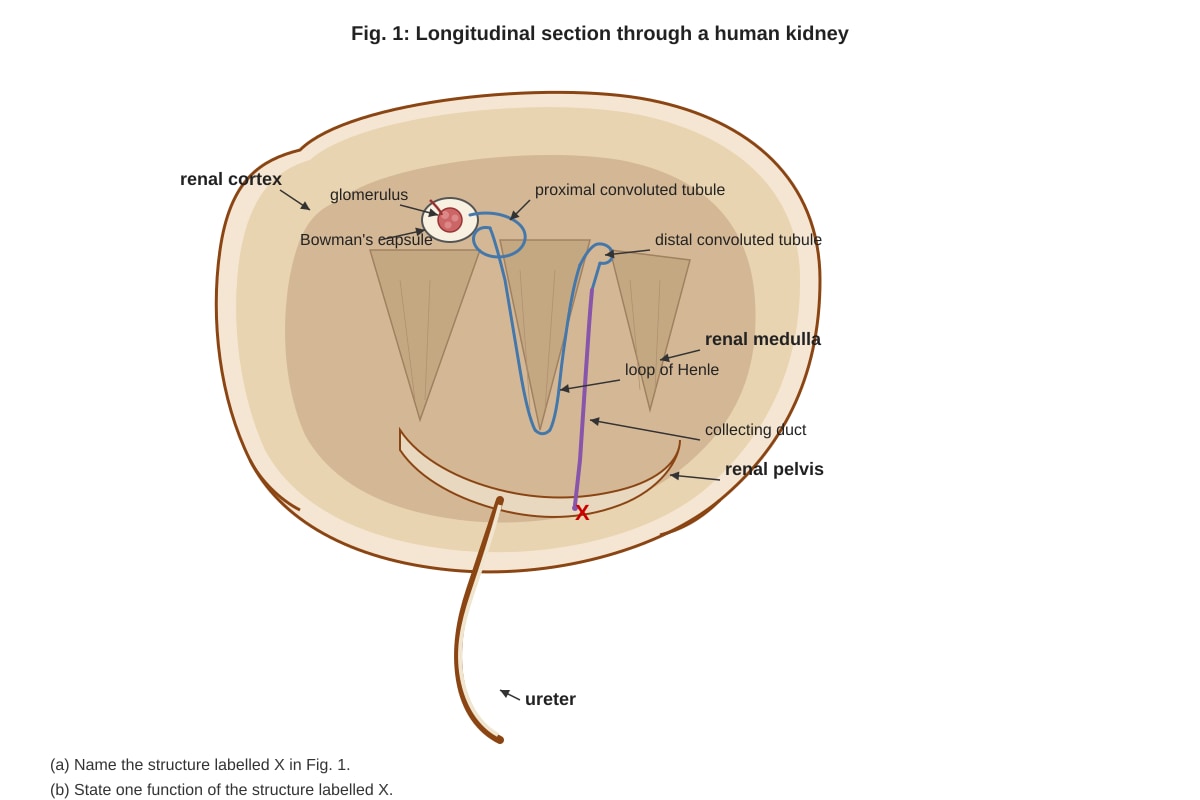
Answer: The structure labelled X is the ureter.
(b) (1 mark)
Answer: The ureter transports urine from the kidney to the urinary bladder (by peristalsis).
Teaching Notes:
- The ureter is a muscular tube that connects each kidney to the bladder.
- Urine is produced in the kidneys and flows down the ureters due to gravity and peristaltic contractions of the smooth muscle in the ureter walls.
- The ureter is not the same as the urethra, which carries urine from the bladder to the outside of the body.
Common Mistakes:
- Confusing the ureter with the urethra or the renal pelvis. The renal pelvis is the funnel-shaped structure inside the kidney that collects urine before it enters the ureter.
Question 3 (3 marks)
Answer:
- The alveoli have a very large total surface area, providing a large area for gas exchange. (1 mark)
- The alveolar walls are extremely thin (one cell thick, squamous epithelium), providing a short diffusion distance for gases. (1 mark)
- The alveoli are surrounded by a dense network of capillaries, maintaining a steep concentration gradient for oxygen and carbon dioxide. (1 mark)
Teaching Notes:
- Gas exchange occurs by simple diffusion. The rate of diffusion is described by Fick's law: rate ∝ (surface area × concentration gradient) / diffusion distance.
- The large surface area of the millions of alveoli maximises the area for diffusion.
- The thin walls (alveolar epithelium and capillary endothelium are each one cell thick) minimise the diffusion distance.
- The continuous blood flow through the capillaries maintains a steep concentration gradient: oxygen is constantly removed (carried away by haemoglobin) and carbon dioxide is constantly delivered.
- Other adaptations include the moist lining of the alveoli (allowing gases to dissolve) and the presence of surfactant (reducing surface tension and preventing collapse).
Common Mistakes:
- Listing adaptations without explaining how they relate to the efficiency of gas exchange.
- Forgetting to mention the concentration gradient maintained by blood flow.
- Confusing the direction of gas movement.
Question 4 (3 marks)
(a) (1 mark)
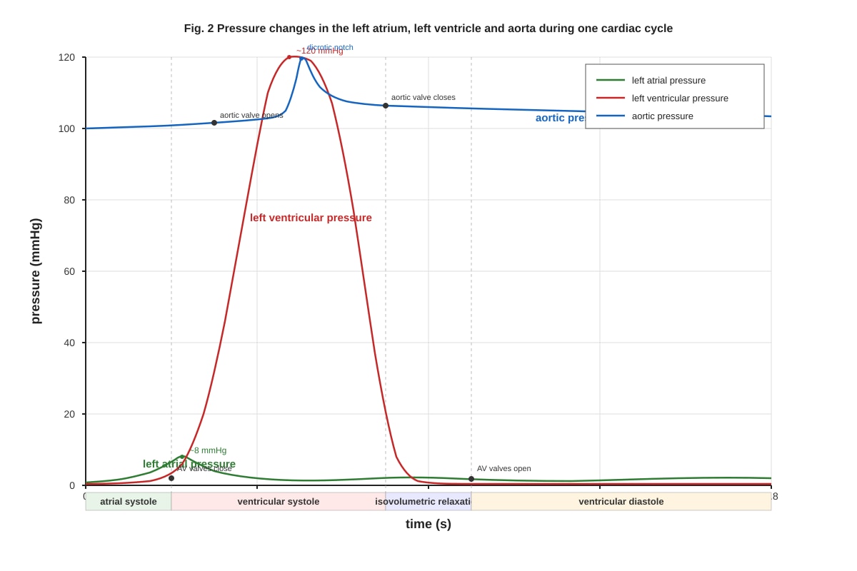
Answer: The atrioventricular valves close at the beginning of ventricular systole.
(b) (2 marks)
Answer:
- During ventricular systole, the ventricles contract, and the pressure inside the ventricles rises rapidly. (1 mark)
- When the ventricular pressure exceeds the atrial pressure, the higher pressure in the ventricles forces the atrioventricular valves (bicuspid and tricuspid valves) shut, preventing backflow of blood into the atria. (1 mark)
Teaching Notes:
- The atrioventricular (AV) valves are the bicuspid (mitral) valve on the left side and the tricuspid valve on the right side.
- They are one-way valves that prevent blood from flowing back into the atria when the ventricles contract.
- The closure of the AV valves produces the first heart sound ("lub").
- The semilunar valves (aortic and pulmonary) close at the beginning of ventricular diastole, producing the second heart sound ("dub").
Common Mistakes:
- Confusing the timing of AV valve closure with semilunar valve closure.
- Stating that the valves close due to muscle contraction rather than pressure differences.
Question 5 (4 marks)
Answer:
- The ascending limb of the loop of Henle actively transports Na⁺ (and Cl⁻) ions out of the filtrate into the medulla, making the medulla hypertonic. (1 mark)
- The ascending limb is impermeable to water, so water cannot follow the ions, and the filtrate becomes more dilute as it ascends. (1 mark)
- The descending limb is permeable to water but impermeable to ions. As the filtrate descends, water moves out by osmosis into the hypertonic medulla, concentrating the filtrate. (1 mark)
- This creates a counter-current multiplier: the flow of filtrate in opposite directions in the two limbs, combined with the active transport of ions and the differential permeability of the limbs, establishes and maintains a concentration gradient in the medulla, with the deepest part of the medulla being most concentrated. (1 mark)
Teaching Notes:
- The loop of Henle is a hairpin-shaped tubule with a descending limb and an ascending limb.
- The counter-current multiplier works because the two limbs are in close proximity and carry fluid in opposite directions.
- The active transport of Na⁺ out of the ascending limb raises the osmolarity of the medulla.
- The descending limb loses water by osmosis, concentrating the filtrate.
- The ascending limb gains ions (from the medulla) and loses water (it is impermeable to water), diluting the filtrate.
- The result is that the medulla has a high solute concentration, which is essential for the kidney's ability to produce concentrated urine via the collecting duct.
Common Mistakes:
- Confusing the permeability of the descending and ascending limbs.
- Forgetting to mention that the ascending limb actively transports ions.
- Not explaining how the counter-current arrangement multiplies the concentration effect.
Section B: Data-Based and Structured Questions (Questions 6–10, 20 marks)
Question 6 (5 marks)
(a) (1 mark)
Answer: The concentration is approximately 0.35 mol dm⁻³ (between 0.2 and 0.4 mol dm⁻³).
Teaching Notes:
- An isotonic solution is one where there is no net movement of water, so the percentage change in mass is zero.
- From the data, the change in mass is +6.0% at 0.2 mol dm⁻³ and -1.5% at 0.4 mol dm⁻³. The zero-change point lies between these two concentrations.
- Interpolating linearly: 0.2 + (6.0 / (6.0 + 1.5)) × 0.2 = 0.2 + 0.16 = 0.36 mol dm⁻³. Accept any value between 0.3 and 0.4 mol dm⁻³.
(b) (2 marks)
Answer:
- The distilled water (0.0 mol dm⁻³) has a lower solute concentration (higher water potential) than the potato cell sap. (1 mark)
- Water moves by osmosis from the distilled water into the potato cells, causing the cells to become turgid and the cylinders to gain mass. (1 mark)
Teaching Notes:
- Osmosis is the net movement of water molecules from a region of higher water potential to a region of lower water potential, across a partially permeable membrane.
- The cell surface membrane of the potato cells is partially permeable.
- In distilled water, the water potential outside the cells is higher (0 kPa) than inside the cells (negative), so water enters the cells.
- As water enters, the cells swell and become turgid, increasing the mass of the potato cylinders.
(c) (2 marks)
Answer:
- The percentage change in mass would become more negative (e.g., approximately -25% or lower). (1 mark)
- This is because the water potential gradient between the cell sap and the 1.0 mol dm⁻³ solution would remain, so water would continue to move out of the cells by osmosis. However, as the cells lose water, the water potential of the cell sap decreases, reducing the gradient over time. The rate of water loss would slow, but the total loss would be greater than at 30 minutes. (1 mark)
Teaching Notes:
- The potato cells are in a hypertonic solution (1.0 mol dm⁻³), so water moves out of the cells by osmosis.
- Over time, the cells lose more water, but the water potential gradient decreases as the cell sap becomes more concentrated.
- The mass loss would not be exactly double because the rate of osmosis decreases as the gradient decreases.
- Accept any reasonable prediction that shows a greater loss of mass than at 30 minutes, with an appropriate explanation.
Common Mistakes:
- Predicting exactly double the mass loss without considering the decreasing gradient.
- Not explaining the mechanism of osmosis.
Question 7 (5 marks)
(a) (1 mark)
Answer: Ultrafiltration (or glomerular filtration).
(b) (2 marks)
Answer: Any two of the following: glucose, amino acids, vitamins, ions (e.g., Na⁺, K⁺, Cl⁻), urea (if present in small amounts). (1 mark each, max 2)
Teaching Notes:
- Glomerular filtrate is similar to blood plasma but without proteins and blood cells.
- Glucose and amino acids are normally completely reabsorbed in the proximal convoluted tubule.
- Some ions are reabsorbed, but the amount excreted depends on the body's needs.
- Urea is present in urine but is also present in filtrate; the question asks for substances "normally absent from urine," so urea is not a correct answer.
(c) (2 marks)
Answer:
- The cells have microvilli on their apical surface, increasing the surface area for reabsorption. (1 mark)
- They are rich in mitochondria, providing ATP for active transport of substances such as glucose and amino acids. (1 mark)
Teaching Notes:
- The proximal convoluted tubule (PCT) is the main site of reabsorption.
- Microvilli increase the surface area for transport proteins and channels.
- The cells have many mitochondria to supply ATP for active transport and for the Na⁺/K⁺ pump, which maintains the Na⁺ gradient that drives co-transport of glucose and amino acids.
- The cells also have many transport proteins in their membranes.
Common Mistakes:
- Mentioning adaptations that are not specific to the PCT (e.g., "thin walls" is more relevant to the loop of Henle).
- Not linking the adaptation to the function (e.g., "mitochondria provide energy" without specifying for active transport).
Question 8 (4 marks)
(a) (1 mark)
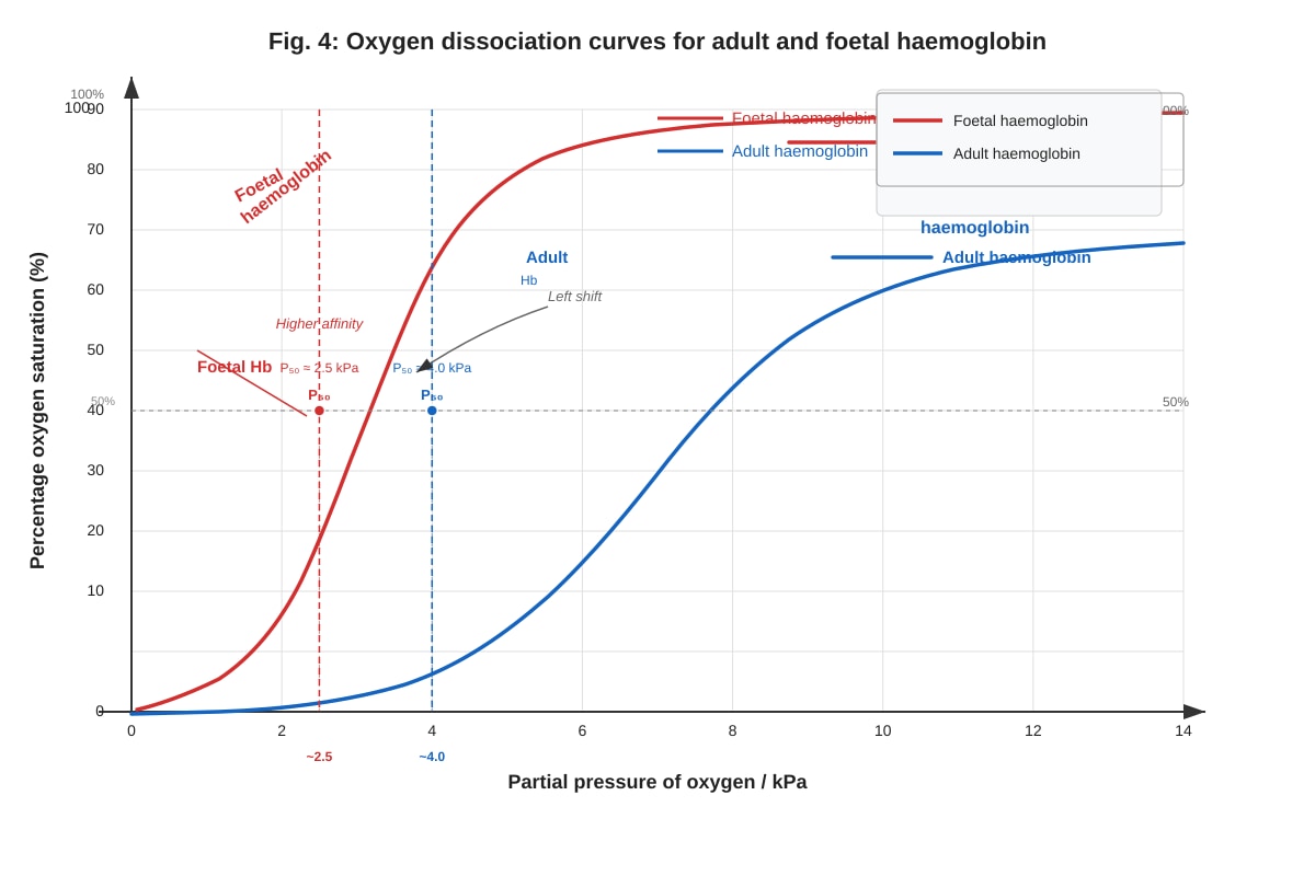
Answer: The P50 value for foetal haemoglobin is approximately 2.5 kPa.
(b) (3 marks)
Answer:
- The P50 value for foetal haemoglobin is lower than that for adult haemoglobin, meaning foetal haemoglobin has a higher affinity for oxygen. (1 mark)
- This allows foetal haemoglobin to take up oxygen from the maternal blood in the placenta, where the partial pressure of oxygen is relatively low. (1 mark)
- This ensures that the foetus receives an adequate supply of oxygen for its metabolic needs. (1 mark)
Teaching Notes:
- P50 is the partial pressure of oxygen at which haemoglobin is 50% saturated. A lower P50 indicates a higher affinity for oxygen.
- Foetal haemoglobin has a different structure (two gamma chains instead of two beta chains), which gives it a higher affinity for oxygen.
- In the placenta, maternal blood has a PO2 of about 4-5 kPa, and foetal blood has a PO2 of about 2-3 kPa. The higher affinity of foetal haemoglobin allows it to load oxygen from the maternal blood.
- This is an example of an adaptation for oxygen transfer across the placenta.
Common Mistakes:
- Confusing higher affinity with lower affinity.
- Not explaining the significance in terms of oxygen transfer in the placenta.
Question 9 (6 marks)
(a) Insulin (3 marks)
Answer:
- Insulin is secreted by the beta cells of the islets of Langerhans in the pancreas in response to high blood glucose concentration. (1 mark)
- It promotes the uptake of glucose by cells, particularly muscle and adipose tissue, by increasing the number of glucose transporter proteins (GLUT4) in the cell membranes. (1 mark)
- It stimulates the conversion of glucose to glycogen (glycogenesis) in the liver and muscles, and promotes the conversion of glucose to fat in adipose tissue, thereby lowering blood glucose concentration. (1 mark)
Teaching Notes:
- Insulin is a peptide hormone that binds to receptors on target cells.
- It has a rapid effect on glucose uptake and a longer-term effect on gene expression.
- Insulin also inhibits gluconeogenesis (the production of glucose from non-carbohydrate sources) in the liver.
(b) Glucagon (3 marks)
Answer:
- Glucagon is secreted by the alpha cells of the islets of Langerhans in the pancreas in response to low blood glucose concentration. (1 mark)
- It stimulates the breakdown of glycogen to glucose (glycogenolysis) in the liver. (1 mark)
- It promotes gluconeogenesis (the synthesis of glucose from amino acids and glycerol) in the liver, thereby raising blood glucose concentration. (1 mark)
Teaching Notes:
- Glucagon is a peptide hormone that acts mainly on the liver.
- It activates enzymes involved in glycogenolysis and gluconeogenesis.
- It also stimulates the release of amino acids from muscle and glycerol from adipose tissue, which are used as substrates for gluconeogenesis.
Common Mistakes:
- Confusing the actions of insulin and glucagon.
- Not mentioning the specific target organs (liver, muscle, adipose tissue).
- Not mentioning the specific processes (glycogenesis, glycogenolysis, gluconeogenesis).
Question 10 (5 marks)
(a) (1 mark)
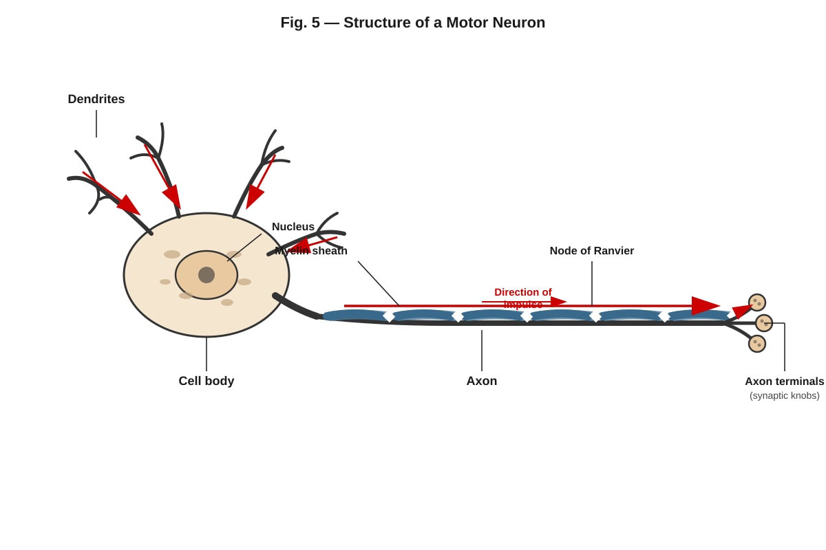
Answer: Dendrites (or the cell body).
(b) (1 mark)
Answer: The myelin sheath insulates the axon and speeds up the transmission of nerve impulses (saltatory conduction).
(c) (3 marks)
Answer:
- An impulse is transmitted as a wave of depolarisation along the axon. (1 mark)
- In myelinated neurons, the myelin sheath acts as an insulator, preventing ion movement across the membrane except at the nodes of Ranvier. (1 mark)
- This causes the impulse to "jump" from node to node (saltatory conduction), which is faster than continuous conduction in unmyelinated neurons. (1 mark)
Teaching Notes:
- The resting potential is maintained by the Na⁺/K⁺ pump and the differential permeability of the membrane.
- When an impulse arrives, voltage-gated Na⁺ channels open, causing depolarisation.
- The depolarisation spreads along the axon, but in myelinated neurons, it can only occur at the nodes of Ranvier, where the myelin sheath is absent.
- This saltatory conduction is faster and more energy-efficient.
Common Mistakes:
- Stating that the myelin sheath speeds up the impulse but not explaining the mechanism (saltatory conduction).
- Confusing the roles of dendrites and axons.
Section C: Extended Response Questions (Questions 11–15, 15 marks)
Question 11 (4 marks)
Answer:
- The heart has four chambers: two atria (left and right) and two ventricles (left and right). The atria receive blood from the veins and pump it into the ventricles; the ventricles pump blood out of the heart. (1 mark)
- The right atrium receives deoxygenated blood from the vena cava, and the right ventricle pumps it to the lungs via the pulmonary artery. The left atrium receives oxygenated blood from the pulmonary veins, and the left ventricle pumps it to the body via the aorta. (1 mark)
- The atrioventricular valves (tricuspid on the right, bicuspid/mitral on the left) prevent backflow of blood from the ventricles to the atria. The semilunar valves (pulmonary and aortic) prevent backflow from the arteries to the ventricles. (1 mark)
- The major blood vessels are the vena cava (returns deoxygenated blood from the body), pulmonary artery (carries deoxygenated blood to the lungs), pulmonary veins (carry oxygenated blood from the lungs), and aorta (carries oxygenated blood to the body). (1 mark)
Teaching Notes:
- The heart is a double pump: the right side pumps blood to the lungs (pulmonary circulation), and the left side pumps blood to the rest of the body (systemic circulation).
- The left ventricle has a thicker muscular wall than the right ventricle because it needs to pump blood at higher pressure to the entire body.
- The valves ensure one-way flow of blood.
Common Mistakes:
- Confusing the functions of the left and right sides of the heart.
- Forgetting to mention the valves or their functions.
- Not naming the major blood vessels correctly.
Question 12 (4 marks)
Answer:
- When the water potential of the blood decreases (e.g., due to dehydration), osmoreceptors in the hypothalamus detect the change. (1 mark)
- The hypothalamus sends nerve impulses to the posterior pituitary gland, which releases antidiuretic hormone (ADH) into the blood. (1 mark)
- ADH travels to the kidneys and increases the permeability of the collecting ducts (and distal convoluted tubules) to water by inserting aquaporins into the cell membranes. (1 mark)
- This allows more water to be reabsorbed from the filtrate back into the blood, producing a smaller volume of more concentrated urine and restoring the water potential of the blood to normal. (1 mark)
Teaching Notes:
- This is an example of negative feedback.
- When the water potential of the blood is too high, less ADH is released, and more water is excreted in the urine.
- The hypothalamus also triggers the thirst sensation, encouraging water intake.
Common Mistakes:
- Confusing the roles of the hypothalamus and the pituitary gland.
- Not mentioning aquaporins or the mechanism of ADH action.
- Not explaining the negative feedback aspect.
Question 13 (4 marks)
Answer:
- Ventilation (breathing) brings air into the lungs (inspiration) and removes air from the lungs (expiration). Inspiration is brought about by the contraction of the diaphragm and external intercostal muscles, increasing the volume of the thoracic cavity and decreasing the pressure, causing air to enter. Expiration is largely passive, with relaxation of these muscles decreasing the volume and increasing the pressure, forcing air out. (1 mark)
- Gas exchange occurs by diffusion across the alveolar-capillary membrane. Oxygen diffuses from the alveoli (high partial pressure) into the blood (low partial pressure), and carbon dioxide diffuses from the blood (high partial pressure) into the alveoli (low partial pressure). (1 mark)
- Oxygen is transported in the blood mainly bound to haemoglobin in red blood cells, forming oxyhaemoglobin. A small amount is dissolved in plasma. (1 mark)
- Carbon dioxide is transported in the blood in three ways: dissolved in plasma, as bicarbonate ions (the majority), and bound to haemoglobin as carbaminohaemoglobin. (1 mark)
Teaching Notes:
- The concentration gradients for oxygen and carbon dioxide are maintained by ventilation and blood flow.
- The rate of gas exchange is affected by the surface area, diffusion distance, and concentration gradient (Fick's law).
Common Mistakes:
- Confusing the direction of diffusion of oxygen and carbon dioxide.
- Not mentioning the role of haemoglobin in oxygen transport.
- Not mentioning the different forms of carbon dioxide transport.
Question 14 (4 marks)
Answer:
- Homeostasis is the maintenance of a constant internal environment despite external changes. It involves negative feedback mechanisms. (1 mark)
- Blood glucose concentration is controlled by insulin and glucagon. When blood glucose is high, insulin is released, promoting glucose uptake and glycogenesis, lowering blood glucose. When blood glucose is low, glucagon is released, promoting glycogenolysis and gluconeogenesis, raising blood glucose. (1 mark)
- Body temperature is controlled by the hypothalamus. When body temperature rises, the hypothalamus triggers vasodilation of skin arterioles, sweating, and a decrease in metabolic rate, lowering body temperature. When body temperature falls, the hypothalamus triggers vasoconstriction, shivering, and an increase in metabolic rate, raising body temperature. (1 mark)
- These are examples of negative feedback, where the response counteracts the change from the set point, maintaining homeostasis. (1 mark)
Teaching Notes:
- Negative feedback is the key mechanism of homeostasis.
- The set point for blood glucose is about 90 mg/100 cm³, and the set point for body temperature is about 37°C.
- The effectors for blood glucose are the liver, muscles, and adipose tissue. The effectors for temperature are the skin arterioles, sweat glands, and skeletal muscles.
Common Mistakes:
- Not mentioning negative feedback.
- Confusing the roles of insulin and glucagon.
- Not mentioning the role of the hypothalamus in temperature regulation.
Question 15 (4 marks)
Answer:
- A synapse is a junction between two neurons (or between a neuron and a muscle cell). It consists of a presynaptic terminal (containing synaptic vesicles), a synaptic cleft, and a postsynaptic membrane (containing receptors). (1 mark)
- When an impulse arrives at the presynaptic terminal, it causes voltage-gated Ca²⁺ channels to open, and Ca²⁺ ions enter the terminal. (1 mark)
- This causes synaptic vesicles to fuse with the presynaptic membrane and release neurotransmitter molecules (e.g., acetylcholine) into the synaptic cleft by exocytosis. (1 mark)
- The neurotransmitter diffuses across the cleft and binds to specific receptors on the postsynaptic membrane, causing ion channels to open. This generates an excitatory postsynaptic potential (EPSP) or an inhibitory postsynaptic potential (IPSP), which may trigger a new impulse in the postsynaptic neuron. (1 mark)
Teaching Notes:
- The neurotransmitter is then broken down by enzymes (e.g., acetylcholinesterase) to prevent continuous stimulation.
- Synapses allow for unidirectional transmission, integration of signals, and modulation of signals.
Common Mistakes:
- Not mentioning the role of Ca²⁺ ions.
- Confusing the direction of transmission.
- Not mentioning the breakdown of the neurotransmitter.
Section D: Application and Analysis Questions (Questions 16–20, 20 marks)
Question 16 (2 marks)
Answer:
- Cardiac output = heart rate × stroke volume (1 mark)
- Cardiac output = 75 beats min⁻¹ × 70 cm³ = 5250 cm³ min⁻¹ (or 5.25 dm³ min⁻¹) (1 mark)
Teaching Notes:
- Cardiac output is the volume of blood pumped by one ventricle per minute.
- The units should be stated (cm³ min⁻¹ or dm³ min⁻¹).
Common Mistakes:
- Forgetting the formula.
- Not including units.
Question 17 (4 marks)
(a) (1 mark)
Answer: Increasing carbon dioxide concentration decreases the affinity of haemoglobin for oxygen (the curve shifts to the right).
(b) (3 marks)
Answer:
- During exercise, muscles produce more carbon dioxide, which diffuses into the blood. (1 mark)
- The increased CO₂ concentration lowers the pH of the blood (Bohr effect), which decreases the affinity of haemoglobin for oxygen. (1 mark)
- This causes haemoglobin to release more oxygen to the actively respiring muscles, which have a high oxygen demand. (1 mark)
Teaching Notes:
- The Bohr effect describes the effect of CO₂ and H⁺ on the oxygen dissociation curve.
- A rightward shift means that at any given PO₂, haemoglobin is less saturated, so more oxygen is released.
- This is beneficial during exercise because it delivers more oxygen to the tissues that need it most.
Common Mistakes:
- Confusing the direction of the shift (right = decreased affinity, left = increased affinity).
- Not explaining the mechanism (pH change/Bohr effect).
Question 18 (6 marks)
(a) (2 marks)
Answer:
- As the environmental temperature increases from 5°C to 25°C, the rate of oxygen consumption decreases. (1 mark)
- From 25°C to 40°C, the rate of oxygen consumption increases again. (1 mark)
Teaching Notes:
- The relationship is U-shaped, with a minimum at around 25°C.
- This is typical for an endotherm (warm-blooded animal).
(b) (2 marks)
Answer:
- Between 5°C and 25°C, the rate of oxygen consumption decreases because the animal needs less energy to maintain its body temperature. (1 mark)
- At lower temperatures, the temperature gradient between the body and the environment is greater, so more heat is lost, and the animal must increase its metabolic rate to generate more heat. As the temperature rises, less heat is needed, so the metabolic rate decreases. (1 mark)
Teaching Notes:
- The animal is an endotherm, so it maintains a constant
<stage5_quiz_answers_md>
A-Level Biology H1 Quiz - Human Physiology - ANSWER KEY
Total Marks: 50
Section A: Short Answer Questions (Questions 1–5, 15 marks)
1. State the precise location of the sinoatrial node in the mammalian heart and outline its role in the cardiac cycle. [3]
Answer:
- Location: The sinoatrial (SA) node is located in the wall of the right atrium, near the opening of the superior vena cava. [1]
- Role: The SA node acts as the heart's natural pacemaker. It generates spontaneous electrical impulses (action potentials) that spread across the atria, causing them to contract (atrial systole). [1] This impulse then reaches the atrioventricular (AV) node, which delays it before passing it to the ventricles. [1]
2. Fig. 1 shows a section through a human kidney.
(a) Name the structure labelled X in Fig. 1. [1]
Answer: Renal pelvis [1]
(b) State one function of the structure labelled X. [1]
Answer: Collects urine from the collecting ducts / Acts as a funnel to channel urine into the ureter. [1]
3. Describe how the structure of the alveolus is adapted for efficient gas exchange. [3]
Answer:
- Large surface area: The alveoli are numerous (millions) and have thin, folded walls, providing a large surface area for diffusion. [1]
- Thin walls: The alveolar walls are only one cell thick (squamous epithelium), creating a short diffusion distance for gases. [1]
- Rich blood supply: Each alveolus is surrounded by a dense network of capillaries, maintaining a steep concentration gradient for oxygen and carbon dioxide. [1]
- Moist lining: The inner surface is moist, allowing gases to dissolve before diffusing. [1]
- Good ventilation: The lungs are constantly ventilated, bringing in fresh oxygen and removing carbon dioxide, maintaining steep concentration gradients. [1]
(Accept any three of the above for 1 mark each)
4. Fig. 2 shows the pressure changes in the left atrium, left ventricle, and aorta during one cardiac cycle.
(a) State the phase of the cardiac cycle during which the atrioventricular valves close. [1]
Answer: The beginning of ventricular systole (when ventricular pressure exceeds atrial pressure). [1]
(b) Explain why the atrioventricular valves close during this phase. [2]
Answer: During ventricular systole, the ventricles contract, causing the pressure inside them to rise rapidly. [1] When the ventricular pressure exceeds the pressure in the atria, the atrioventricular valves (bicuspid and tricuspid) are forced shut to prevent backflow of blood into the atria. [1]
5. Explain how the counter-current multiplier mechanism in the loop of Henle establishes a concentration gradient in the medulla of the kidney. [4]
Answer:
- The descending limb is permeable to water but not to salts. As filtrate descends, water moves out by osmosis into the increasingly concentrated medulla. [1]
- The ascending limb is impermeable to water but actively transports sodium ions (Na⁺) and chloride ions (Cl⁻) out into the medulla. [1]
- The thin ascending limb is permeable to salts, which diffuse out. The thick ascending limb actively pumps out salts, making the medulla very concentrated. [1]
- The flow of filtrate in opposite directions (descending and ascending) creates a counter-current multiplier. The constant pumping of salts out of the ascending limb and the loss of water from the descending limb multiply the concentration gradient, making the medulla progressively more concentrated towards the tip. [1]
Section B: Data-Based and Structured Questions (Questions 6–10, 20 marks)
6. A student investigated the effect of different concentrations of a solution on the rate of osmosis in potato tissue.
(a) State the concentration of the solution that is approximately isotonic to the potato cell sap. [1]
Answer: Approximately 0.4 mol dm⁻³ (where there is no net change in mass, or the change is close to zero). [1]
(b) Explain why the potato cylinders gained mass in the 0.0 mol dm⁻³ solution. [2]
Answer: The 0.0 mol dm⁻³ solution (distilled water) has a higher water potential than the potato cell sap. [1] Water enters the potato cells by osmosis, causing them to swell and increase in mass. [1]
(c) Predict the percentage change in mass of the potato cylinders if they were left in the 1.0 mol dm⁻³ solution for 60 minutes instead of 30 minutes. Explain your prediction. [2]
Answer: The percentage change in mass would be more negative (e.g., -25% or lower). [1] This is because the potato cylinders would continue to lose water by osmosis for a longer period, until the water potential inside the cells reaches equilibrium with the external solution, or until the cells are fully plasmolysed. [1]
7. Fig. 3 shows the structure of a nephron and its associated blood supply.
(a) Name the process by which fluid is filtered from the glomerulus into Bowman's capsule. [1]
Answer: Ultrafiltration [1]
(b) State two substances that are present in the glomerular filtrate but are normally absent from urine. [2]
Answer: Glucose and amino acids (or any other small, useful substances that are reabsorbed, such as vitamins or some ions). [2]
(c) Explain how the cells lining the proximal convoluted tubule are adapted for reabsorption. [2]
Answer:
- Microvilli: The cells have a brush border of microvilli on their apical surface, which greatly increases the surface area for reabsorption. [1]
- Many mitochondria: The cells contain numerous mitochondria to provide ATP for active transport of substances (e.g., glucose, amino acids, ions) from the filtrate into the blood. [1]
- Tight junctions: The cells are connected by tight junctions, preventing leakage of reabsorbed substances back into the tubule lumen. [1]
(Accept any two of the above for 1 mark each)
8. Fig. 4 shows the oxygen dissociation curves for adult haemoglobin and foetal haemoglobin.
(a) State the P50 value for foetal haemoglobin. [1]
Answer: Approximately 2.5 kPa [1]
(b) Explain the significance of the difference in P50 values between adult and foetal haemoglobin. [3]
Answer:
- Foetal haemoglobin has a higher affinity for oxygen than adult haemoglobin (its curve is to the left). [1]
- This means that at the same partial pressure of oxygen, foetal haemoglobin will be more saturated with oxygen than adult haemoglobin. [1]
- This allows the foetus to efficiently extract oxygen from the mother's blood across the placenta, where the partial pressure of oxygen is relatively low. [1]
9. Describe the roles of the following hormones in the control of blood glucose concentration:
(a) Insulin [3]
Answer:
- Insulin is secreted by the beta cells of the islets of Langerhans in the pancreas when blood glucose concentration is high. [1]
- It stimulates the uptake of glucose by body cells (especially muscle and liver cells) by increasing the number of glucose transporter proteins (GLUT4) in their cell membranes. [1]
- It also promotes the conversion of glucose to glycogen (glycogenesis) in the liver and muscles, and inhibits the breakdown of glycogen and gluconeogenesis. [1]
(b) Glucagon [3]
Answer:
- Glucagon is secreted by the alpha cells of the islets of Langerhans in the pancreas when blood glucose concentration is low. [1]
- It stimulates the breakdown of glycogen to glucose (glycogenolysis) in the liver. [1]
- It also promotes the production of glucose from non-carbohydrate sources (gluconeogenesis) in the liver. [1]
10. Fig. 5 shows the structure of a motor neuron.
(a) Name the part of the neuron that receives impulses from other neurons. [1]
Answer: Dendrites [1]
(b) State the function of the myelin sheath. [1]
Answer: To insulate the axon and speed up the transmission of nerve impulses (saltatory conduction). [1]
(c) Explain how an impulse is transmitted along a myelinated neuron. [3]
Answer:
- The myelin sheath acts as an electrical insulator, preventing the flow of ions across the axon membrane in the myelinated regions. [1]
- Depolarisation (the action potential) can only occur at the nodes of Ranvier, where the axon membrane is exposed. [1]
- The local currents generated by the action potential at one node flow to the next node, triggering depolarisation there. This is called saltatory conduction, and it allows the impulse to "jump" from node to node, which is much faster than continuous conduction along an unmyelinated axon. [1]
Section C: Extended Response Questions (Questions 11–15, 15 marks)
11. Describe the structure and function of the human heart, including the roles of the four chambers, the valves, and the major blood vessels. [4]
Answer:
- Four chambers: The heart has four chambers: the right atrium (receives deoxygenated blood from the body), the right ventricle (pumps deoxygenated blood to the lungs), the left atrium (receives oxygenated blood from the lungs), and the left ventricle (pumps oxygenated blood to the body). [1]
- Valves: The atrioventricular valves (tricuspid on the right, bicuspid/mitral on the left) prevent backflow of blood into the atria during ventricular systole. The semilunar valves (pulmonary and aortic) prevent backflow of blood into the ventricles during diastole. [1]
- Major blood vessels: The vena cava brings deoxygenated blood to the right atrium. The pulmonary artery carries deoxygenated blood from the right ventricle to the lungs. The pulmonary veins bring oxygenated blood from the lungs to the left atrium. The aorta carries oxygenated blood from the left ventricle to the body. [1]
- Overall function: The heart acts as a double pump, ensuring that deoxygenated blood is sent to the lungs for oxygenation and that oxygenated blood is circulated to the rest of the body. [1]
12. Explain how the kidney regulates the water potential of the blood, with reference to the roles of the hypothalamus, posterior pituitary gland, and antidiuretic hormone (ADH). [4]
Answer:
- Detection: Osmoreceptors in the hypothalamus detect a decrease in blood water potential (increased solute concentration). [1]
- Response: The hypothalamus sends nerve impulses to the posterior pituitary gland, stimulating it to release more ADH into the bloodstream. [1]
- Action of ADH: ADH increases the permeability of the collecting ducts and distal convoluted tubules to water by inserting aquaporin channels into their cell membranes. [1]
- Effect: More water is reabsorbed from the filtrate back into the blood, producing a smaller volume of more concentrated urine and increasing the water potential of the blood back towards normal. [1]
13. Describe the process of gas exchange in the lungs, including the roles of ventilation, diffusion, and the transport of oxygen and carbon dioxide in the blood. [4]
Answer:
- Ventilation: Breathing (ventilation) moves air in and out of the alveoli, maintaining a high concentration of oxygen and a low concentration of carbon dioxide in the alveolar air. [1]
- Diffusion: Oxygen diffuses down its concentration gradient from the alveoli into the blood capillaries. Carbon dioxide diffuses down its concentration gradient from the blood into the alveoli. [1]
- Oxygen transport: Most oxygen (about 98%) is transported in the blood bound to haemoglobin in red blood cells, forming oxyhaemoglobin. A small amount is dissolved in plasma. [1]
- Carbon dioxide transport: Carbon dioxide is transported in three ways: dissolved in plasma, as bicarbonate ions (HCO₃⁻) in plasma (the majority), and bound to haemoglobin as carbaminohaemoglobin. [1]
14. Explain how the body maintains a constant internal environment (homeostasis) with reference to negative feedback. [4]
Answer:
- Definition: Homeostasis is the maintenance of a stable internal environment despite external changes. [1]
- Negative feedback: This is the primary mechanism of homeostasis. A change in a factor (e.g., temperature, blood glucose) triggers a response that reverses the change, bringing the factor back to its set point. [1]
- Example (temperature): If body temperature rises, receptors in the skin and hypothalamus detect the change. The hypothalamus (coordinator) triggers effectors (e.g., sweat glands, blood vessels) to cause cooling (sweating, vasodilation). This reduces temperature, and the response is switched off. [1]
- Example (blood glucose): If blood glucose rises, the pancreas releases insulin, which lowers blood glucose. If blood glucose falls, the pancreas releases glucagon, which raises blood glucose. [1]
15. Describe the structure and function of a synapse. [3]
Answer:
- Structure: A synapse is a junction between two neurons (or a neuron and an effector). It consists of a presynaptic knob (containing synaptic vesicles), a synaptic cleft (a narrow gap), and a postsynaptic membrane (containing receptor proteins). [1]
- Function: The synapse transmits nerve impulses from one neuron to the next. [1]
- Process: An action potential arrives at the presynaptic knob, causing calcium ions to enter. This triggers the release of neurotransmitter (e.g., acetylcholine) from synaptic vesicles into the synaptic cleft. The neurotransmitter diffuses across the cleft and binds to receptors on the postsynaptic membrane, causing ion channels to open and generating a new action potential in the postsynaptic neuron. [1]