AI Generated Quiz
A Level H1 Biology Cells Biomolecules Quiz
Free A Level H1 Biology Cells Biomolecules quiz, LongCat AI version, with questions, answers, and A Level-style practice for Singapore students.
These static practice materials are generated from the site's syllabus and paper-generation workflow, with source and model context shown so students and parents can evaluate the material before use.
Questions
A-Level Biology H1 Quiz - Cells Biomolecules
Name: ___________________________
Class: ___________________________
Date: ___________________________
Score: ________ / 50
Duration: 60 minutes
Total Marks: 50
Instructions
- Answer ALL questions.
- Write your answers in the spaces provided.
- The number of marks for each question is shown in brackets [ ].
- Where a question refers to a figure, study the figure carefully before answering.
- You may use a calculator where necessary.
- Marks are awarded for clear, well-structured answers that use correct biological terminology.
Section A: Multiple Choice (Questions 1–5) [10 marks]
Each question is worth 2 marks. Choose the one best answer.
1. Which of the following organelles is found in eukaryotic cells but NOT in prokaryotic cells?
A. Ribosomes
B. Cell membrane
C. Golgi apparatus
D. Cytoplasm
Answer: ___________
2. A student tested four unknown food samples with different reagents. The results are shown in the table below.
| Sample | Benedict's test (after heating) | Iodine test | Biuret test | Ethanol emulsion test |
|---|---|---|---|---|
| W | Blue | Brown-yellow | Purple | Cloudy white |
| X | Orange-red | Brown-yellow | Blue | Clear |
| Y | Blue | Blue-black | Blue | Clear |
| Z | Blue | Brown-yellow | Blue | Cloudy white |
Which sample contains protein AND lipid?
A. Sample W
B. Sample X
C. Sample Y
D. Sample Z
Answer: ___________
3. Which property of water is most directly responsible for its ability to act as a transport medium in living organisms?
A. High specific heat capacity
B. Cohesion and adhesion
C. High latent heat of vaporisation
D. Lower density as a solid than as a liquid
Answer: ___________
4. Fig. 1 shows a section through a cell membrane.
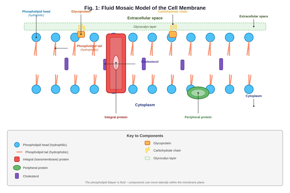
Generated diagram for Q4.
With reference to Fig. 1, which component is responsible for maintaining membrane fluidity at low temperatures?
A. Integral protein
B. Peripheral protein
C. Cholesterol
D. Glycoprotein
Answer: ___________
5. During an experiment, a cell is placed in a solution with a higher solute concentration than the cell's cytoplasm. Which of the following correctly describes what happens?
A. Water moves into the cell by osmosis, causing the cell to swell.
B. Water moves out of the cell by osmosis, causing the cell to shrink.
C. Solute moves into the cell by diffusion, causing the cell to swell.
D. Solute moves out of the cell by active transport, causing the cell to shrink.
Answer: ___________
Section B: Structured Questions (Questions 6–15) [25 marks]
6. State two structural differences between prokaryotic and eukaryotic cells. [2]
7. Fig. 2 shows the structure of a monosaccharide.
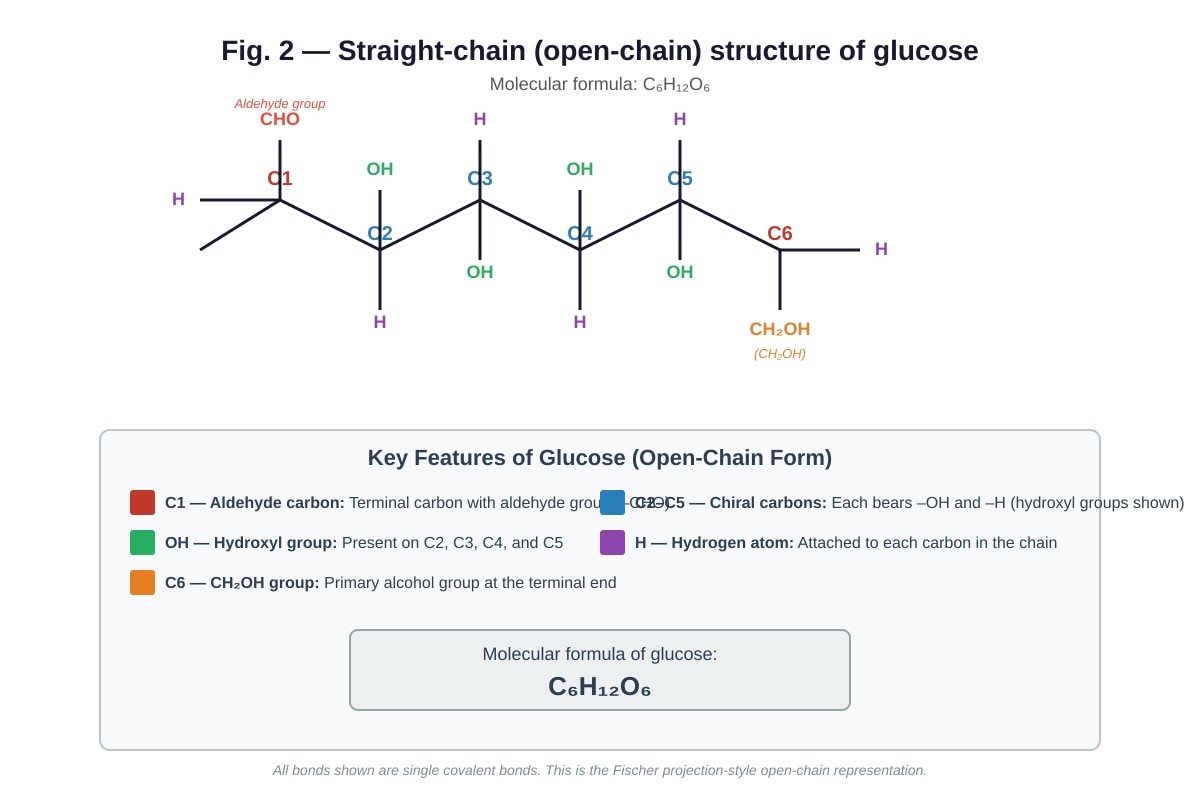
Generated diagram for Q7.
(a) With reference to Fig. 2, name the type of reaction that occurs when two glucose molecules join to form a disaccharide. [1]
(b) State the molecular formula of the disaccharide formed when two glucose molecules react. Show your working. [2]
8. Describe the structure of a triglyceride and explain how its properties make it suitable as an energy storage molecule. [3]
9. Fig. 3 shows the effect of temperature on the rate of an enzyme-catalysed reaction.
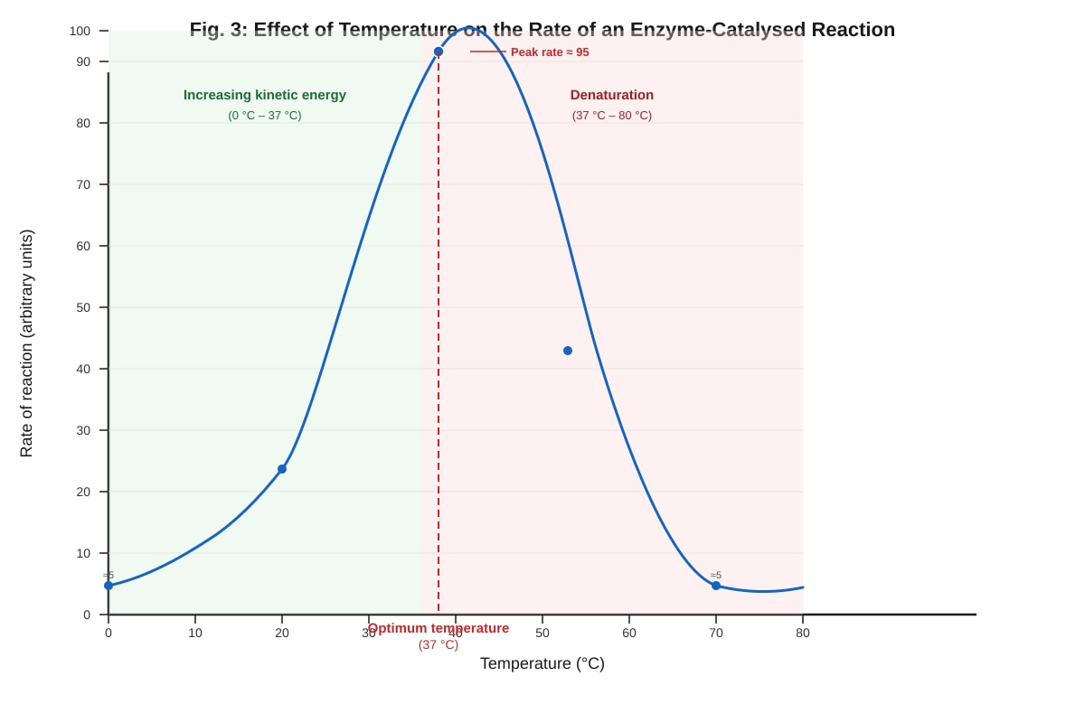
Generated graph for Q9.
(a) With reference to Fig. 3, state the optimum temperature for this enzyme. [1]
(b) Explain the shape of the curve between 0 °C and 37 °C. [2]
(c) Explain the shape of the curve between 37 °C and 80 °C. [2]
10. Explain why the enzyme pepsin, which functions in the stomach at pH 2, would not function effectively in the small intestine at pH 8. [3]
11. Table 1 shows the results of an experiment to investigate the effect of pH on the activity of enzyme X.
| pH | Rate of reaction (μmol min⁻¹) |
|---|---|
| 2 | 5 |
| 4 | 22 |
| 6 | 48 |
| 7 | 55 |
| 8 | 50 |
| 10 | 12 |
| 12 | 2 |
(a) Plot a graph of the data in Table 1 on the grid provided. [3]
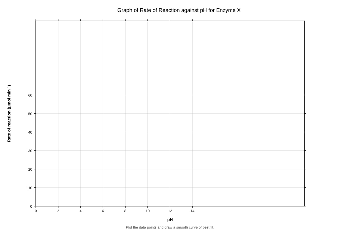
Generated graph for Q11.
(b) From your graph, estimate the optimum pH for enzyme X. [1]
(c) Suggest why enzyme X shows very low activity at pH 2 and pH 12. [2]
12. Describe the process of facilitated diffusion. In your answer, include the role of membrane proteins and the direction of movement relative to the concentration gradient. [3]
13. Fig. 4 shows two molecules that join together in a condensation reaction.
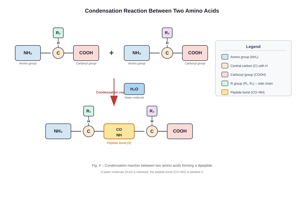
Generated diagram for Q13.
(a) With reference to Fig. 4, name the bond labelled X that forms between the two amino acids. [1]
(b) State the name of the reaction shown in Fig. 4. [1]
(c) How many water molecules are required to completely hydrolyse a polypeptide made of 12 amino acids? Explain your answer. [2]
14. Compare and contrast active transport and osmosis. Include in your answer: the direction of movement relative to the concentration gradient, the requirement for energy, and the role of the cell membrane. [4]
15. A student carried out an experiment to test the effect of enzyme concentration on the rate of reaction. The enzyme catalase was added to hydrogen peroxide and the volume of oxygen gas produced was measured over 5 minutes. The results are shown in Table 2.
| Catalase concentration (% v/v) | Volume of O₂ produced in 5 min (cm³) |
|---|---|
| 0 | 0 |
| 10 | 18 |
| 20 | 34 |
| 30 | 48 |
| 40 | 55 |
| 50 | 55 |
| 60 | 55 |
(a) Describe the trend shown in Table 2. [2]
(b) Explain why the volume of oxygen produced remains constant at catalase concentrations of 40% and above. [2]
(c) State one variable that should be kept constant in this experiment to ensure the results are valid. [1]
Section C: Data-Based and Free Response Questions (Questions 16–20) [15 marks]
16. Fig. 5 shows a transmission electron micrograph (TEM) of a cell.
Image pending generation: figure for Q16.
(a) With reference to Fig. 5, identify the organelle responsible for the following functions. [3]
(i) Synthesis of lipids: _______________________________________________________
(ii) Synthesis of proteins that will be secreted from the cell: _________________________
(iii) Production of ATP during aerobic respiration: _________________________________
(b) State one piece of evidence from Fig. 5 that indicates this is a eukaryotic cell and not a prokaryotic cell. [1]
17. Read the following passage and answer the questions that follow.
The cell surface membrane is described by the fluid mosaic model. The basic structure is a phospholipid bilayer in which the hydrophilic phosphate heads face the aqueous environments on either side of the membrane, while the hydrophobic fatty acid tails face inwards, away from water. Proteins are embedded within or attached to the surface of the bilayer. Some proteins span the entire membrane (integral proteins) while others are found only on one surface (peripheral proteins). Cholesterol molecules are found between the phospholipid tails and help to regulate membrane fluidity. Glycoproteins, which have carbohydrate chains attached, are found on the extracellular surface and play important roles in cell recognition and signalling.
(a) Explain why the phospholipid bilayer arrangement is described as a "fluid" mosaic. [2]
(b) Describe the role of cholesterol in the cell membrane. [2]
(c) Explain the biological significance of glycoproteins on the cell surface. [2]
18. A student investigated the permeability of beetroot cell membranes at different temperatures. Beetroot cylinders were placed in distilled water at various temperatures for 10 minutes. The colour of the solution was then measured using a colorimeter, which recorded the absorbance of light at 540 nm. Higher absorbance indicates more pigment (betacyanin) has leaked from the cells, indicating greater membrane damage.
The results are shown in Table 3.
| Temperature (°C) | Absorbance at 540 nm (arbitrary units) |
|---|---|
| 10 | 0.05 |
| 20 | 0.08 |
| 30 | 0.12 |
| 40 | 0.25 |
| 50 | 0.58 |
| 60 | 0.92 |
| 70 | 0.95 |
(a) Describe the relationship between temperature and membrane permeability as shown in Table 3. [2]
(b) Explain the results obtained at 60 °C and 70 °C. [3]
(c) Suggest why the student used distilled water rather than a sugar solution in this experiment. [1]
19. Fig. 6 shows the structure of a section of DNA.
Image pending generation: diagram for Q19.
(a) With reference to Fig. 6, state the type of bond that holds the two strands of DNA together. [1]
(b) Name the sugar molecule in DNA. [1]
(c) Explain how the structure of DNA enables it to carry genetic information. [3]
20. A student carried out food tests on three different food samples (P, Q, and R). The results are shown in Table 4.
| Test | Sample P | Sample Q | Sample R |
|---|---|---|---|
| Benedict's test (after heating) | Blue | Brick-red precipitate | Blue |
| Iodine test | Blue-black | Brown-yellow | Brown-yellow |
| Biuret test | Blue | Blue | Purple |
| Ethanol emulsion test | Clear | Clear | Cloudy white |
(a) Identify the biological molecule(s) present in each sample. [3]
Sample P: _________________________________________________________________
Sample Q: _________________________________________________________________
Sample R: _________________________________________________________________
(b) Explain why the Benedict's test requires heating in a water bath. [2]
(c) Describe how the Biuret test is carried out. [2]
END OF QUIZ
Answers
A-Level Biology H1 Quiz - Cells Biomolecules
Answer Key
Section A: Multiple Choice [10 marks]
1. C — Golgi apparatus [2]
The Golgi apparatus is a membrane-bound organelle found only in eukaryotic cells. Prokaryotic cells lack membrane-bound organelles. Ribosomes (A) are found in both prokaryotic and eukaryotic cells (though they differ in size: 70S in prokaryotes, 80S in eukaryotes). The cell membrane (B) and cytoplasm (D) are present in both cell types.
Common mistake: Students may select ribosomes because they associate them primarily with eukaryotic cells, but ribosomes are universal to all cells.
2. A — Sample W [2]
- Sample W: Biuret test is purple (positive for protein) AND ethanol emulsion test is cloudy white (positive for lipid). ✓
- Sample X: Benedict's test is orange-red (positive for reducing sugar), Biuret test is blue (negative for protein). ✗
- Sample Y: Iodine test is blue-black (positive for starch), Biuret test is blue (negative for protein). ✗
- Sample Z: Ethanol emulsion test is cloudy white (positive for lipid), but Biuret test is blue (negative for protein). ✗
Teaching note: Students should systematically check each sample against both required criteria (protein AND lipid) rather than looking for just one positive result.
3. B — Cohesion and adhesion [2]
Cohesion (attraction between water molecules) and adhesion (attraction between water molecules and other surfaces) enable water to form continuous columns in blood vessels and xylem, making it an effective transport medium. High specific heat capacity (A) relates to temperature regulation. High latent heat of vaporisation (C) relates to cooling by evaporation. Lower density as ice (D) relates to insulation of aquatic habitats.
Common mistake: Students may select high specific heat capacity because it is a well-known property of water, but it does not directly explain transport function.
4. C — Cholesterol [2]
Cholesterol molecules are positioned between the phospholipid tails in the hydrophobic region of the bilayer. At low temperatures, cholesterol prevents the phospholipid tails from packing too closely together, thereby maintaining membrane fluidity. At high temperatures, cholesterol restricts excessive movement of phospholipids, providing stability. Integral proteins (A) and peripheral proteins (B) are not primarily responsible for regulating fluidity. Glycoproteins (D) are involved in cell recognition.
Expected visual features for Q4-fig1: The diagram must clearly show cholesterol molecules positioned between the phospholipid tails in the interior of the bilayer, distinct from the integral and peripheral proteins.
5. B — Water moves out of the cell by osmosis, causing the cell to shrink [2]
Osmosis is the net movement of water molecules from a region of higher water potential (lower solute concentration) to a region of lower water potential (higher solute concentration) across a selectively permeable membrane. Since the external solution has a higher solute concentration (lower water potential) than the cytoplasm, water moves out of the cell, causing it to shrink (crenation in animal cells, plasmolysis in plant cells).
Common mistake: Students may confuse the direction of water movement with the direction of solute movement. Osmosis specifically refers to water movement, not solute movement.
Section B: Structured Questions [25 marks]
6. [2]
Any two of the following (1 mark each):
- Prokaryotic cells lack a nucleus / have genetic material in the cytoplasm (nucleoid region), whereas eukaryotic cells have a membrane-bound nucleus.
- Prokaryotic cells lack membrane-bound organelles, whereas eukaryotic cells have membrane-bound organelles (e.g., mitochondria, ER, Golgi).
- Prokaryotic cells have 70S ribosomes, whereas eukaryotic cells have 80S ribosomes.
- Prokaryotic cells have a cell wall made of murein (peptidoglycan), whereas eukaryotic plant cells have a cell wall made of cellulose.
- Prokaryotic cells are generally smaller (1–5 μm) than eukaryotic cells (10–100 μm).
Marking note: Answers must compare prokaryotic and eukaryotic cells directly. Stating only one cell type without comparison scores 0.
7. (a) Condensation [1]
A condensation reaction is one in which two molecules join together with the elimination of a water molecule. When two monosaccharides join to form a disaccharide, a glycosidic bond is formed and a molecule of water is released.
Common mistake: Students may write "hydrolysis" — this is the reverse reaction (breaking down using water).
(b) C₁₂H₂₂O₁₁ [2]
Working:
- Glucose molecular formula: C₆H₁₂O₆
- Two glucose molecules: 2 × C₆H₁₂O₆ = C₁₂H₂₄O₁₂
- One water molecule is removed during condensation: C₁₂H₂₄O₁₂ − H₂O = C₁₂H₂₂O₁₁
Marking: 1 mark for correct molecular formula, 1 mark for showing the working (subtraction of H₂O).
Teaching note: This is a common exam question. Students should remember that a condensation reaction removes one H₂O molecule per bond formed.
8. [3]
A triglyceride consists of one glycerol molecule bonded to three fatty acid molecules via ester bonds formed by condensation reactions.
Properties that make triglycerides suitable as energy storage molecules:
- High energy content: The long hydrocarbon chains of fatty acid tails contain many C–H bonds, which release large amounts of energy when oxidised during respiration. Triglycerides store more energy per gram than carbohydrates.
- Insoluble in water: Triglycerides are non-polar and hydrophobic, so they do not affect the water potential of cells. This means they can be stored in large quantities without causing osmotic problems (unlike glucose, which would draw water into cells by osmosis).
- Low density: Lipids are less dense than water, making them lightweight energy reserves — important for mobile organisms.
Marking: 1 mark for correct description of structure (glycerol + 3 fatty acids + ester bonds), 1 mark for high energy content / many C–H bonds, 1 mark for insolubility / no osmotic effect. Maximum 3 marks.
9. (a) 37 °C [1]
The optimum temperature is the temperature at which the rate of reaction is highest, which from Fig. 3 is 37 °C.
(b) [2]
As temperature increases from 0 °C to 37 °C, the kinetic energy of both enzyme and substrate molecules increases. This causes the molecules to move faster, resulting in more frequent successful collisions between the active site of the enzyme and the substrate molecules. The rate of formation of enzyme–substrate complexes increases, so the rate of reaction increases.
Marking: 1 mark for increased kinetic energy / faster molecular movement, 1 mark for more frequent collisions / more enzyme–substrate complexes formed.
(c) [2]
Above 37 °C, the enzyme molecules gain too much kinetic energy, which disrupts the hydrogen bonds and other weak interactions (e.g., ionic bonds, hydrophobic interactions) that maintain the tertiary structure of the enzyme. The active site changes shape (denaturation), so the substrate can no longer fit into it. Enzyme–substrate complexes can no longer form, and the rate of reaction decreases sharply.
Marking: 1 mark for disruption of hydrogen bonds / weak interactions / tertiary structure, 1 mark for active site changes shape / denaturation / substrate no longer fits.
Common mistake: Students may say "the enzyme is killed" — enzymes are not living organisms and cannot be "killed." The correct term is "denatured."
10. [3]
Enzymes have an optimum pH at which their active site has the correct shape and charge to bind the substrate effectively. Pepsin's optimum pH is approximately pH 2, which matches the highly acidic environment of the stomach.
At pH 8 (the pH of the small intestine), the higher concentration of OH⁻ ions alters the ionic charges on the amino acid residues in the active site and throughout the enzyme's tertiary structure. This disrupts the ionic bonds and hydrogen bonds that maintain the three-dimensional shape of the enzyme. As a result, the active site becomes denatured — its shape changes so that the substrate can no longer bind effectively. The enzyme loses its catalytic function.
Additionally, the ionisation state of the substrate molecules may also change at pH 8, further reducing the enzyme's ability to catalyse the reaction.
Marking: 1 mark for reference to optimum pH / pH 2 for pepsin, 1 mark for disruption of bonds (ionic/hydrogen) at pH 8, 1 mark for active site shape change / denaturation / substrate cannot bind.
11. (a) [3]
Marking criteria for graph:
- 1 mark: Correctly labelled axes with units (x-axis: pH, y-axis: Rate of reaction in μmol min⁻¹)
- 1 mark: Appropriate scale used on both axes (data points use at least half the grid)
- 1 mark: All 7 points plotted correctly and a smooth curve drawn through the points
Expected plotted points: (2, 5), (4, 22), (6, 48), (7, 55), (8, 50), (10, 12), (12, 2)
Expected visual features for Q11-fig1: The blank grid must have pH on the x-axis (0–14) and Rate of reaction on the y-axis (0–60), with clear gridlines for accurate plotting.
(b) pH 7 [1]
From the graph, the peak of the curve occurs at pH 7, which is the optimum pH for enzyme X.
(c) [2]
At very low pH (pH 2) and very high pH (pH 12), the enzyme is denatured. The extreme H⁺ or OH⁻ ion concentrations disrupt the ionic bonds and hydrogen bonds that maintain the enzyme's tertiary structure. The active site changes shape so that the substrate can no longer bind to it, and the rate of reaction drops dramatically.
Marking: 1 mark for denaturation / bonds broken, 1 mark for active site shape change / substrate cannot bind.
12. [3]
Facilitated diffusion is the net movement of molecules or ions across a cell membrane from a region of higher concentration to a region of lower concentration (down the concentration gradient), with the assistance of membrane transport proteins.
The process involves:
- Channel proteins: These form hydrophilic pores or tunnels across the membrane that allow specific ions or small polar molecules (e.g., Na⁺, K⁺, Cl⁻) to pass through. Some channel proteins are gated and open or close in response to specific signals.
- Carrier proteins: These bind to specific molecules (e.g., glucose, amino acids) on one side of the membrane, undergo a conformational (shape) change, and release the molecule on the other side.
Facilitated diffusion does not require energy (ATP) because the movement is passive — it occurs down the concentration gradient. The cell membrane's selectively permeable nature means that without these transport proteins, the molecules would be unable to cross the hydrophobic core of the phospholipid bilayer.
Marking: 1 mark for movement down concentration gradient, 1 mark for role of membrane proteins (channel or carrier), 1 mark for no energy required / passive process.
13. (a) Peptide bond [1]
The bond formed between the carboxyl group (–COOH) of one amino acid and the amino group (–NH₂) of another amino acid is called a peptide bond. It is a covalent bond.
(b) Condensation reaction [1]
A condensation reaction joins two molecules together with the elimination of a water molecule. In this case, the –OH from the carboxyl group and the –H from the amino group combine to form H₂O.
(c) 11 water molecules [2]
A polypeptide of 12 amino acids contains 11 peptide bonds (since each bond joins two amino acids: 12 − 1 = 11). During hydrolysis, one water molecule is required to break each peptide bond. Therefore, 11 water molecules are needed to completely hydrolyse the polypeptide into 12 individual amino acids.
Working: Number of peptide bonds = 12 − 1 = 11. Each peptide bond requires 1 H₂O to break. Total = 11 water molecules.
Marking: 1 mark for correct answer (11), 1 mark for correct reasoning/working.
Common mistake: Students may answer 12, confusing the number of amino acids with the number of bonds.
14. [4]
| Feature | Active Transport | Osmosis |
|---|---|---|
| Direction relative to concentration gradient | Moves substances against (up) the concentration gradient, from low to high concentration | Moves water down the water potential gradient, from high to low water potential |
| Energy requirement | Requires energy (ATP) to power the conformational change in carrier proteins | Does not require energy — it is a passive process |
| Role of cell membrane | Uses specific carrier proteins (pumps) embedded in the membrane to transport substances | Occurs across a selectively permeable membrane (phospholipid bilayer); may involve aquaporins |
| Type of substance transported | Ions, large molecules (e.g., glucose, amino acids, Na⁺, K⁺) | Water molecules only |
Marking: 1 mark for each correctly compared feature (direction, energy, membrane role, substance). Maximum 4 marks. Answers must compare both processes for each feature to score the mark.
15. (a) [2]
As the concentration of catalase increases from 0% to 40%, the volume of oxygen produced increases. The rate of increase is steepest between 0% and 30%, then begins to level off between 30% and 40%. Above 40%, the volume of oxygen produced remains constant at 55 cm³.
Marking: 1 mark for describing the increase, 1 mark for describing the levelling off / plateau.
(b) [2]
At catalase concentrations of 40% and above, the enzyme concentration is no longer the limiting factor. All the hydrogen peroxide (substrate) molecules are already bound to enzyme active sites — the enzyme is working at its maximum rate (Vmax). Adding more enzyme does not increase the rate because there is no additional substrate available for the extra enzyme molecules to act upon. The substrate concentration has become the limiting factor.
Marking: 1 mark for enzyme no longer limiting / substrate is limiting, 1 mark for all substrate bound / enzyme working at maximum rate.
(c) [1]
Any one of the following:
- Temperature
- Volume of hydrogen peroxide
- Concentration of hydrogen peroxide
- pH of the solution
- Size/length of beetroot cylinders (if applicable)
Marking note: The variable must be a valid controlled variable for this experiment.
Section C: Data-Based and Free Response Questions [15 marks]
16. (a) [3]
(i) Smooth endoplasmic reticulum (SER) [1]
The SER is the site of lipid synthesis, including phospholipids and steroids. It lacks ribosomes on its surface, distinguishing it from the RER.
(ii) Rough endoplasmic reticulum (RER) [1]
The RER has ribosomes attached to its surface. Ribosomes synthesise proteins, and those attached to the RER produce proteins destined for secretion from the cell or for incorporation into membranes. The proteins are folded and modified within the RER before being transported to the Golgi apparatus for further processing.
(iii) Mitochondrion [1]
Mitochondria are the sites of aerobic respiration. The Krebs cycle occurs in the mitochondrial matrix, and the electron transport chain occurs on the inner membrane (cristae), where the majority of ATP is produced through oxidative phosphorylation.
Expected visual features for Q16-fig1: The TEM must clearly show the nuclear envelope as a double membrane with visible nuclear pores, cristae inside mitochondria, ribosomes as dark dots on the RER, and a scale bar for reference.
(b) [1]
Any one of the following:
- The cell has a membrane-bound nucleus (visible in the TEM).
- The cell contains membrane-bound organelles (e.g., mitochondria, ER, Golgi apparatus).
- The cell has 80S ribosomes (visible as small dark dots in the cytoplasm).
Marking note: The answer must refer to evidence visible in Fig. 5.
17. (a) [2]
The term "fluid" refers to the fact that the phospholipid molecules (and some proteins) are not fixed in place but can move laterally within the bilayer, giving the membrane a flexible, fluid consistency. The term "mosaic" refers to the scattered arrangement of proteins within the phospholipid bilayer, which resembles a mosaic pattern when viewed from above. The proteins are of different types, sizes, and shapes, and they are embedded at various positions within or on the surface of the membrane.
Marking: 1 mark for explaining "fluid" (lateral movement of phospholipids/proteins), 1 mark for explaining "mosaic" (scattered arrangement of different proteins).
(b) [2]
Cholesterol molecules are positioned between the phospholipid tails in the hydrophobic region of the bilayer. They regulate membrane fluidity in two ways:
- At low temperatures, cholesterol prevents the phospholipid tails from packing too closely together, maintaining membrane fluidity and preventing the membrane from becoming too rigid.
- At high temperatures, cholesterol restricts excessive movement of phospholipid molecules, preventing the membrane from becoming too fluid or losing its structural integrity.
Marking: 1 mark for position between phospholipid tails, 1 mark for regulating fluidity at both low and high temperatures.
(c) [2]
Glycoproteins have carbohydrate chains attached to proteins on the extracellular surface of the cell membrane. They are biologically significant because:
- Cell recognition: Glycoproteins act as molecular markers (antigens) that allow cells to recognise each other. This is important for immune responses, where the body distinguishes between "self" and "non-self" cells.
- Cell signalling: Glycoproteins can act as receptors for chemical signals (e.g., hormones), allowing cells to receive and respond to messages from other cells.
- Protection: The glycocalyx (carbohydrate coat) formed by glycoproteins, glycolipids, and other molecules protects the cell surface from mechanical damage and chemical attack.
Marking: 1 mark for cell recognition / antigens, 1 mark for cell signalling / receptors / protection. Maximum 2 marks.
18. (a) [2]
As temperature increases from 10 °C to 70 °C, the absorbance at 540 nm increases, indicating that more betacyanin pigment leaks out of the beetroot cells. This shows that membrane permeability increases with temperature. The increase is gradual between 10 °C and 40 °C, but becomes much more rapid between 40 °C and 60 °C, after which it levels off between 60 °C and 70 °C.
Marking: 1 mark for stating that membrane permeability increases with temperature, 1 mark for describing the pattern (gradual then rapid increase / levelling off at high temperatures).
(b) [3]
At 60 °C and 70 °C, the high temperature causes significant damage to the cell membrane:
- The phospholipid bilayer gains excessive kinetic energy, causing the phospholipids to move more rapidly and creating gaps in the membrane structure. This increases permeability.
- The membrane proteins (including transport proteins and channel proteins) become denatured. The high temperature disrupts the hydrogen bonds and other weak interactions that maintain their tertiary structure, causing them to lose their specific shapes. Denatured proteins cannot regulate transport across the membrane effectively.
- The tonoplast (vacuole membrane) is also damaged, allowing the betacyanin pigment stored in the cell vacuole to leak out into the surrounding distilled water.
- The absorbance values at 60 °C and 70 °C are very similar (0.92 and 0.95), suggesting that the membrane is almost completely disrupted at 60 °C, and further temperature increase causes little additional damage.
Marking: 1 mark for increased kinetic energy of phospholipids / gaps in bilayer, 1 mark for denaturation of membrane proteins, 1 mark for damage to tonoplast / vacuole membrane / pigment release. Maximum 3 marks.
(c) [1]
Distilled water has a very high water potential (approximately 0 kPa), which ensures that any colour change in the solution is due to pigment leaking from the cells through damaged membranes, and not due to osmotic effects. If a sugar solution were used, water might move into or out of the cells by osmosis, which could independently affect membrane integrity or cell volume, confounding the results. Using distilled water isolates temperature as the only variable affecting membrane permeability.
Marking note: Any valid explanation that addresses the need to control osmotic effects scores the mark.
19. (a) Hydrogen bonds [1]
The two strands of DNA are held together by hydrogen bonds between complementary base pairs. Adenine pairs with thymine via 2 hydrogen bonds, and guanine pairs with cytosine via 3 hydrogen bonds.
(b) Deoxyribose [1]
The sugar in DNA is deoxyribose, a pentose (5-carbon) sugar. It differs from ribose (found in RNA) by the absence of an oxygen atom on carbon 2 (hence "deoxy-").
(c) [3]
The structure of DNA enables it to carry genetic information in the following ways:
-
Sequence of bases: The genetic information is encoded in the linear sequence of nucleotide bases (A, T, G, C) along the DNA strand. The vast number of possible sequences allows DNA to store enormous amounts of information. A sequence of three bases (a codon) codes for a specific amino acid.
-
Complementary base pairing: The specific pairing of adenine with thymine and guanine with cytosine (complementary base pairing) ensures that the information in one strand is complementary to the other. This allows DNA to be accurately replicated during cell division and enables transcription of the correct mRNA sequence.
-
Double helix stability: The double-stranded helical structure, held together by hydrogen bonds and protected by the sugar-phosphate backbone, provides chemical stability. This ensures that the genetic information is preserved and protected from damage over time.
Marking: 1 mark for sequence of bases encoding information, 1 mark for complementary base pairing enabling accurate replication/transcription, 1 mark for double helix stability / protection of information.
Expected visual features for Q19-fig1: The diagram must clearly show antiparallel strands with 5' and 3' ends labelled, hydrogen bonds as dashed lines (2 for A-T, 3 for G-C), and the sugar-phosphate backbone.
20. (a) [3]
- Sample P: Starch [1] — Iodine test is blue-black (positive for starch). All other tests are negative.
- Sample Q: Reducing sugar (e.g., glucose) [1] — Benedict's test produces a brick-red precipitate (positive for reducing sugar). All other tests are negative.
- Sample R: Protein and lipid [1] — Biuret test is purple (positive for protein) AND ethanol emulsion test is cloudy white (positive for lipid).
Marking: 1 mark per correctly identified sample. Students must name the specific molecule(s), not just state which tests were positive.
(b) [2]
The Benedict's test requires heating in a water bath because the reduction of copper(II) sulfate (blue Cu²⁺ ions) to copper(I) oxide (brick-red Cu₂O precipitate) by reducing sugars is a chemical reaction that requires heat energy to proceed at a measurable rate. Heating provides the activation energy needed for the redox reaction to occur. The aldehyde or ketone group of the reducing sugar reduces Cu²⁺ ions in the alkaline Benedict's reagent to Cu⁺ ions, which form an insoluble brick-red precipitate of copper(I) oxide.
Marking: 1 mark for providing activation energy / speeding up the reaction, 1 mark for redox reaction / reduction of Cu²⁺ to Cu⁺ / formation of Cu₂O.
(c) [2]
The Biuret test is carried out as follows:
- Add sodium hydroxide solution (NaOH) to the test sample to create an alkaline environment.
- Add a few drops of copper(II) sulfate solution (CuSO₄) to the mixture.
- If protein is present, the mixture turns from blue to purple/violet. The colour change occurs because the Cu²⁺ ions form a complex with the peptide bonds in the protein under alkaline conditions.
Marking: 1 mark for adding NaOH (alkaline conditions), 1 mark for adding CuSO₄ and observing purple colour change with peptide bonds.
Common mistake: Students may confuse the Biuret test with the Benedict's test. The Biuret test does NOT require heating, and it uses dilute CuSO₄ (not the alkaline copper sulfate solution used in Benedict's).
END OF ANSWER KEY
Total: 50 marks
Free quiz and exam paper access
Enter your details to view this paper
Your access is remembered on this device.