From Real Exams Quiz
A Level H1 Biology Cells Biomolecules Quiz
Free A Level H1 Biology Cells Biomolecules quiz, LongCat Exam version, with questions, answers, and A Level-style practice for Singapore students.
These static practice materials are generated from the site's syllabus and paper-generation workflow, with source and model context shown so students and parents can evaluate the material before use.
Questions
A-Level Biology H1 Quiz - Cells Biomolecules
Name: ______________________________
Class: ______________________________
Date: ______________________________
Score: ________ / 50
Duration: 60 minutes
Total Marks: 50
Instructions:
- Answer ALL questions.
- Write your answers in the spaces provided.
- The number of marks for each question is shown in brackets [ ].
- Where a question requires an explanation, answers should be written in clear, concise sentences.
- The use of diagrams is encouraged where appropriate to support your answer.
Section A: Multiple Choice & Short Answer (Questions 1–10)
Questions 1–5: Multiple Choice. Circle the single best answer. [1 mark each]
1. Which of the following organelles is found in eukaryotic cells but NOT in prokaryotic cells?
A. Ribosomes
B. Cell membrane
C. Golgi apparatus
D. Cytoplasm
2. A student tested four unknown solutions with Benedict's reagent and iodine solution. The results are shown below.
| Solution | Benedict's test (after heating) | Iodine test |
|---|---|---|
| W | Blue | Blue-black |
| X | Brick-red precipitate | Blue-brown |
| Y | Brick-red precipitate | Blue-brown |
| Z | Blue | Blue-brown |
Which solution contains starch only?
A. Solution W
B. Solution X
C. Solution Y
D. Solution Z
3. Which property of water is most directly responsible for its role as a transport medium in living organisms?
A. High specific heat capacity
B. High latent heat of vaporisation
C. Cohesion between water molecules
D. Lower density as a solid than as a liquid
4. During an experiment, radioactive uracil was introduced into a culture of growing cells. In which organelle would radioactivity first be detected?
A. Mitochondrion
B. Ribosome
C. Nucleus
D. Golgi apparatus
5. Which of the following best describes the role of cholesterol in the cell membrane?
A. It provides energy for active transport across the membrane.
B. It increases membrane fluidity at all temperatures.
C. It regulates membrane fluidity by restricting phospholipid movement at high temperatures and preventing tight packing at low temperatures.
D. It acts as a receptor for cell signalling molecules.
Questions 6–10: Short Answer. [2–3 marks each]
6. State two structural differences between prokaryotic and eukaryotic cells. [2]
7. Describe the structure of a phospholipid molecule and explain how phospholipids arrange themselves in the cell membrane. [3]
8. Explain why enzymes are described as having a "specific" active site. In your answer, refer to the lock-and-key hypothesis. [2]
9. Distinguish between a monomer and a polymer, using one named example of each from biological molecules. [2]
10. A student placed red blood cells in three solutions of different salt concentrations. The results are shown below.
| Solution | Observation |
|---|---|
| A | Cells appear swollen and some have burst |
| B | Cells appear normal |
| C | Cells appear shrunken and crenated |
(a) Which solution is hypertonic to the red blood cells? [1]
(b) Explain your answer to part (a) in terms of water potential. [2]
Section B: Structured Response (Questions 11–17)
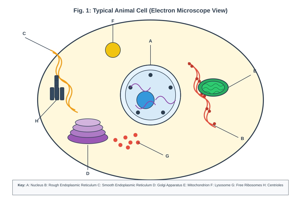
11. Figure 1 shows the structure of a typical animal cell as seen under an electron microscope.
Generated diagram for Q11.
Fig. 1
(a) Identify the organelles labelled A–H. [4]
A: ______________________________
B: ______________________________
C: ______________________________
D: ______________________________
E: ______________________________
F: ______________________________
G: ______________________________
H: ______________________________
(b) State one function of the organelle labelled C. [1]
(c) Explain why the organelle labelled E is abundant in cells that secrete large amounts of protein. [2]
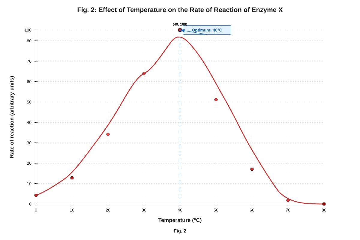
12. Figure 2 shows the results of an experiment to investigate the effect of temperature on the activity of enzyme X.
Generated graph for Q12.
Fig. 2
(a) State the optimum temperature for enzyme X. [1]
(b) Explain the shape of the curve between 0°C and 40°C. [2]
(c) Explain the shape of the curve between 40°C and 70°C. [2]
(d) A student repeated the experiment using a fixed concentration of a non-competitive inhibitor. Sketch on the axes of Fig. 2 the curve you would expect. Label this curve "I". [2]
(Sketch on the graph above)
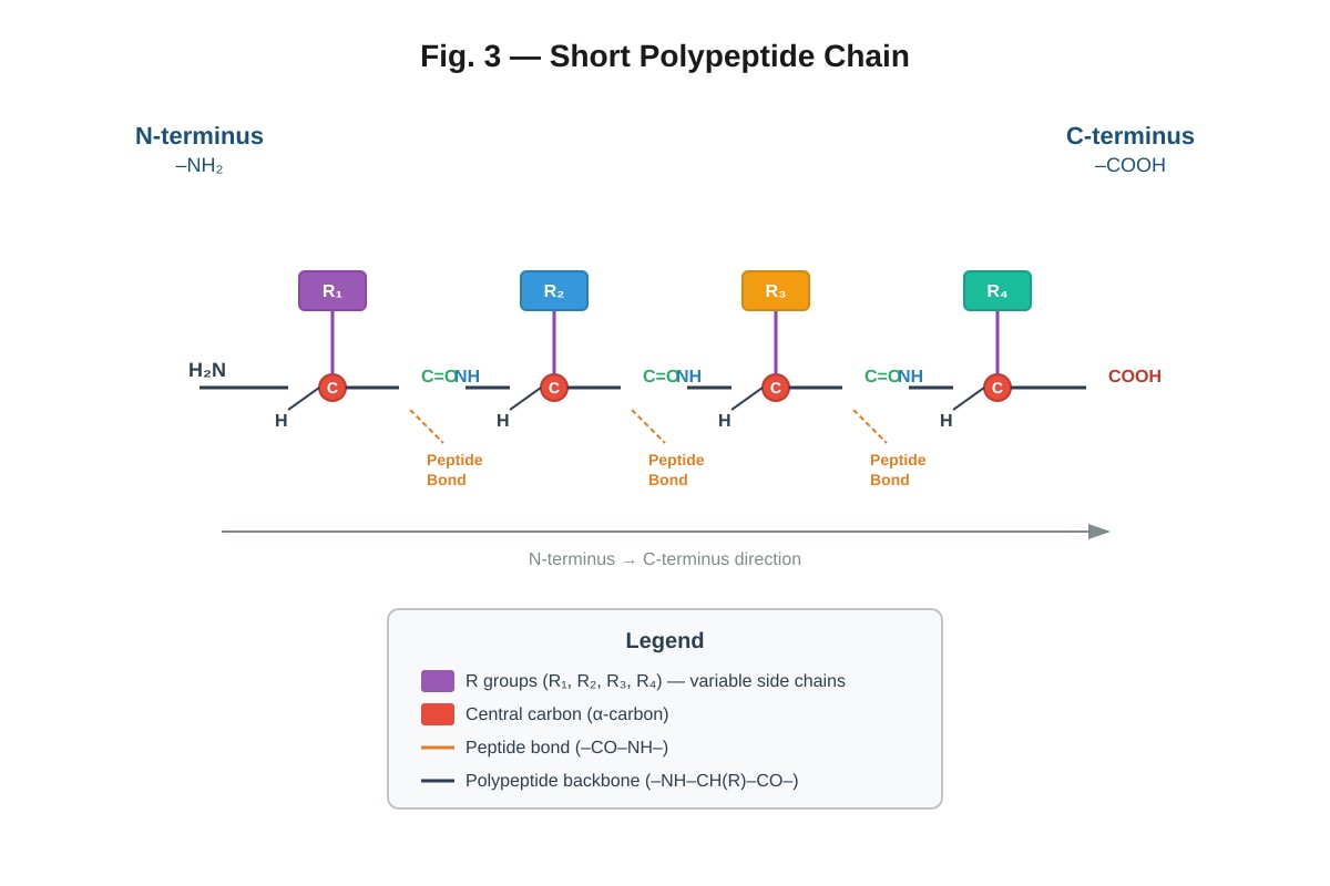
13. Figure 3 shows a molecule of a biological polymer.
Generated diagram for Q13.
Fig. 3
(a) Name the type of bond labelled X in Fig. 3. [1]
(b) Name the reaction that forms this bond. [1]
(c) State the general formula for an amino acid. [1]
(d) Explain how the R group of an amino acid affects the structure and function of a protein. [2]
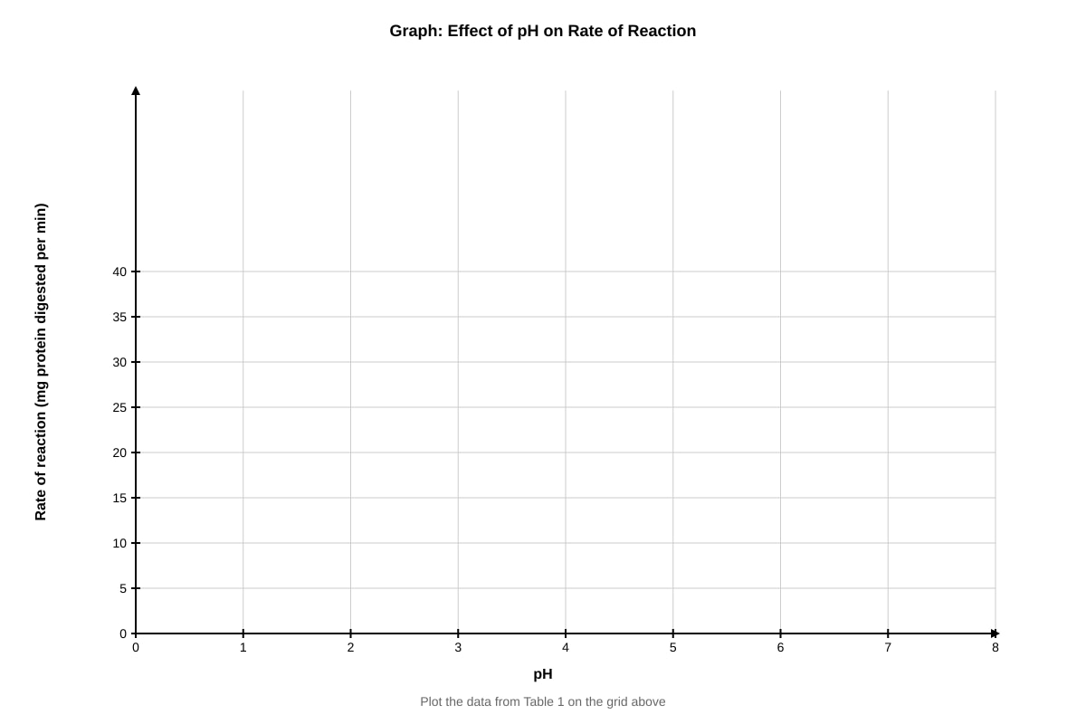
14. A student investigated the effect of pH on the activity of the enzyme pepsin. The rate of reaction was measured at different pH values and the results are shown in Table 1.
Table 1
| pH | Rate of reaction (mg protein digested per min) |
|---|---|
| 1.0 | 8 |
| 1.5 | 22 |
| 2.0 | 35 |
| 2.5 | 30 |
| 3.0 | 18 |
| 4.0 | 5 |
| 5.0 | 1 |
| 7.0 | 0 |
(a) Plot a graph of the results shown in Table 1 on the grid below. [3]
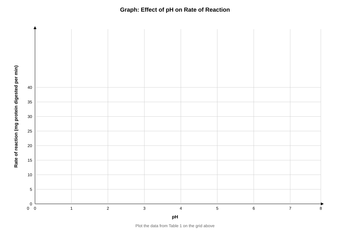
Generated graph for Q14.
(b) From your graph, estimate the optimum pH for pepsin. [1]
(c) Explain why the rate of reaction decreases significantly above pH 3.0. [2]
(d) Predict the rate of reaction at pH 8.0 and give a reason for your prediction. [2]
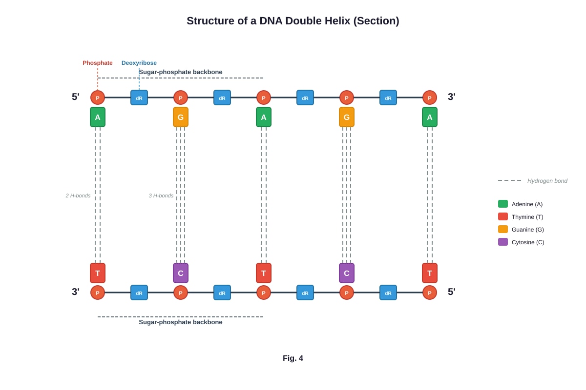
15. Figure 4 shows the structure of a section of DNA.
Image pending generation: diagram for Q15.
Fig. 4
(a) Name the bond that joins the two strands of DNA together. [1]
(b) State the type of sugar found in DNA. [1]
(c) Explain how the structure of DNA allows it to carry out its function of storing genetic information. [3]
16. A student carried out an experiment to test for the presence of biological molecules in three food samples (P, Q, and R). The results are shown in Table 2.
Table 2
| Test | Food sample P | Food sample Q | Food sample R |
|---|---|---|---|
| Benedict's test (after heating) | Blue | Brick-red precipitate | Blue |
| Iodine test | Blue-brown | Blue-brown | Blue-black |
| Biuret test | Purple | Blue | Blue |
| Ethanol emulsion test | Milky emulsion | Clear | Clear |
(a) Identify which food sample contains: [3]
(i) Protein: ______________
(ii) Reducing sugar: ______________
(iii) Starch: ______________
(b) Describe how the ethanol emulsion test is carried out. [2]
(c) Explain why the Biuret test produces a purple colour in the presence of protein. [1]
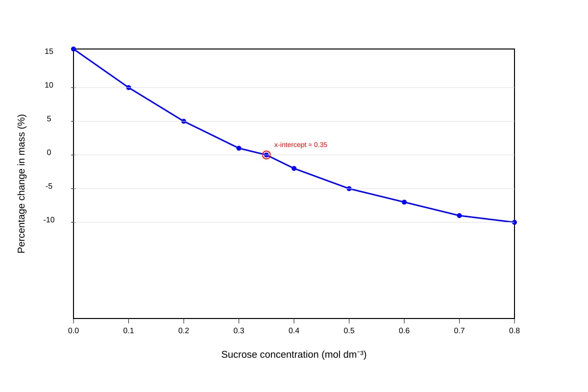
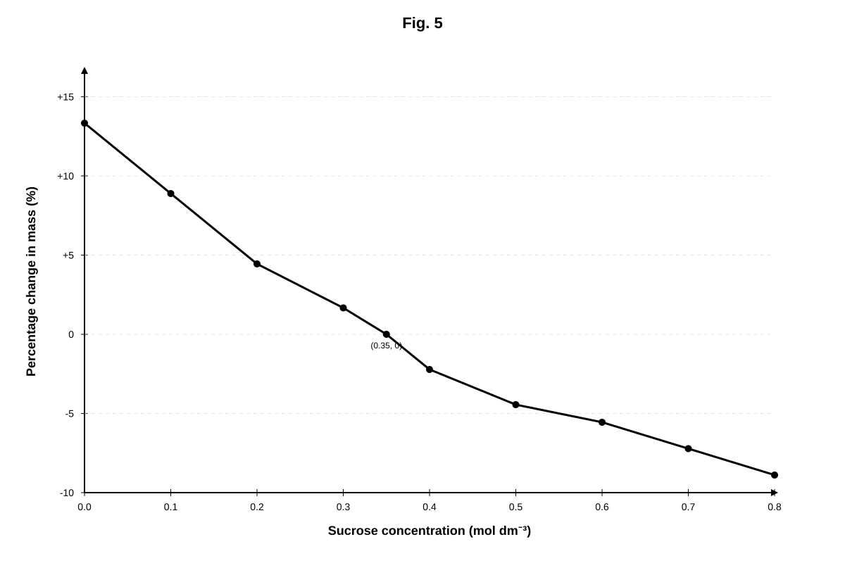
17. Figure 5 shows a graph of the change in mass of potato cylinders placed in sucrose solutions of different concentrations after 30 minutes.
Generated graph for Q17.
Fig. 5
(a) Explain why the potato cylinders gained mass in 0.1 mol dm⁻³ sucrose solution. [2]
(b) Estimate the water potential of the potato cells. Explain how you obtained your answer. [2]
(c) Explain why the potato cylinders lost mass in 0.6 mol dm⁻³ sucrose solution. [2]
Section C: Data Interpretation & Extended Response (Questions 18–20)
18. Read the following passage and answer the questions that follow.
The fluid mosaic model, proposed by Singer and Nicolson in 1972, describes the cell membrane as a dynamic structure. The phospholipid bilayer forms the basic framework, with phospholipid molecules able to move laterally within the layer. Proteins are embedded within or attached to the bilayer at various positions — some span the entire membrane (integral proteins), while others are found on the surface (peripheral proteins). Cholesterol molecules are interspersed among the phospholipids, helping to regulate membrane fluidity. Glycoproteins and glycolipids on the cell surface play important roles in cell recognition and signalling.
(a) Explain why the model is described as "fluid mosaic". [2]
(b) Describe two functions of cholesterol in the cell membrane. [2]
(c) Explain the role of glycoproteins in the immune response. [2]
(d) Explain how the structure of the phospholipid bilayer makes it a effective barrier to the passage of ions. [2]
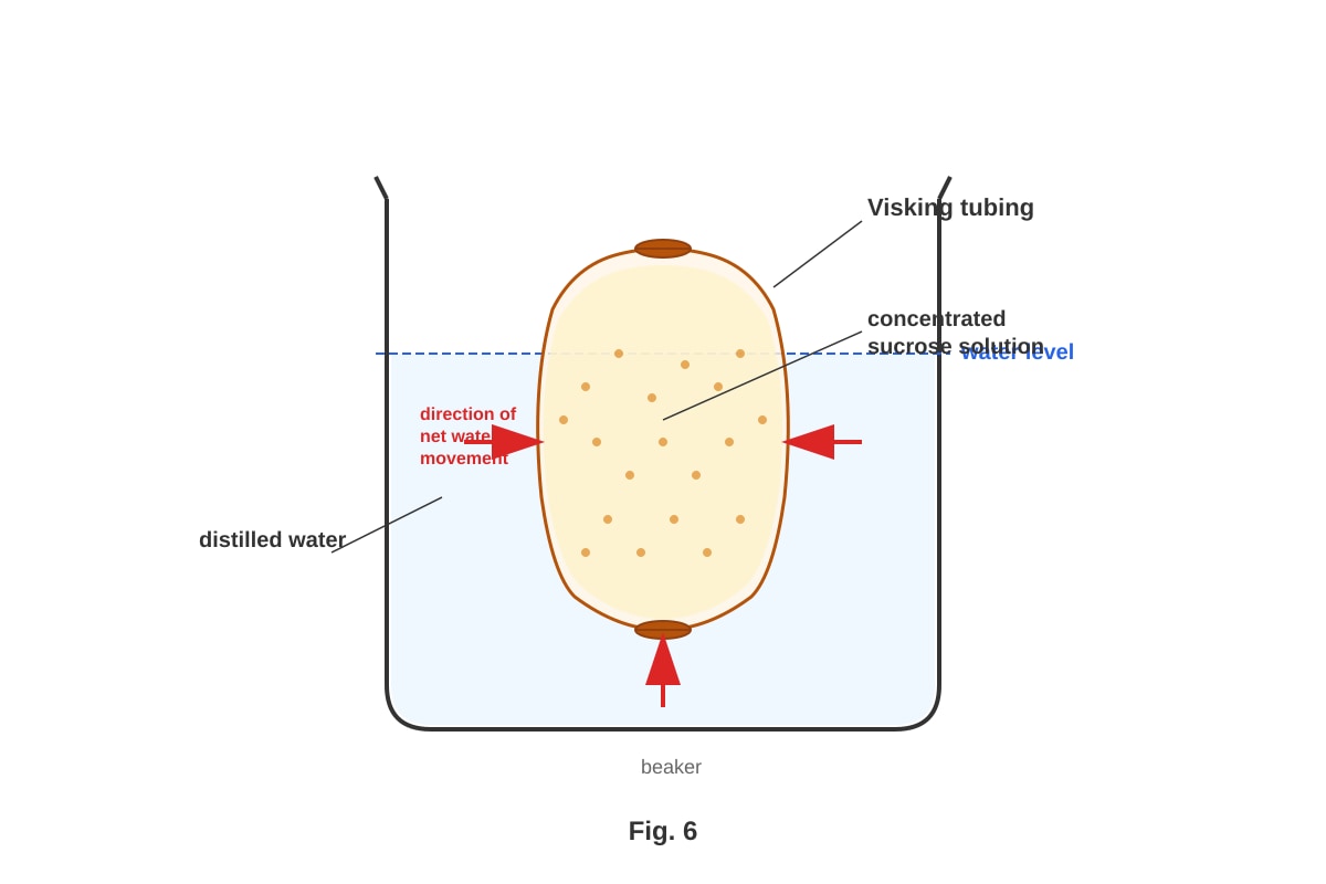
19. Figure 6 shows an experiment to investigate osmosis using Visking tubing.
Generated experimental_setup for Q19.
Fig. 6
(a) Explain why water moves into the Visking tubing bag. [3]
(b) Predict what would happen to the water level in the beaker over time. Explain your answer. [2]
(c) If the experiment was repeated with Visking tubing containing distilled water placed in a concentrated sucrose solution, describe and explain what would happen. [3]
20. Compare and contrast the structure and function of starch and glycogen. In your answer, refer to the following:
- The type of glycosidic bond present
- The degree of branching
- The solubility in water
- The biological role in living organisms
[6]
<stage3_quiz_answers_md>
A-Level Biology H1 Quiz - Cells Biomolecules
Answer Key
Section A: Multiple Choice & Short Answer (Questions 1–10)
1. C — Golgi apparatus [1]
Explanation: Prokaryotic cells lack membrane-bound organelles. Ribosomes, cell membranes, and cytoplasm are present in both prokaryotic and eukaryotic cells. The Golgi apparatus is a membrane-bound organelle found only in eukaryotic cells. This is a common exam trap — students often forget that ribosomes (70S in prokaryotes, 80S in eukaryotes) are present in both cell types.
2. A — Solution W [1]
Explanation: Benedict's test detects reducing sugars (positive = brick-red precipitate). Iodine test detects starch (positive = blue-black). Solution W is negative for reducing sugar (Benedict's remains blue) but positive for starch (iodine turns blue-black), so it contains starch only. Solution Z contains neither. Solutions X and Y contain reducing sugar but no starch.
3. C — Cohesion between water molecules [1]
Explanation: Cohesion (hydrogen bonding between water molecules) allows water to form continuous columns in xylem vessels and enables bulk flow in transport systems. While high specific heat capacity (A) and high latent heat (B) are important for temperature regulation, they are not directly responsible for water's role as a transport medium. Option D relates to habitat provision, not transport.
4. C — Nucleus [1]
Explanation: Uracil is a nitrogenous base found in RNA but not DNA. Radioactive uracil is incorporated during transcription, which occurs in the nucleus. Although ribosomes use RNA (mRNA, tRNA, rRNA) for translation, the RNA is first synthesised in the nucleus. This question tests the link between the marker (uracil → RNA) and the site of RNA synthesis (nucleus).
5. C — It regulates membrane fluidity by restricting phospholipid movement at high temperatures and preventing tight packing at low temperatures. [1]
Explanation: Cholesterol acts as a fluidity buffer. At high temperatures, it restricts the movement of phospholipid fatty acid tails, reducing fluidity. At low temperatures, it prevents the fatty acid tails from packing too closely together, maintaining fluidity. It does not provide energy (A) or act primarily as a receptor (D). Option B is incorrect because cholesterol does not simply increase fluidity — it regulates it in both directions.
6. [2 marks — 1 mark per correct difference]
Any two of the following:
- Prokaryotic cells lack a nucleus (DNA is in the nucleoid region), whereas eukaryotic cells have a membrane-bound nucleus.
- Prokaryotic cells lack membrane-bound organelles, whereas eukaryotic cells have membrane-bound organelles (e.g., mitochondria, ER, Golgi).
- Prokaryotic cells have 70S ribosomes, whereas eukaryotic cells have 80S ribosomes.
- Prokaryotic cells have a cell wall made of murein (peptidoglycan), whereas eukaryotic plant cells have a cell wall made of cellulose.
- Prokaryotic cells are generally smaller (1–5 μm) than eukaryotic cells (10–100 μm).
Marking note: Answers must compare the same feature in both cell types to earn the mark. Simply stating "prokaryotes have no nucleus" without reference to eukaryotes may not earn the mark in some marking schemes.
7. [3 marks]
A phospholipid consists of a glycerol molecule bonded to two fatty acid chains (hydrophobic tails) and one phosphate group (hydrophilic head). [1]
In the cell membrane, phospholipids arrange themselves into a bilayer. [1]
The hydrophilic phosphate heads face outwards towards the aqueous environment (both extracellular fluid and cytoplasm), while the hydrophobic fatty acid tails face inwards, away from water. [1]
Teaching note: This arrangement is spontaneous (thermodynamically favourable) and forms the basic structure of all cell membranes. Students should be able to draw a labelled diagram of a phospholipid and the bilayer arrangement.
8. [2 marks]
The active site of an enzyme has a specific three-dimensional shape that is complementary to the shape of its substrate. [1]
According to the lock-and-key hypothesis, the substrate fits into the active site of the enzyme like a key fits into a lock, forming an enzyme-substrate complex. [1]
Teaching note: This specificity arises from the unique arrangement of amino acid R groups in the active site, which creates a specific shape and chemical environment. Students should distinguish this from the induced-fit model, which states that the active site changes shape slightly to accommodate the substrate.
9. [2 marks]
A monomer is a small, single unit that can join with other similar molecules to form a larger molecule. [1]
A polymer is a large molecule made up of many repeating monomer units joined together. [1]
Example: Glucose is a monomer (monosaccharide); starch is a polymer made up of many glucose monomers joined by glycosidic bonds. (Other valid examples: amino acid → protein; nucleotide → DNA/RNA; fatty acid + glycerol → lipid.)
Marking note: Students must provide a named example of both a monomer and a polymer for full marks.
10. (a) Solution C [1]
(b) [2 marks]
Solution C has a lower water potential (more negative) than the inside of the red blood cells. [1]
Water moves by osmosis from a region of higher water potential (inside the cell) to a region of lower water potential (the solution) across the partially permeable cell membrane, causing the cells to lose water and shrink (crenation). [1]
Teaching note: Solution A is hypotonic (higher water potential than the cell), causing cells to swell and burst (haemolysis). Solution B is isotonic (same water potential as the cell), so there is no net movement of water. Students must use the term "water potential" correctly — water moves from high (less negative) to low (more negative) water potential.
Section B: Structured Response (Questions 11–17)
11. (a) [4 marks — ½ mark per correct identification]
Possible identifications (depending on diagram labelling):
- A: Nucleus
- B: Rough endoplasmic reticulum
- C: Golgi apparatus
- D: Mitochondrion
- E: Ribosome (free)
- F: Smooth endoplasmic reticulum
- G: Lysosome
- H: Cell membrane
(Accept any reasonable set of 8 organelles consistent with a typical animal cell diagram.)
(b) [1 mark]
If C = Golgi apparatus: It modifies, sorts, and packages proteins for secretion or transport to other organelles. (Accept: forms lysosomes; adds carbohydrate groups to proteins — glycosylation.)
(c) [2 marks]
Cells that secrete large amounts of protein require many ribosomes to synthesise these proteins. [1]
Ribosomes are the site of protein synthesis (translation), where mRNA is used as a template to assemble amino acids into polypeptide chains. [1]
Teaching note: Secretory proteins are synthesised by ribosomes on the rough ER, then transported to the Golgi apparatus for modification and packaging into vesicles for secretion. Students should understand the pathway: ribosome → rough ER → Golgi → vesicle → cell membrane (exocytosis).
12. (a) 40°C [1]
(b) [2 marks]
As temperature increases from 0°C to 40°C, the kinetic energy of both enzyme and substrate molecules increases. [1]
This leads to more frequent successful collisions between enzyme and substrate molecules, forming more enzyme-substrate complexes per unit time, so the rate of reaction increases. [1]
(c) [2 marks]
Above 40°C, the enzyme begins to denature. [1]
The hydrogen bonds and other weak interactions that maintain the tertiary structure of the enzyme are broken, causing the active site to change shape so that the substrate can no longer fit, and the rate of reaction decreases. [1]
(d) [2 marks]
The curve should show:
- A lower maximum rate of reaction (lower peak) than the original curve [1]
- The optimum temperature remains at 40°C [1]
Explanation: A non-competitive inhibitor binds to a site other than the active site, changing the shape of the enzyme. This reduces the number of functional enzyme molecules, lowering the maximum rate of reaction (Vmax is reduced). The optimum temperature is not affected because it is a property of the enzyme's structure, not the inhibitor.
13. (a) Peptide bond [1]
(b) Condensation reaction (dehydration synthesis) [1]
(c) The general formula for an amino acid is: NH₂–CH(R)–COOH [1]
Where R represents the variable side chain (R group) that differs between amino acids.
(d) [2 marks]
The R group determines the chemical properties of each amino acid (e.g., polar, non-polar, charged, hydrophilic, hydrophobic). [1]
Different R groups interact with each other (through hydrogen bonds, ionic bonds, disulphide bridges, and hydrophobic interactions) to determine the tertiary structure of the protein, which in turn determines its function. [1]
Teaching note: For example, if R groups that form disulphide bonds are present, the protein may have a more rigid structure (e.g., in structural proteins). Hydrophobic R groups tend to be found in the interior of globular proteins, away from water. A change in even one R group (e.g., in sickle cell anaemia, glutamic acid → valine) can dramatically alter protein function.
14. (a) [3 marks]
- Correctly plotted points (all 8 points within tolerance) [1]
- Correct line/curve drawn (smooth curve through points) [1]
- Axes correctly labelled with units [1]
Expected graph: A bell-shaped curve peaking at pH 2.0 with a rate of 35 mg/min, declining sharply to 0 at pH 7.0.
(b) pH 2.0 [1]
(c) [2 marks]
Above pH 3.0, the hydrogen ion concentration decreases, disrupting the ionic and hydrogen bonds that maintain the tertiary structure of pepsin. [1]
The active site changes shape (denaturation), so the substrate can no longer bind effectively, and the rate of reaction decreases. [1]
(d) [2 marks]
The rate of reaction would be 0 (or very close to 0). [1]
At pH 8.0 (alkaline conditions), pepsin would be completely denatured because it is adapted to function in the highly acidic environment of the stomach (pH ~2). The enzyme's tertiary structure would be irreversibly disrupted. [1]
Teaching note: Pepsin is an extremophile enzyme adapted to the stomach's acidic pH. Students should note that most enzymes have an optimum near neutral pH (e.g., salivary amylase, pH 6.8–7.0), but pepsin is an important exception.
15. (a) Hydrogen bonds [1]
(b) Deoxyribose [1]
(c) [3 marks]
- DNA has a double helix structure with complementary base pairing (A-T, G-C), which allows accurate replication during cell division. [1]
- The sequence of bases along the DNA strand codes for the sequence of amino acids in proteins, allowing genetic information to be stored. [1]
- The sugar-phosphate backbone is strong and stable (covalent bonds), protecting the genetic information stored in the bases. The double-stranded structure also provides a template for repair if one strand is damaged. [1]
Additional acceptable points:
- The large number of possible base sequences allows vast amounts of information to be stored.
- Hydrogen bonds between base pairs allow the strands to be separated for replication and transcription.
16. (a) [3 marks — 1 mark each]
(i) Protein: P (Biuret test positive — purple)
(ii) Reducing sugar: Q (Benedict's test positive — brick-red precipitate)
(iii) Starch: R (Iodine test positive — blue-black)
(b) [2 marks]
Add ethanol to the food sample and shake to dissolve any lipids present. [1]
Then pour the mixture into water. If lipids are present, a milky white emulsion forms. [1]
(c) [1 mark]
The Biuret test detects peptide bonds. Copper(II) ions in the alkaline solution form a violet/purple complex with the peptide bonds (–CO–NH–) in proteins.
Teaching note: Students should remember the colour changes for all four food tests:
- Benedict's: blue → green → yellow → orange → brick-red (reducing sugars)
- Iodine: brown/yellow → blue-black (starch)
- Biuret: blue → purple (proteins)
- Ethanol emulsion: clear → milky white (lipids)
17. (a) [2 marks]
The 0.1 mol dm⁻³ sucrose solution has a higher water potential (less negative) than the water potential inside the potato cells. [1]
Water moves by osmosis from the solution (higher water potential) into the potato cells (lower water potential) across the partially permeable cell membrane, causing the potato cylinders to gain mass. [1]
(b) [2 marks]
The water potential of the potato cells is approximately −0.35 mol dm⁻³ (the sucrose concentration at which there is no change in mass). [1]
At this point, the water potential of the external solution equals the water potential of the potato cells, so there is no net movement of water. [1]
(c) [2 marks]
The 0.6 mol dm⁻³ sucrose solution has a lower water potential (more negative) than the water potential inside the potato cells. [1]
Water moves by osmosis from the potato cells (higher water potential) into the solution (lower water potential), causing the potato cylinders to lose mass (plasmolysis occurs). [1]
Section C: Data Interpretation & Extended Response (Questions 18–20)
18. (a) [2 marks]
"Fluid" refers to the fact that the phospholipid bilayer is not rigid — phospholipid molecules can move laterally (sideways) within the layer, giving the membrane a flexible, fluid nature. [1]
"Mosaic" refers to the scattered arrangement of proteins within the bilayer, which resembles a mosaic pattern when viewed from above. [1]
(b) [2 marks]
- At high temperatures, cholesterol restricts the movement of phospholipid fatty acid tails, reducing membrane fluidity and preventing the membrane from becoming too permeable. [1]
- At low temperatures, cholesterol prevents the fatty acid tails from packing too closely together, maintaining membrane fluidity and preventing the membrane from becoming too rigid. [1]
(c) [2 marks]
Glycoproteins on the cell surface act as antigens (cell surface markers). [1]
They are recognised by immune cells (e.g., T cells and B cells) as "self" or "non-self", enabling the immune system to identify and target foreign cells (e.g., pathogens, transplanted organs) while leaving the body's own cells unharmed. [1]
(d) [2 marks]
The interior of the phospholipid bilayer consists of hydrophobic (non-polar) fatty acid tails. [1]
Ions are charged (hydrophilic) particles that cannot pass through the hydrophobic interior of the bilayer. They require channel proteins or carrier proteins to cross the membrane. [1]
19. (a) [3 marks]
The concentrated sucrose solution inside the Visking tubing has a lower water potential (more negative) than the distilled water in the beaker. [1]
The Visking tubing is partially permeable — it allows small water molecules to pass through but not larger sucrose molecules. [1]
Water moves by osmosis from the distilled water (higher water potential) through the Visking tubing into the sucrose solution (lower water potential). [1]
(b) [2 marks]
The water level in the beaker would decrease. [1]
Water is moving from the beaker into the Visking tubing bag by osmosis, so the volume of water in the beaker decreases over time. [1]
(c) [3 marks]
The distilled water inside the Visking tubing has a higher water potential than the concentrated sucrose solution outside. [1]
Water would move by osmosis out of the Visking tubing (from high water potential to low water potential) into the surrounding sucrose solution. [1]
The Visking tubing bag would shrink/deflate, and the water level in the beaker would rise as water leaves the bag. [1]
20. [6 marks]
| Feature | Starch | Glycogen |
|---|---|---|
| Glycosidic bond | α-1,4 glycosidic bonds (in amylose and amylopectin); α-1,6 bonds at branch points in amylopectin | α-1,4 glycosidic bonds; α-1,6 bonds at branch points |
| Degree of branching | Amylose is unbranched (helical); amylopectin is branched (every 24–30 glucose units) | Highly branched (every 8–12 glucose units) — more branched than amylopectin |
| Solubility | Insoluble in water (does not affect water potential of cells) | Insoluble in water |
| Biological role | Energy storage in plants | Energy storage in animals (mainly in liver and muscle cells) |
Marking descriptors (6 marks):
- Correct description of glycosidic bond type in both [1]
- Correct comparison of degree of branching (glycogen more highly branched) [1]
- Correct statement about solubility (both insoluble) [1]
- Correct biological role of starch in plants [1]
- Correct biological role of glycogen in animals [1]
- Clear, well-organised comparison with appropriate use of biological terminology [1]
Teaching note: Both starch and glycogen are polymers of α-glucose. The high degree of branching in glycogen means more ends are available for enzymes to act on simultaneously, allowing rapid release of glucose when energy is needed — important for active animals. Starch's lower branching is sufficient for the less urgent energy demands of plants. Both are insoluble, which is important because soluble glucose would affect the water potential of cells, causing water to enter by osmosis.
<stage3_quiz_md>
A-Level Biology H1 Quiz - Cells Biomolecules
Name: ______________________________ Class: ______________________________ Date: ______________________________ Score: ________ / 50
Duration: 60 minutes Total Marks: 50
Instructions:
- Answer ALL questions.
- Write your answers in the spaces provided.
- The number of marks for each question is shown in brackets [ ].
- Where a question requires an explanation, answers should be written in clear, concise sentences.
- The use of diagrams is encouraged where appropriate to support your answer.
Section A: Multiple Choice & Short Answer (Questions 1–10)
Questions 1–5: Multiple Choice. Circle the single best answer. [1 mark each]
1. Which of the following organelles is found in eukaryotic cells but NOT in prokaryotic cells?
A. Ribosomes B. Cell membrane C. Golgi apparatus D. Cytoplasm
2. A student tested four unknown solutions with Benedict's reagent and iodine solution. The results are shown below.
| Solution | Benedict's test (after heating) | Iodine test |
|---|---|---|
| W | Blue | Blue-black |
| X | Brick-red precipitate | Blue-brown |
| Y | Brick-red precipitate | Blue-brown |
| Z | Blue | Blue-brown |
Which solution contains starch only?
A. Solution W B. Solution X C. Solution Y D. Solution Z
3. Which property of water is most directly responsible for its role as a transport medium in living organisms?
A. High specific heat capacity B. High latent heat of vaporisation C. Cohesion between water molecules D. Lower density as a solid than as a liquid
4. During an experiment, radioactive uracil was introduced into a culture of growing cells. In which organelle would radioactivity first be detected?
A. Mitochondrion B. Ribosome C. Nucleus D. Golgi apparatus
5. Which of the following best describes the role of cholesterol in the cell membrane?
A. It provides energy for active transport across the membrane. B. It increases membrane fluidity at all temperatures. C. It regulates membrane fluidity by restricting phospholipid movement at high temperatures and preventing tight packing at low temperatures. D. It acts as a receptor for cell signalling molecules.
Questions 6–10: Short Answer. [2–3 marks each]
6. State two structural differences between prokaryotic and eukaryotic cells. [2]
7. Describe the structure of a phospholipid molecule and explain how phospholipids arrange themselves in the cell membrane. [3]
8. Explain why enzymes are described as having a "specific" active site. In your answer, refer to the lock-and-key hypothesis. [2]
9. Distinguish between a monomer and a polymer, using one named example of each from biological molecules. [2]
10. A student placed red blood cells in three solutions of different salt concentrations. The results are shown below.
| Solution | Observation |
|---|---|
| A | Cells appear swollen and some have burst |
| B | Cells appear normal |
| C | Cells appear shrunken and crenated |
(a) Which solution is hypertonic to the red blood cells? [1]
(b) Explain your answer to part (a) in terms of water potential. [2]
Section B: Structured Response (Questions 11–17)
11. Figure 1 shows the structure of a typical animal cell as seen under an electron microscope.
Image pending generation: diagram for Q11.
Fig. 1
(a) Identify the organelles labelled A–H. [4]
A: ______________________________ B: ______________________________ C: ______________________________ D: ______________________________ E: ______________________________ F: ______________________________ G: ______________________________ H: ______________________________
(b) State one function of the organelle labelled C. [1]
(c) Explain why the organelle labelled E is abundant in cells that secrete large amounts of protein. [2]
12. Figure 2 shows the results of an experiment to investigate the effect of temperature on the activity of enzyme X.
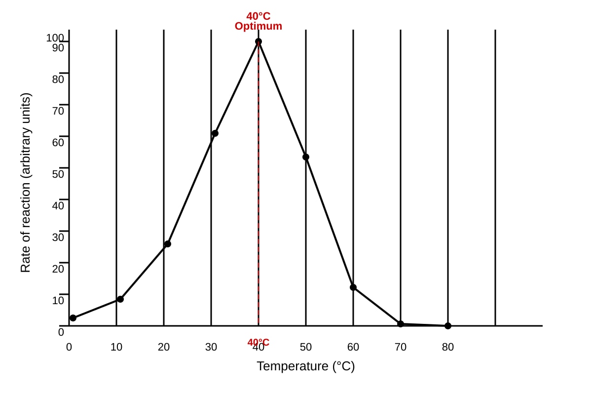
Generated graph for Q12.
Fig. 2
(a) State the optimum temperature for enzyme X. [1]
(b) Explain the shape of the curve between 0°C and 40°C. [2]
(c) Explain the shape of the curve between 40°C and 70°C. [2]
(d) A student repeated the experiment using a fixed concentration of a non-competitive inhibitor. Sketch on the axes of Fig. 2 the curve you would expect. Label this curve "I". [2] (Sketch on the graph above)
13. Figure 3 shows a molecule of a biological polymer.
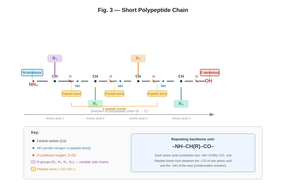
Generated diagram for Q13.
Fig. 3
(a) Name the type of bond labelled X in Fig. 3. [1]
(b) Name the reaction that forms this bond. [1]
(c) State the general formula for an amino acid. [1]
(d) Explain how the R group of an amino acid affects the structure and function of a protein. [2]
14. A student investigated the effect of pH on the activity of the enzyme pepsin. The rate of reaction was measured at different pH values and the results are shown in Table 1.
Table 1
| pH | Rate of reaction (mg protein digested per min) |
|---|---|
| 1.0 | 8 |
| 1.5 | 22 |
| 2.0 | 35 |
| 2.5 | 30 |
| 3.0 | 18 |
| 4.0 | 5 |
| 5.0 | 1 |
| 7.0 | 0 |
(a) Plot a graph of the results shown in Table 1 on the grid below. [3]
Generated graph for Q14.
(b) From your graph, estimate the optimum pH for pepsin. [1]
(c) Explain why the rate of reaction decreases significantly above pH 3.0. [2]
(d) Predict the rate of reaction at pH 8.0 and give a reason for your prediction. [2]
15. Figure 4 shows the structure of a section of DNA.
Generated diagram for Q15.
Fig. 4
(a) Name the bond that joins the two strands of DNA together. [1]
(b) State the type of sugar found in DNA. [1]
(c) Explain how the structure of DNA allows it to carry out its function of storing genetic information. [3]
16. A student carried out an experiment to test for the presence of biological molecules in three food samples (P, Q, and R). The results are shown in Table 2.
Table 2
| Test | Food sample P | Food sample Q | Food sample R |
|---|---|---|---|
| Benedict's test (after heating) | Blue | Brick-red precipitate | Blue |
| Iodine test | Blue-brown | Blue-brown | Blue-black |
| Biuret test | Purple | Blue | Blue |
| Ethanol emulsion test | Milky emulsion | Clear | Clear |
(a) Identify which food sample contains: [3]
(i) Protein: ______________ (ii) Reducing sugar: ______________ (iii) Starch: ______________
(b) Describe how the ethanol emulsion test is carried out. [2]
(c) Explain why the Biuret test produces a purple colour in the presence of protein. [1]
17. Figure 5 shows a graph of the change in mass of potato cylinders placed in sucrose solutions of different concentrations after 30 minutes.
Generated graph for Q17.
Fig. 5
(a) Describe the trend shown in Fig. 5. [2]
(b) Estimate the sucrose concentration at which there is no net change in mass of the potato cylinders. [1]
(c) Explain why the potato cylinders gain mass in dilute sucrose solutions. [2]
(d) A student repeated the experiment using boiled potato cylinders. Predict and explain the results you would expect. [2]
END OF PAPER
Answers
A-Level Biology H1 Quiz - Cells Biomolecules
Answer Key
Section A: Multiple Choice & Short Answer (Questions 1–10)
1. C — Golgi apparatus [1]
Explanation: Prokaryotic cells lack membrane-bound organelles. Ribosomes, cell membranes, and cytoplasm are present in both prokaryotic and eukaryotic cells. The Golgi apparatus is a membrane-bound organelle found only in eukaryotic cells. This is a common exam trap — students often forget that ribosomes (70S in prokaryotes, 80S in eukaryotes) are present in both cell types.
2. A — Solution W [1]
Explanation: Benedict's test detects reducing sugars (positive = brick-red precipitate). Iodine test detects starch (positive = blue-black). Solution W is negative for reducing sugar (Benedict's remains blue) but positive for starch (iodine turns blue-black), so it contains starch only. Solution Z contains neither. Solutions X and Y contain reducing sugar but no starch.
3. C — Cohesion between water molecules [1]
Explanation: Cohesion (hydrogen bonding between water molecules) allows water to form continuous columns in xylem vessels and enables bulk flow in transport systems. While high specific heat capacity (A) and high latent heat (B) are important for temperature regulation, they are not directly responsible for water's role as a transport medium. Option D relates to habitat provision, not transport.
4. C — Nucleus [1]
Explanation: Uracil is a nitrogenous base found in RNA but not DNA. Radioactive uracil is incorporated during transcription, which occurs in the nucleus. Although ribosomes use RNA (mRNA, tRNA, rRNA) for translation, the RNA is first synthesised in the nucleus. This question tests the link between the marker (uracil → RNA) and the site of RNA synthesis (nucleus).
5. C — It regulates membrane fluidity by restricting phospholipid movement at high temperatures and preventing tight packing at low temperatures. [1]
Explanation: Cholesterol acts as a fluidity buffer. At high temperatures, it restricts the movement of phospholipid fatty acid tails, reducing fluidity. At low temperatures, it prevents the fatty acid tails from packing too closely together, maintaining fluidity. It does not provide energy (A) or act primarily as a receptor (D). Option B is incorrect because cholesterol does not simply increase fluidity — it regulates it in both directions.
6. [2 marks — 1 mark per correct difference]
Any two of the following:
- Prokaryotic cells lack a nucleus (DNA is in the nucleoid region), whereas eukaryotic cells have a membrane-bound nucleus.
- Prokaryotic cells lack membrane-bound organelles, whereas eukaryotic cells have membrane-bound organelles (e.g., mitochondria, ER, Golgi).
- Prokaryotic cells have 70S ribosomes, whereas eukaryotic cells have 80S ribosomes.
- Prokaryotic cells have a cell wall made of murein (peptidoglycan), whereas eukaryotic plant cells have a cell wall made of cellulose.
- Prokaryotic cells are generally smaller (1–5 μm) than eukaryotic cells (10–100 μm).
Marking note: Answers must compare the same feature in both cell types to earn the mark. Simply stating "prokaryotes have no nucleus" without reference to eukaryotes may not earn the mark in some marking schemes.
7. [3 marks]
A phospholipid consists of a glycerol molecule bonded to two fatty acid chains (hydrophobic tails) and one phosphate group (hydrophilic head). [1]
In the cell membrane, phospholipids arrange themselves into a bilayer. [1]
The hydrophilic phosphate heads face outwards towards the aqueous environment (both extracellular fluid and cytoplasm), while the hydrophobic fatty acid tails face inwards, away from water. [1]
Teaching note: This arrangement is spontaneous (thermodynamically favourable) and forms the basic structure of all cell membranes. Students should be able to draw a labelled diagram of a phospholipid and the bilayer arrangement.
8. [2 marks]
The active site of an enzyme has a specific three-dimensional shape that is complementary to the shape of its substrate. [1]
According to the lock-and-key hypothesis, the substrate fits into the active site of the enzyme like a key fits into a lock, forming an enzyme-substrate complex. [1]
Teaching note: This specificity arises from the unique arrangement of amino acid R groups in the active site, which creates a specific shape and chemical environment. Students should distinguish this from the induced-fit model, which states that the active site changes shape slightly to accommodate the substrate.
9. [2 marks]
A monomer is a small, single unit that can join with other similar molecules to form a larger molecule. [1]
A polymer is a large molecule made up of many repeating monomer units joined together. [1]
Example: Glucose is a monomer (monosaccharide); starch is a polymer made up of many glucose monomers joined by glycosidic bonds. (Other valid examples: amino acid → protein; nucleotide → DNA/RNA; fatty acid + glycerol → lipid.)
Marking note: Students must provide a named example of both a monomer and a polymer for full marks.
10. (a) Solution C [1]
(b) [2 marks]
Solution C has a lower water potential (more negative) than the inside of the red blood cells. [1]
Water moves by osmosis from a region of higher water potential (inside the cell) to a region of lower water potential (the solution) across the partially permeable cell membrane, causing the cells to lose water and shrink (crenation). [1]
Teaching note: Solution A is hypotonic (higher water potential than the cell), causing cells to swell and burst (haemolysis). Solution B is isotonic (same water potential as the cell), so there is no net movement of water. Students must use the term "water potential" correctly — water moves from high (less negative) to low (more negative) water potential.
Section B: Structured Response (Questions 11–17)
11. (a) [4 marks — ½ mark per correct identification]
Possible identifications (depending on diagram labelling):
- A: Nucleus
- B: Rough endoplasmic reticulum
- C: Golgi apparatus
- D: Mitochondrion
- E: Ribosome (free)
- F: Smooth endoplasmic reticulum
- G: Lysosome
- H: Cell membrane
(Accept any reasonable set of 8 organelles consistent with a typical animal cell diagram.)
(b) [1 mark]
If C = Golgi apparatus: It modifies, sorts, and packages proteins for secretion or transport to other organelles. (Accept: forms lysosomes; adds carbohydrate groups to proteins — glycosylation.)
(c) [2 marks]
Cells that secrete large amounts of protein require many ribosomes to synthesise these proteins. [1]
Ribosomes are the site of protein synthesis (translation), where mRNA is used as a template to assemble amino acids into polypeptide chains. [1]
Teaching note: Secretory proteins are synthesised by ribosomes on the rough ER, then transported to the Golgi apparatus for modification and packaging into vesicles for secretion. Students should understand the pathway: ribosome → rough ER → Golgi → vesicle → cell membrane (exocytosis).
12. (a) 40°C [1]
(b) [2 marks]
As temperature increases from 0°C to 40°C, the kinetic energy of both enzyme and substrate molecules increases. [1]
This leads to more frequent successful collisions between enzyme and substrate molecules, forming more enzyme-substrate complexes per unit time, so the rate of reaction increases. [1]
(c) [2 marks]
Above 40°C, the enzyme begins to denature. [1]
The hydrogen bonds and other weak interactions that maintain the tertiary structure of the enzyme are broken, causing the active site to change shape so that the substrate can no longer fit, and the rate of reaction decreases. [1]
(d) [2 marks]
The curve should show:
- A lower maximum rate of reaction (lower peak) than the original curve [1]
- The optimum temperature remains at 40°C [1]
Explanation: A non-competitive inhibitor binds to a site other than the active site, changing the shape of the enzyme. This reduces the number of functional enzyme molecules, lowering the maximum rate of reaction (Vmax is reduced). The optimum temperature is not affected because it is a property of the enzyme's structure, not the inhibitor.
13. (a) Peptide bond [1]
(b) Condensation reaction (dehydration synthesis) [1]
(c) The general formula for an amino acid is: NH₂–CH(R)–COOH [1]
Where R represents the variable side chain (R group) that differs between amino acids.
(d) [2 marks]
The R group determines the chemical properties of each amino acid (e.g., polar, non-polar, charged, hydrophilic, hydrophobic). [1]
Different R groups interact with each other (through hydrogen bonds, ionic bonds, disulphide bridges, and hydrophobic interactions) to determine the tertiary structure of the protein, which in turn determines its function. [1]
Teaching note: For example, if R groups that form disulphide bonds are present, the protein may have a more rigid structure (e.g., in structural proteins). Hydrophobic R groups tend to be found in the interior of globular proteins, away from water. A change in even one R group (e.g., in sickle cell anaemia, glutamic acid → valine) can dramatically alter protein function.
14. (a) [3 marks]
- Correctly plotted points (all 8 points within tolerance) [1]
- Correct line/curve drawn (smooth curve through points) [1]
- Axes correctly labelled with units [1]
Expected graph: A bell-shaped curve peaking at pH 2.0 with a rate of 35 mg/min, declining sharply to 0 at pH 7.0.
(b) pH 2.0 [1]
(c) [2 marks]
Above pH 3.0, the hydrogen ion concentration decreases, disrupting the ionic and hydrogen bonds that maintain the tertiary structure of pepsin. [1]
The active site changes shape (denaturation), so the substrate can no longer bind effectively, and the rate of reaction decreases. [1]
(d) [2 marks]
The rate of reaction would be 0 (or very close to 0). [1]
At pH 8.0 (alkaline conditions), pepsin would be completely denatured because it is adapted to function in the highly acidic environment of the stomach (pH ~2). The enzyme's tertiary structure would be irreversibly disrupted. [1]
Teaching note: Pepsin is an extremophile enzyme adapted to the stomach's acidic pH. Students should note that most enzymes have an optimum near neutral pH (e.g., salivary amylase, pH 6.8–7.0), but pepsin is an important exception.
15. (a) Hydrogen bonds [1]
(b) Deoxyribose [1]
(c) [3 marks]
- DNA has a double helix structure with complementary base pairing (A-T, G-C), which allows accurate replication during cell division. [1]
- The sequence of bases along the DNA strand codes for the sequence of amino acids in proteins, allowing genetic information to be stored. [1]
- The sugar-phosphate backbone is strong and stable (covalent bonds), protecting the genetic information stored in the bases. The double-stranded structure also provides a template for repair if one strand is damaged. [1]
Additional acceptable points:
- The large number of possible base sequences allows vast amounts of information to be stored.
- Hydrogen bonds between base pairs allow the strands to be separated for replication and transcription.
16. (a) [3 marks — 1 mark each]
(i) Protein: P (Biuret test positive — purple)
(ii) Reducing sugar: Q (Benedict's test positive — brick-red precipitate)
(iii) Starch: R (Iodine test positive — blue-black)
(b) [2 marks]
Add ethanol to the food sample and shake to dissolve any lipids present. [1]
Then pour the mixture into water. If lipids are present, a milky white emulsion forms. [1]
(c) [1 mark]
The Biuret test detects peptide bonds. Copper(II) ions in the alkaline solution form a violet/purple complex with the peptide bonds (–CO–NH–) in proteins.
Teaching note: Students should remember the colour changes for all four food tests:
- Benedict's: blue → green → yellow → orange → brick-red (reducing sugars)
- Iodine: brown/yellow → blue-black (starch)
- Biuret: blue → purple (proteins)
- Ethanol emulsion: clear → milky white (lipids)
17. (a) [2 marks]
The 0.1 mol dm⁻³ sucrose solution has a higher water potential (less negative) than the water potential inside the potato cells. [1]
Water moves by osmosis from the solution (higher water potential) into the potato cells (lower water potential) across the partially permeable cell membrane, causing the potato cylinders to gain mass. [1]
(b) [2 marks]
The water potential of the potato cells is approximately −0.35 mol dm⁻³ (the sucrose concentration at which there is no change in mass). [1]
At this point, the water potential of the external solution equals the water potential of the potato cells, so there is no net movement of water. [1]
(c) [2 marks]
The 0.6 mol dm⁻³ sucrose solution has a lower water potential (more negative) than the water potential inside the potato cells. [1]
Water moves by osmosis from the potato cells (higher water potential) into the solution (lower water potential), causing the potato cylinders to lose mass (plasmolysis occurs). [1]
Section C: Data Interpretation & Extended Response (Questions 18–20)
18. (a) [2 marks]
"Fluid" refers to the fact that the phospholipid bilayer is not rigid — phospholipid molecules can move laterally (sideways) within the layer, giving the membrane a flexible, fluid nature. [1]
"Mosaic" refers to the scattered arrangement of proteins within the bilayer, which resembles a mosaic pattern when viewed from above. [1]
(b) [2 marks]
- At high temperatures, cholesterol restricts the movement of phospholipid fatty acid tails, reducing membrane fluidity and preventing the membrane from becoming too permeable. [1]
- At low temperatures, cholesterol prevents the fatty acid tails from packing too closely together, maintaining membrane fluidity and preventing the membrane from becoming too rigid. [1]
(c) [2 marks]
Glycoproteins on the cell surface act as antigens (cell surface markers). [1]
They are recognised by immune cells (e.g., T cells and B cells) as "self" or "non-self", enabling the immune system to identify and target foreign cells (e.g., pathogens, transplanted organs) while leaving the body's own cells unharmed. [1]
(d) [2 marks]
The interior of the phospholipid bilayer consists of hydrophobic (non-polar) fatty acid tails. [1]
Ions are charged (hydrophilic) particles that cannot pass through the hydrophobic interior of the bilayer. They require channel proteins or carrier proteins to cross the membrane. [1]
19. (a) [3 marks]
The concentrated sucrose solution inside the Visking tubing has a lower water potential (more negative) than the distilled water in the beaker. [1]
The Visking tubing is partially permeable — it allows small water molecules to pass through but not larger sucrose molecules. [1]
Water moves by osmosis from the distilled water (higher water potential) through the Visking tubing into the sucrose solution (lower water potential). [1]
(b) [2 marks]
The water level in the beaker would decrease. [1]
Water is moving from the beaker into the Visking tubing bag by osmosis, so the volume of water in the beaker decreases over time. [1]
(c) [3 marks]
The distilled water inside the Visking tubing has a higher water potential than the concentrated sucrose solution outside. [1]
Water would move by osmosis out of the Visking tubing (from high water potential to low water potential) into the surrounding sucrose solution. [1]
The Visking tubing bag would shrink/deflate, and the water level in the beaker would rise as water leaves the bag. [1]
20. [6 marks]
| Feature | Starch | Glycogen |
|---|---|---|
| Glycosidic bond | α-1,4 glycosidic bonds (in amylose and amylopectin); α-1,6 bonds at branch points in amylopectin | α-1,4 glycosidic bonds; α-1,6 bonds at branch points |
| Degree of branching | Amylose is unbranched (helical); amylopectin is branched (every 24–30 glucose units) | Highly branched (every 8–12 glucose units) — more branched than amylopectin |
| Solubility | Insoluble in water (does not affect water potential of cells) | Insoluble in water |
| Biological role | Energy storage in plants | Energy storage in animals (mainly in liver and muscle cells) |
Marking descriptors (6 marks):
- Correct description of glycosidic bond type in both [1]
- Correct comparison of degree of branching (glycogen more highly branched) [1]
- Correct statement about solubility (both insoluble) [1]
- Correct biological role of starch in plants [1]
- Correct biological role of glycogen in animals [1]
- Clear, well-organised comparison with appropriate use of biological terminology [1]
Teaching note: Both starch and glycogen are polymers of α-glucose. The high degree of branching in glycogen means more ends are available for enzymes to act on simultaneously, allowing rapid release of glucose when energy is needed — important for active animals. Starch's lower branching is sufficient for the less urgent energy demands of plants. Both are insoluble, which is important because soluble glucose would affect the water potential of cells, causing water to enter by osmosis.
<stage3_quiz_md>
A-Level Biology H1 Quiz - Cells Biomolecules
Name: ______________________________ Class: ______________________________ Date: ______________________________ Score: ________ / 50
Duration: 60 minutes Total Marks: 50
Instructions:
- Answer ALL questions.
- Write your answers in the spaces provided.
- The number of marks for each question is shown in brackets [ ].
- Where a question requires an explanation, answers should be written in clear, concise sentences.
- The use of diagrams is encouraged where appropriate to support your answer.
Section A: Multiple Choice & Short Answer (Questions 1–10)
Questions 1–5: Multiple Choice. Circle the single best answer. [1 mark each]
1. Which of the following organelles is found in eukaryotic cells but NOT in prokaryotic cells?
A. Ribosomes B. Cell membrane C. Golgi apparatus D. Cytoplasm
2. A student tested four unknown solutions with Benedict's reagent and iodine solution. The results are shown below.
| Solution | Benedict's test (after heating) | Iodine test |
|---|---|---|
| W | Blue | Blue-black |
| X | Brick-red precipitate | Blue-brown |
| Y | Brick-red precipitate | Blue-brown |
| Z | Blue | Blue-brown |
Which solution contains starch only?
A. Solution W B. Solution X C. Solution Y D. Solution Z
3. Which property of water is most directly responsible for its role as a transport medium in living organisms?
A. High specific heat capacity B. High latent heat of vaporisation C. Cohesion between water molecules D. Lower density as a solid than as a liquid
4. During an experiment, radioactive uracil was introduced into a culture of growing cells. In which organelle would radioactivity first be detected?
A. Mitochondrion B. Ribosome C. Nucleus D. Golgi apparatus
5. Which of the following best describes the role of cholesterol in the cell membrane?
A. It provides energy for active transport across the membrane. B. It increases membrane fluidity at all temperatures. C. It regulates membrane fluidity by restricting phospholipid movement at high temperatures and preventing tight packing at low temperatures. D. It acts as a receptor for cell signalling molecules.
Questions 6–10: Short Answer. [2–3 marks each]
6. State two structural differences between prokaryotic and eukaryotic cells. [2]
7. Describe the structure of a phospholipid molecule and explain how phospholipids arrange themselves in the cell membrane. [3]
8. Explain why enzymes are described as having a "specific" active site. In your answer, refer to the lock-and-key hypothesis. [2]
9. Distinguish between a monomer and a polymer, using one named example of each from biological molecules. [2]
10. A student placed red blood cells in three solutions of different salt concentrations. The results are shown below.
| Solution | Observation |
|---|---|
| A | Cells appear swollen and some have burst |
| B | Cells appear normal |
| C | Cells appear shrunken and crenated |
(a) Which solution is hypertonic to the red blood cells? [1]
(b) Explain your answer to part (a) in terms of water potential. [2]
Section B: Structured Response (Questions 11–17)
11. Figure 1 shows the structure of a typical animal cell as seen under an electron microscope.
<image_placeholder> id: Q11-fig1 type: diagram linked_question: Q11 description: A labelled diagram of a typical animal cell as seen under an electron microscope, showing the following organelles: nucleus (with nuclear envelope, nuclear pores, nucleolus, and chromatin), rough endoplasmic reticulum (with ribosomes attached), smooth endoplasmic reticulum, Golgi apparatus (with cis and trans faces), mitochondrion (with cristae visible), lysosome, ribosomes (free), cell membrane, centrioles. labels: A, B, C, D, E, F, G, H (eight labels pointing to eight different organelles) values: N/A must_show: All eight organelles must be clearly visible and labelled with letters A–H. The nucleus should show nuclear envelope, pores, and nucleolus. The mitochondrion should show cristae. The rough ER should show ribosomes. The Golgi apparatus should show stacked cisternae. </image_placeholder>
Fig. 1
(a) Identify the organelles labelled A–H. [4]
A: ______________________________ B: ______________________________ C: ______________________________ D: ______________________________ E: ______________________________ F: ______________________________ G: ______________________________ H: ______________________________
(b) State one function of the organelle labelled C. [1]
(c) Explain why the organelle labelled E is abundant in cells that secrete large amounts of protein. [2]
12. Figure 2 shows the results of an experiment to investigate the effect of temperature on the activity of enzyme X.
<image_placeholder> id: Q12-fig1 type: graph linked_question: Q12 description: A line graph showing the effect of temperature (°C) on the rate of reaction of enzyme X. The x-axis is Temperature (°C), ranging from 0 to 80. The y-axis is Rate of reaction (arbitrary units), ranging from 0 to 100. The curve rises steeply from 0°C to an optimum at 40°C (rate = 100), then declines sharply from 40°C to 80°C (rate approaches 0 at 70°C). The curve is a smooth bell shape. labels: x-axis: Temperature (°C); y-axis: Rate of reaction (arbitrary units); optimum temperature marked at 40°C values: Key data points: (0, 5), (10, 15), (20, 40), (30, 75), (40, 100), (50, 60), (60, 20), (70, 2), (80, 0) must_show: Clear bell-shaped curve, labelled axes with units, optimum temperature at 40°C clearly identifiable, curve declining to near zero by 70°C. </image_placeholder>
Fig. 2
(a) State the optimum temperature for enzyme X. [1]
(b) Explain the shape of the curve between 0°C and 40°C. [2]
(c) Explain the shape of the curve between 40°C and 70°C. [2]
(d) A student repeated the experiment using a fixed concentration of a non-competitive inhibitor. Sketch on the axes of Fig. 2 the curve you would expect. Label this curve "I". [2] (Sketch on the graph above)
13. Figure 3 shows a molecule of a biological polymer.
<image_placeholder> id: Q13-fig1 type: diagram linked_question: Q13 description: A diagram showing a short polypeptide chain of four amino acids linked by peptide bonds. The backbone shows repeating units of –NH–CH(R)–CO–, with four different R groups (R1, R2, R3, R4) extending from the central carbon of each amino acid. Three peptide bonds are shown as –CO–NH– linkages. The N-terminus (–NH₂) is on the left and the C-terminus (–COOH) is on the right. labels: Peptide bonds labelled, R groups labelled R1, R2, R3, R4, N-terminus, C-terminus values: N/A must_show: Four amino acids linked by three peptide bonds, R groups clearly shown as different, N-terminus and C-terminus labelled, peptide bonds clearly indicated. </image_placeholder>
Fig. 3
(a) Name the type of bond labelled X in Fig. 3. [1]
(b) Name the reaction that forms this bond. [1]
(c) State the general formula for an amino acid. [1]
(d) Explain how the R group of an amino acid affects the structure and function of a protein. [2]
14. A student investigated the effect of pH on the activity of the enzyme pepsin. The rate of reaction was measured at different pH values and the results are shown in Table 1.
Table 1
| pH | Rate of reaction (mg protein digested per min) |
|---|---|
| 1.0 | 8 |
| 1.5 | 22 |
| 2.0 | 35 |
| 2.5 | 30 |
| 3.0 | 18 |
| 4.0 | 5 |
| 5.0 | 1 |
| 7.0 | 0 |
(a) Plot a graph of the results shown in Table 1 on the grid below. [3]
<image_placeholder> id: Q14-fig1 type: graph linked_question: Q14 description: A blank grid provided for the student to plot a graph. The x-axis is pH, ranging from 0 to 8. The y-axis is Rate of reaction (mg protein digested per min), ranging from 0 to 40. Grid lines are present at intervals of 1 on the x-axis and 5 on the y-axis. labels: x-axis: pH; y-axis: Rate of reaction (mg protein digested per min) values: N/A (student plots data from Table 1) must_show: Blank grid with labelled axes and appropriate scale for the data range. </image_placeholder>
(b) From your graph, estimate the optimum pH for pepsin. [1]
(c) Explain why the rate of reaction decreases significantly above pH 3.0. [2]
(d) Predict the rate of reaction at pH 8.0 and give a reason for your prediction. [2]
15. Figure 4 shows the structure of a section of DNA.
<image_placeholder> id: Q15-fig1 type: diagram linked_question: Q15 description: A diagram showing a short double-stranded DNA molecule. The two antiparallel strands are shown with sugar-phosphate backbones. One strand runs 5' to 3' (top), the other 3' to 5' (bottom). Four base pairs are shown: adenine-thymine (A-T) connected by 2 hydrogen bonds, and guanine-cytosine (G-C) connected by 3 hydrogen bonds. The deoxyribose sugars and phosphate groups are shown in the backbone. Hydrogen bonds are shown as dashed lines between complementary bases. labels: 5' and 3' ends labelled on both strands, A, T, G, C bases labelled, hydrogen bonds shown as dashed lines, sugar-phosphate backbone indicated, deoxyribose and phosphate labelled values: N/A must_show: Antiparallel strands with correct 5' and 3' orientation, at least one A-T pair (2 H-bonds) and one G-C pair (3 H-bonds), sugar-phosphate backbone visible, hydrogen bonds shown between base pairs. </image_placeholder>
Fig. 4
(a) Name the bond that joins the two strands of DNA together. [1]
(b) State the type of sugar found in DNA. [1]
(c) Explain how the structure of DNA allows it to carry out its function of storing genetic information. [3]
16. A student carried out an experiment to test for the presence of biological molecules in three food samples (P, Q, and R). The results are shown in Table 2.
Table 2
| Test | Food sample P | Food sample Q | Food sample R |
|---|---|---|---|
| Benedict's test (after heating) | Blue | Brick-red precipitate | Blue |
| Iodine test | Blue-brown | Blue-brown | Blue-black |
| Biuret test | Purple | Blue | Blue |
| Ethanol emulsion test | Milky emulsion | Clear | Clear |
(a) Identify which food sample contains: [3]
(i) Protein: ______________ (ii) Reducing sugar: ______________ (iii) Starch: ______________
(b) Describe how the ethanol emulsion test is carried out. [2]
(c) Explain why the Biuret test produces a purple colour in the presence of protein. [1]
17. Figure 5 shows a graph of the change in mass of potato cylinders placed in sucrose solutions of different concentrations after 30 minutes.
<image_placeholder> id: Q17-fig1 type: graph linked_question: Q17 description: A line graph showing the effect of sucrose concentration on the percentage change in mass of potato cylinders. The x-axis is Sucrose concentration (mol dm⁻³), ranging from 0.0 to 0.8. The y-axis is Percentage change in mass (%), ranging from -10 to +15. The line starts at approximately +12% at 0.0 mol dm⁻³, decreases linearly, crosses the x-axis at approximately 0.35 mol dm⁻³, and continues to approximately -8% at 0.8 mol dm⁻³. labels: x-axis: Sucrose concentration (mol dm⁻³); y-axis: Percentage change in mass (%); point where line crosses x-axis marked values: Key data points: (0.0, +12), (0.1, +8), (0.2, +4), (0.3, +1), (0.35, 0), (0.4, -2), (0.5, -4), (0.6, -5), (0.7, -6.5), (0.8, -8) must_show: Clear straight line with negative gradient, labelled axes with units, line crossing x-axis at approximately 0.35 mol dm⁻³, positive percentage change at low concentrations, negative percentage change at high concentrations. </image_placeholder>
Fig. 5
(a) Describe the trend shown in Fig. 5. [2]
(b) Estimate the sucrose concentration at which there is no net change in mass of the potato cylinders. [1]
(c) Explain why the potato cylinders gain mass in dilute sucrose solutions. [2]
(d) A student repeated the experiment using boiled potato cylinders. Predict and explain the results you would expect. [2]
END OF PAPER
</stage3_quiz_md>
<stage3_quiz_answers_md>
A-Level Biology H1 Quiz - Cells Biomolecules: ANSWER KEY
Section A: Multiple Choice & Short Answer (Questions 1–10)
1. C — Golgi apparatus [1] (Prokaryotic cells lack membrane-bound organelles such as the Golgi apparatus. Ribosomes, cell membranes, and cytoplasm are present in both prokaryotic and eukaryotic cells.)
2. A — Solution W [1] (Solution W tests negative with Benedict's (blue = no reducing sugar) and positive with iodine (blue-black = starch present). Therefore it contains starch only.)
3. C — Cohesion between water molecules [1] (Cohesion allows water to form continuous columns in xylem vessels and enables bulk flow in transport systems. While the other properties are important, cohesion is most directly responsible for water's role as a transport medium.)
4. C — Nucleus [1] (Uracil is a base found in RNA, not DNA. Transcription of RNA occurs in the nucleus, so radioactivity from radioactive uracil would first be detected in the nucleus where mRNA is synthesised.)
5. C — It regulates membrane fluidity by restricting phospholipid movement at high temperatures and preventing tight packing at low temperatures. [1] (Cholesterol acts as a buffer: at high temperatures it restricts phospholipid movement reducing fluidity; at low temperatures it prevents phospholipids from packing too closely, maintaining fluidity.)
6. Any two of the following, 1 mark each (max 2 marks):
- Prokaryotic cells lack a nucleus / have DNA free in the cytoplasm, whereas eukaryotic cells have a true nucleus enclosed by a nuclear envelope. [1]
- Prokaryotic cells lack membrane-bound organelles (e.g., mitochondria, ER, Golgi), whereas eukaryotic cells possess membrane-bound organelles. [1]
- Prokaryotic cells have smaller ribosomes (70S) compared to eukaryotic cells (80S). [1]
- Prokaryotic cells have a cell wall made of murein/peptidoglycan, whereas eukaryotic plant cells have a cell wall made of cellulose. [1]
- Prokaryotic cells have circular DNA, whereas eukaryotic cells have linear DNA associated with histone proteins. [1]
7. Structure of a phospholipid (up to 2 marks):
A phospholipid consists of a glycerol molecule bonded to two fatty acid chains and one phosphate group. [1] The phosphate head is hydrophilic (water-loving) and the fatty acid tails are hydrophobic (water-hating). [1]
Arrangement in the membrane (1 mark):
In the cell membrane, phospholipids arrange themselves into a bilayer. [1] The hydrophilic heads face outwards towards the aqueous environment (both extracellular fluid and cytoplasm), while the hydrophobic tails face inwards, away from water. This arrangement forms the basic structure of the fluid mosaic model.
8. Enzymes have a specific active site because the shape of the active site is complementary to the shape of their specific substrate. [1] According to the lock-and-key hypothesis, the substrate fits into the active site of the enzyme like a key fits into a lock — only a substrate with the correct shape can bind to the active site. [1] This means each enzyme catalyses only one specific reaction or type of reaction.
9. A monomer is a small, single molecule that can join with other similar molecules to form a larger molecule. [1] A polymer is a large molecule made up of many repeating monomer units joined together. [1]
Example: Glucose is a monomer (monosaccharide) and starch is a polymer made up of many glucose monomers joined by glycosidic bonds. [1] (Accept other valid examples, e.g., amino acid → protein/polypeptide; nucleotide → DNA/RNA.)
10. (a) Solution C [1]
(b) Solution C has a lower water potential (more negative) than the inside of the red blood cells. [1] Water moves by osmosis from a region of higher water potential (inside the cell) to a region of lower water potential (the solution) across the partially permeable membrane. [1] This causes water to leave the cells, resulting in the cells shrinking and becoming crenated.
Section B: Structured Response (Questions 11–17)
11. (a) Identification of organelles A–H (1 mark each, max 4 marks — accept any 4 correctly identified):
- Nucleus (with nuclear envelope, nuclear pores, nucleolus, chromatin) [1]
- Rough endoplasmic reticulum (with ribosomes attached) [1]
- Golgi apparatus (with cis and trans faces / stacked cisternae) [1]
- Mitochondrion (with cristae visible) [1]
- Smooth endoplasmic reticulum [1]
- Lysosome [1]
- Ribosomes (free) [1]
- Cell membrane [1]
- Centrioles [1]
(Award 1 mark for each correct identification up to a maximum of 4 marks. The specific letters A–H may be assigned to any of the above organelles depending on the diagram.)
(b) One function of organelle C (depending on labelling):
If C = Golgi apparatus: Modifies, sorts, and packages proteins for secretion / transport to other organelles. [1] (Accept any valid function of whichever organelle is labelled C.)
(c) If E = Rough endoplasmic reticulum (RER): The RER has ribosomes attached to its surface. [1] These ribosomes are the site of protein synthesis. [1] Cells that secrete large amounts of protein require extensive RER to synthesise and transport these proteins (e.g., digestive enzymes, hormones). The RER provides a large surface area for ribosomes and a pathway for transporting proteins to the Golgi apparatus for processing and secretion.
12. (a) 40°C [1]
(b) Between 0°C and 40°C, as temperature increases, the kinetic energy of both enzyme and substrate molecules increases. [1] This leads to more frequent successful collisions between enzyme and substrate molecules, forming more enzyme-substrate complexes per unit time, so the rate of reaction increases. [1]
(c) Between 40°C and 70°C, the temperature exceeds the optimum. The enzyme's active site begins to denature — the hydrogen bonds and other bonds maintaining the tertiary structure break. [1] The shape of the active site changes so that the substrate can no longer fit, fewer enzyme-substrate complexes are formed, and the rate of reaction decreases. At very high temperatures the enzyme is completely denatured and the reaction stops. [1]
(d) Curve I should be sketched on the same axes showing: [2]
- A bell-shaped curve with the same optimum temperature (40°C) but with a lower maximum rate of reaction than the original curve. [1]
- The curve should lie entirely below the original curve at all temperatures. [1] (Non-competitive inhibitors bind to a site other than the active site, changing the shape of the active site. They reduce the rate of reaction at all substrate concentrations but do not affect the optimum temperature. The Vmax is reduced.)
13. (a) Peptide bond [1]
(b) Condensation reaction [1]
(c) The general formula for an amino acid is: NH₂–CH(R)–COOH [1] (Accept a description: an amino group (–NH₂), a carboxyl group (–COOH), a hydrogen atom, and a variable R group, all bonded to a central carbon atom.)
(d) The R group determines the chemical properties of each amino acid (e.g., whether it is polar, non-polar, acidic, or basic). [1] Different R groups interact with each other through various bonds (hydrogen bonds, ionic bonds, disulphide bridges, hydrophobic interactions) to determine the tertiary structure of the protein, which in turn determines its specific shape and function. [1]
14. (a) Graph plotting (3 marks):
- Correctly labelled axes with units (x-axis: pH; y-axis: Rate of reaction (mg protein digested per min)) [1]
- Appropriate scale used and all points correctly plotted [1]
- Smooth curve drawn through the points, showing a peak [1]
(b) Optimum pH ≈ 2.0 [1]
(c) Above pH 3.0, the hydrogen ion concentration decreases. This disrupts the ionic bonds and hydrogen bonds that maintain the tertiary structure of the enzyme. [1] The active site begins to denature (change shape), so fewer enzyme-substrate complexes can form and the rate of reaction decreases. At pH 7.0 the enzyme is completely denatured. [1]
(d) At pH 8.0, the predicted rate of reaction would be 0 (or very close to 0). [1] Pepsin is adapted to work in the highly acidic environment of the stomach (pH ~2). At pH 8.0 (alkaline conditions), the enzyme would be permanently denatured — the active site shape is irreversibly changed so the substrate cannot bind. [1]
15. (a) Hydrogen bonds [1]
(b) Deoxyribose [1]
(c) Any three of the following points (max 3 marks):
- DNA has a double-stranded structure with complementary base pairing (A-T and G-C), which allows accurate replication / copying of genetic information. [1]
- The sequence of bases along the DNA strand codes for the sequence of amino acids in proteins (genetic code). [1]
- The sugar-phosphate backbone is strong and stable (covalent bonds), which protects the genetic information stored in the bases. [1]
- DNA is a very long molecule so it can store a large amount of genetic information. [1]
- The double helix structure is compact, allowing large amounts of information to be stored in a small space within the nucleus. [1]
- Hydrogen bonds between base pairs allow the strands to separate during replication and transcription. [1]
16. (a) (i) Protein: Sample P [1] (ii) Reducing sugar: Sample Q [1] (iii) Starch: Sample R [1]
(b) The food sample is mixed with ethanol (or dissolved in ethanol). [1] The mixture is then shaken/poured into water. If lipids are present, a cloudy white emulsion/milky emulsion forms. [1]
(c) The Biuret reagent (sodium hydroxide and copper(II) sulfate) reacts with peptide bonds in proteins. [1] The copper ions form a violet/purple complex with the peptide bonds in alkaline solution.
17. (a) As the sucrose concentration increases from 0.0 to 0.8 mol dm⁻³, the percentage change in mass of the potato cylinders decreases. [1] At low sucrose concentrations the potato cylinders gain mass (positive percentage change), and at high sucrose concentrations they lose mass (negative percentage change). The relationship is linear/negative correlation. [1]
(b) Approximately 0.35 mol dm⁻³ [1]
(c) In dilute sucrose solutions, the water potential of the solution is higher (less negative) than the water potential inside the potato cells. [1] Water moves by osmosis from the solution (higher water potential) into the potato cells (lower water potential) across the partially permeable cell membrane, causing the potato cylinders to gain mass. [1]
(d) The potato cylinders would show no change in mass (or very little change) at any sucrose concentration. [1] Boiling kills the cells and denatures the proteins in the cell membrane, destroying its selectively permeable / partially permeable nature. The membrane becomes fully permeable, so osmosis cannot occur — sucrose and water can both move freely in and out of the cells. [1]
Mark Summary
| Question | Marks |
|---|---|
| 1 | 1 |
| 2 | 1 |
| 3 | 1 |
| 4 | 1 |
| 5 | 1 |
| 6 | 2 |
| 7 | 3 |
| 8 | 2 |
| 9 | 2 |
| 10 | 3 |
| 11 | 7 |
| 12 | 6 |
| 13 | 5 |
| 14 | 8 |
| 15 | 5 |
| 16 | 6 |
| 17 | 7 |
| Total | 50 |
Free quiz and exam paper access
Enter your details to view this paper
Your access is remembered on this device.