AI Generated Exam Paper
A Level H1 Biology Practice Paper 3
Free A Level H1 Biology Practice Paper 3, LongCat AI version, with questions, answers, and A Level-style practice for Singapore students.
These static practice materials are generated from the site's syllabus and paper-generation workflow, with source and model context shown so students and parents can evaluate the material before use.
Questions
TuitionGoWhere Practice Paper - Biology H1 A-Level
TuitionGoWhere Practice Paper (AI)
Subject: Biology Level: A-Level H1 Paper: Practice Paper — Cells & Biomolecules Version: 3 of 5 Duration: 1 hour 30 minutes Total Marks: 60
Name: ___________________________ Class: ___________________________ Date: ___________________________
Instructions
- Answer all questions in the spaces provided.
- Write your answers in the blank spaces or on lined pages as appropriate.
- The number of marks for each question or part-question is shown in brackets [ ].
- You are advised to spend no more than 1 hour 30 minutes on this paper.
- Where a question requires explanation or reasoning, answers should be written in clear, concise biological language.
- The use of diagrams is encouraged where they support your answer, but they are not required unless stated.
Section A: Structured Questions [30 marks]
Questions 1–6
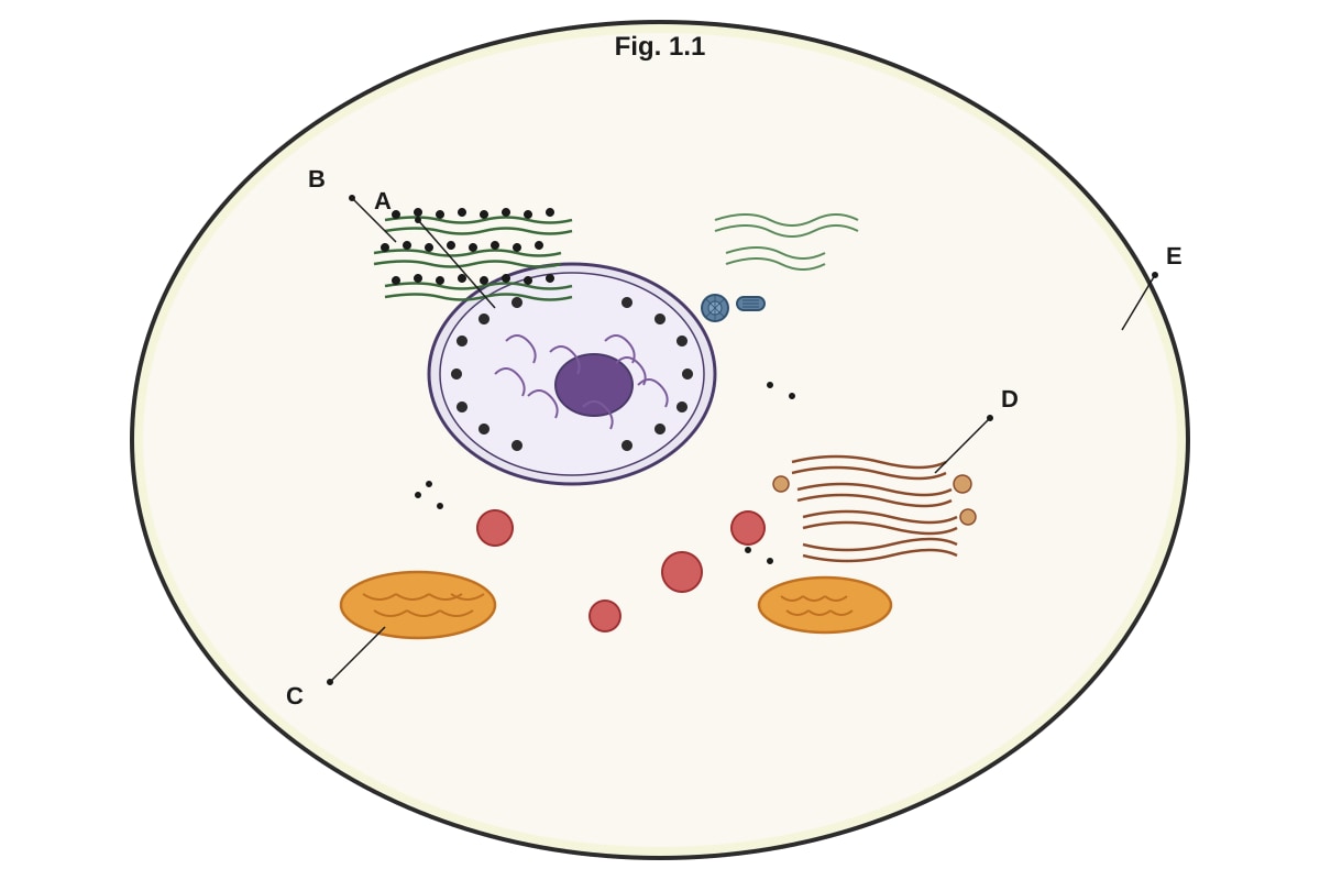
1. Fig. 1.1 shows the structure of a typical animal cell as seen under an electron microscope.
Generated diagram for Q1.
(a) Identify the organelles labelled A, B, C, and D in Fig. 1.1. [4]
A: ___________________________ B: ___________________________ C: ___________________________ D: ___________________________
(b) State one function of the organelle labelled B. [1]
(c) Explain why the organelle labelled C is described as having a "double membrane" and how this structure is related to its function. [3]
2. Table 2.1 shows the results of a food test carried out on three unknown food samples P, Q, and R.
Table 2.1
| Food sample | Benedict's test (after boiling) | Iodine test | Biuret test | Emulsion test |
|---|---|---|---|---|
| P | Blue | Brown-yellow | Pale blue | Translucent |
| Q | Brick-red precipitate | Brown-yellow | Pale blue | Translucent |
| R | Blue | Blue-black | Purple | Translucent |
(a) Identify the biological molecule(s) present in each food sample. [3]
P: ___________________________ Q: ___________________________ R: ___________________________
(b) Explain why Benedict's test requires heating in a water bath before a positive result can be observed. [2]
(c) A student claims that sample P contains no biological molecules. Evaluate this claim. [2]
3. Fig. 3.1 shows the structure of a phospholipid molecule.
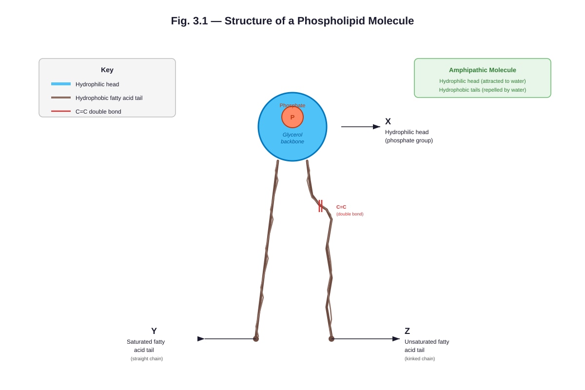
Generated diagram for Q3.
(a) Using Fig. 3.1, explain why phospholipids are described as amphipathic molecules. [2]
(b) Describe how phospholipids arrange themselves when placed in water, and explain the biological significance of this arrangement. [3]
(c) Explain how the presence of the unsaturated fatty acid tail (labelled Z) affects membrane fluidity. [2]
4. Fig. 4.1 shows the effect of temperature on the rate of an enzyme-catalysed reaction.
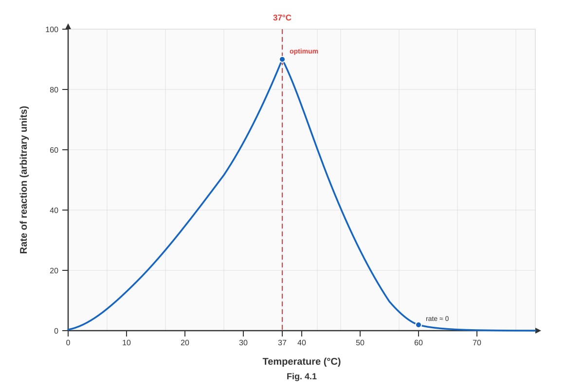
Generated graph for Q4.
(a) With reference to Fig. 4.1, describe the effect of temperature on the rate of this enzyme-catalysed reaction between 0 °C and 37 °C. [2]
(b) Explain the decrease in the rate of reaction above 37 °C. [3]
(c) A student repeated the experiment using the same enzyme but at pH 2. Sketch on Fig. 4.1 the curve you would expect to obtain. Label this curve "pH 2". [2]
5. Fig. 5.1 shows a section through a cell surface membrane, illustrating the fluid mosaic model.
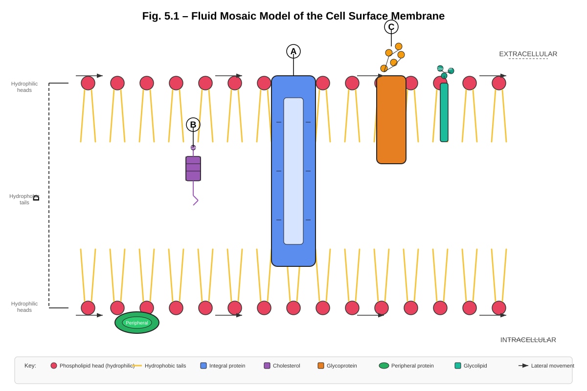
Generated diagram for Q5.
(a) With reference to Fig. 5.1, name the components labelled A, B, and C. [3]
A: ___________________________ B: ___________________________ C: ___________________________
(b) Explain why the model shown in Fig. 5.1 is described as "fluid" and "mosaic". [3]
(c) Describe the role of component B in maintaining membrane stability. [2]
6. A student carried out an experiment to investigate osmosis in potato cylinders. Five potato cylinders of equal length (40 mm) were placed in separate sucrose solutions of different concentrations for 30 minutes. The results are shown in Table 6.1.
Table 6.1
| Sucrose concentration / mol dm⁻³ | Initial length of potato cylinder / mm | Final length of potato cylinder / mm | Change in length / mm |
|---|---|---|---|
| 0.0 | 40 | 43.2 | +3.2 |
| 0.2 | 40 | 41.0 | +1.0 |
| 0.4 | 40 | 40.0 | 0.0 |
| 0.6 | 40 | 38.4 | −1.6 |
| 0.8 | 40 | 36.8 | −3.2 |
(a) Calculate the percentage change in length for the potato cylinder placed in 0.6 mol dm⁻³ sucrose solution. Show your working. [2]
(b) Estimate the sucrose concentration that is approximately equal to the water potential of the potato cells. Explain your reasoning. [2]
(c) Explain why the potato cylinder placed in 0.0 mol dm⁻³ sucrose solution increased in length. [3]
Section B: Data and Source-Based Questions [20 marks]
Questions 7–9
Read the following passage and answer Questions 7–9.
The Discovery and Significance of Aquaporins
Water movement across cell membranes was long thought to occur solely by simple diffusion through the phospholipid bilayer. However, in the 1980s, Peter Agre and colleagues identified a family of integral membrane proteins that facilitated the rapid movement of water across membranes. These proteins, named aquaporins, form channels that allow water molecules to pass through in single file while excluding ions and other solutes.
Aquaporins are found in many tissues, including the kidneys, where they play a crucial role in water reabsorption. In the collecting ducts of the nephron, aquaporin-2 (AQP2) channels are inserted into the apical membrane in response to the hormone antidiuretic hormone (ADH). When ADH binds to receptors on the basolateral membrane of collecting duct cells, a signalling cascade is triggered that causes vesicles containing AQP2 to fuse with the apical membrane, increasing the permeability of the membrane to water.
In the absence of ADH, AQP2 channels are removed from the membrane by endocytosis, reducing water permeability. This mechanism allows the body to regulate urine concentration and maintain water balance. Mutations in the AQP2 gene can cause nephrogenic diabetes insipidus, a condition characterised by the production of large volumes of dilute urine.
Aquaporins are tetrameric proteins — each functional channel is composed of four identical or similar subunits, each containing a central pore. The pore contains a conserved NPA motif (asparagine-proline-alanine) that acts as a selectivity filter, ensuring that only water molecules and not protons (H⁺ ions) pass through. This is critical because the movement of protons would disrupt the electrochemical gradient across the membrane, which is essential for processes such as ATP synthesis in mitochondria.
7. (a) With reference to the passage, explain how the insertion of AQP2 channels into the apical membrane of collecting duct cells is regulated by ADH. [3]
(b) Suggest why the exclusion of H⁺ ions by aquaporins is important for mitochondrial function. [2]
8. (a) Using information from the passage, explain why a person with nephrogenic diabetes insipidus produces large volumes of dilute urine. [3]
(b) Explain the significance of aquaporins being described as "tetrameric" proteins. [2]
9. A student made the following statement: "Aquaporins allow active transport of water across the membrane." Evaluate this statement using information from the passage and your knowledge of membrane transport. [4]
Section C: Extended Response [10 marks]
Question 10
10. Compare and contrast the structure and function of prokaryotic and eukaryotic cells. In your answer, include:
- At least three structural differences between the two cell types
- How these structural differences relate to functional differences
- An example of how compartmentalisation in eukaryotic cells provides an advantage over prokaryotic cells
[10]
End of Paper
Total Marks: 60
Answers
TuitionGoWhere Practice Paper — Biology H1 A-Level
Answer Key & Marking Scheme
Paper: Practice Paper — Cells & Biomolecules Version: 3 of 5 Total Marks: 60
Section A: Structured Questions [30 marks]
Question 1 [8 marks]
(a) Identify the organelles labelled A, B, C, and D. [4]
- A: Nucleus [1]
- B: Rough endoplasmic reticulum (rough ER) [1]
- C: Mitochondrion (plural: mitochondria) [1]
- D: Golgi apparatus (also accepted: Golgi body / Golgi complex) [1]
Marking note: Award 1 mark per correct identification. Accept "rough ER" in full. Do not accept "endoplasmic reticulum" alone without "rough" for B, as the ribosomes are visible in the diagram.
(b) State one function of the organelle labelled B. [1]
- Synthesis of proteins (for secretion / for export from the cell) [1]
Acceptable alternatives:
- Folding of newly synthesised proteins [1]
- Transport of proteins within the cell [1]
- Adding carbohydrate groups to proteins (glycosylation) [1]
Marking note: The key idea is that rough ER is involved in protein synthesis/processing due to the presence of ribosomes. Do not accept "protein synthesis and transport" as two separate points — only 1 mark is available.
(c) Explain why the organelle labelled C is described as having a "double membrane" and how this structure is related to its function. [3]
- The mitochondrion has an outer membrane and a highly folded inner membrane (cristae) [1]
- The inner membrane provides a large surface area for the attachment of electron carriers / enzymes involved in the electron transport chain [1]
- This increases the efficiency of ATP production via oxidative phosphorylation [1]
Marking note: Award 1 mark for identifying the two membranes (outer + inner/cristae). Award 1 mark for linking the inner membrane/cristae to a larger surface area for electron transport chain components. Award 1 mark for linking this to ATP production. Students must connect structure to function for full marks.
Common mistake: Students may state that the double membrane "protects" the mitochondrion — this is not creditworthy at A-Level. The answer must relate to the function of ATP synthesis.
Question 2 [7 marks]
(a) Identify the biological molecule(s) present in each food sample. [3]
- P: Lipid (fats/oil) [1] — positive emulsion test (translucent), negative for reducing sugars, starch, and proteins
- Q: Reducing sugar AND lipid [1] — positive Benedict's test (brick-red precipitate) and positive emulsion test; negative for starch and protein
- R: Starch AND protein [1] — positive iodine test (blue-black) and positive Biuret test (purple); negative for reducing sugars and lipids
Marking note: For Q, the student must identify BOTH reducing sugar and lipid for 1 mark. For R, the student must identify BOTH starch and protein for 1 mark. Partial credit is not available within each sub-part.
(b) Explain why Benedict's test requires heating in a water bath before a positive result can be observed. [2]
- Heating provides the activation energy needed for the redox reaction between the reducing sugar and copper(II) sulfate in Benedict's reagent [1]
- The reducing sugar reduces blue Cu²⁺ ions (copper(II)) to orange/red Cu⁺ ions (copper(I)), forming a brick-red precipitate of copper(I) oxide [1]
Marking note: Award 1 mark for mentioning activation energy / the need for energy to drive the reaction. Award 1 mark for describing the reduction of Cu²⁺ to Cu⁺ (or the formation of copper(I) oxide precipitate). Students who only state "to speed up the reaction" without mentioning activation energy receive only 1 mark.
(c) A student claims that sample P contains no biological molecules. Evaluate this claim. [2]
- The claim is incorrect / not valid [1]
- Sample P tested positive in the emulsion test (translucent), which indicates the presence of lipids / fats [1]
Marking note: Award 1 mark for rejecting the claim. Award 1 mark for citing the evidence (positive emulsion test → lipids present). Students must use the data from the table to support their evaluation.
Question 3 [7 marks]
(a) Using Fig. 3.1, explain why phospholipids are described as amphipathic molecules. [2]
- Phospholipids have a hydrophilic (water-loving) phosphate head [1] and two hydrophobic (water-hating) fatty acid tails [1]
- "Amphipathic" means the molecule has both hydrophilic and hydrophobic regions [1]
Marking note: Maximum 2 marks. Award 1 mark for identifying the hydrophilic head, 1 mark for identifying the hydrophobic tails. The definition of amphipathic can be awarded as part of either mark if clearly stated.
(b) Describe how phospholipids arrange themselves when placed in water, and explain the biological significance of this arrangement. [3]
- Phospholipids spontaneously arrange into a bilayer in water, with hydrophilic heads facing the aqueous environment (outward) and hydrophobic tails facing inward, away from water [1]
- This forms the basic structure of the cell membrane / plasma membrane [1]
- This arrangement creates a selectively permeable barrier that controls the movement of substances into and out of the cell [1]
Marking note: Award 1 mark for describing the bilayer arrangement (heads outward, tails inward). Award 1 mark for identifying this as the basis of the cell membrane. Award 1 mark for explaining the biological significance (selective permeability / barrier function).
(c) Explain how the presence of the unsaturated fatty acid tail (labelled Z) affects membrane fluidity. [2]
- The kink/bend in the unsaturated fatty acid tail (caused by the double bond) prevents the phospholipid molecules from packing closely together [1]
- This increases membrane fluidity / makes the membrane more fluid [1]
Marking note: Award 1 mark for explaining that the kink prevents tight packing. Award 1 mark for stating that this increases fluidity. Students must link the structural feature (kink from C=C double bond) to the effect on packing and then to fluidity for full marks.
Question 4 [7 marks]
(a) With reference to Fig. 4.1, describe the effect of temperature on the rate of this enzyme-catalysed reaction between 0 °C and 37 °C. [2]
- As temperature increases from 0 °C to 37 °C, the rate of reaction increases [1]
- This is because increasing temperature increases the kinetic energy of both enzyme and substrate molecules, leading to more frequent and more energetic successful collisions / more enzyme-substrate complexes formed per unit time [1]
Marking note: Award 1 mark for describing the trend (rate increases with temperature). Award 1 mark for the explanation involving kinetic energy and collision frequency. Students must refer to the data range (0–37°C) for the first mark.
(b) Explain the decrease in the rate of reaction above 37 °C. [3]
- Above 37 °C, the enzyme molecules gain excessive kinetic energy that disrupts the hydrogen bonds and other weak interactions (e.g., ionic bonds, hydrophobic interactions) that maintain the tertiary structure of the enzyme [1]
- This causes the active site to change shape / the enzyme to denature [1]
- Substrate molecules can no longer bind effectively to the altered active site, so fewer enzyme-substrate complexes are formed and the rate of reaction decreases [1]
Marking note: Award 1 mark for identifying disruption of bonds maintaining tertiary structure. Award 1 mark for stating that the active site changes shape (denaturation). Award 1 mark for linking this to reduced substrate binding / fewer enzyme-substrate complexes. All three points are required for full marks.
(c) A student repeated the experiment using the same enzyme but at pH 2. Sketch on Fig. 4.1 the curve you would expect to obtain. Label this curve "pH 2". [2]
- The curve should be drawn below the original curve at all temperatures [1]
- The curve should show a lower optimum rate and may show a lower optimum temperature; the curve should drop to zero at a lower temperature than the original (or the rate should be very low across the entire range) [1]
Marking note: Award 1 mark for a curve that lies entirely below the original curve. Award 1 mark for showing a reduced maximum rate (lower peak). The curve does not need to be perfectly shaped, but it must be clearly lower than the original. If the student draws a curve that crosses above the original at any point, do not award the first mark.
Teaching note: At pH 2, the enzyme is likely to be partially or fully denatured due to the highly acidic conditions disrupting its tertiary structure. This means the enzyme's catalytic efficiency is reduced across all temperatures, resulting in a lower overall rate of reaction.
Question 5 [8 marks]
(a) With reference to Fig. 5.1, name the components labelled A, B, and C. [3]
- A: Integral protein / transmembrane protein [1]
- B: Cholesterol [1]
- C: Glycoprotein [1]
Marking note: Award 1 mark per correct identification. For A, accept "intrinsic protein" or "channel protein" or "carrier protein" if the diagram shows a transmembrane protein. For C, accept "glycoprotein" only — not "protein" alone, as the carbohydrate chain is visible.
(b) Explain why the model shown in Fig. 5.1 is described as "fluid" and "mosaic". [3]
- "Fluid": The phospholipid molecules (and some proteins) can move laterally / sideways within the membrane, giving the membrane a flexible, fluid nature [1]
- "Mosaic": The membrane is composed of many different components (phospholipids, proteins, cholesterol, glycoproteins, glycolipids) arranged in a pattern resembling a mosaic [1]
- The combination of these two properties means the membrane is a dynamic structure with various components able to move within the fluid phospholipid bilayer [1]
Marking note: Award 1 mark for explaining "fluid" (lateral movement of components). Award 1 mark for explaining "mosaic" (variety of different components). Award 1 mark for a clear synthesis statement linking both terms to the dynamic nature of the membrane.
(c) Describe the role of component B (cholesterol) in maintaining membrane stability. [2]
- Cholesterol molecules are positioned between the phospholipid tails in the hydrophobic region of the bilayer [1]
- At high temperatures, cholesterol reduces membrane fluidity by restricting the movement of phospholipid tails; at low temperatures, it prevents the membrane from becoming too rigid by preventing close packing of phospholipids [1]
Marking note: Award 1 mark for describing the location of cholesterol within the bilayer. Award 1 mark for explaining its dual role in regulating fluidity at both high and low temperatures. Students who only mention one temperature effect receive 1 mark.
Question 6 [7 marks]
(a) Calculate the percentage change in length for the potato cylinder placed in 0.6 mol dm⁻³ sucrose solution. Show your working. [2]
Working:
Percentage change=Initial lengthFinal length−Initial length×100%
Percentage change=40.038.4−40.0×100%
Percentage change=40.0−1.6×100%=−4.0%
Answer: −4.0% (or 4.0% decrease) [1] for correct answer with working [1]
Marking note: Award 1 mark for correct working (formula + substitution). Award 1 mark for the correct final answer (−4.0%). If the student shows correct working but makes an arithmetic error, award 1 mark for the method. If the student writes the correct answer without working, award 1 mark only.
(b) Estimate the sucrose concentration that is approximately equal to the water potential of the potato cells. Explain your reasoning. [2]
- Approximately 0.4 mol dm⁻³ [1]
- At this concentration, there is no change in length (0.0 mm), meaning there is no net movement of water into or out of the potato cells — the water potential of the sucrose solution equals the water potential of the potato cell sap [1]
Marking note: Award 1 mark for identifying 0.4 mol dm⁻³. Award 1 mark for explaining that no net osmosis occurs when the external solution water potential equals the cell's water potential.
(c) Explain why the potato cylinder placed in 0.0 mol dm⁻³ sucrose solution increased in length. [3]
- The 0.0 mol dm⁻³ sucrose solution is effectively distilled water, which has a higher water potential (less negative / closer to zero) than the potato cell sap [1]
- Water molecules move by osmosis from the solution (higher water potential) into the potato cells (lower water potential) across the selectively permeable cell membrane [1]
- The influx of water causes the cells to become turgid / swell, resulting in an increase in the overall length of the potato cylinder [1]
Marking note: Award 1 mark for comparing water potentials (external > internal). Award 1 mark for describing osmosis as the mechanism of water movement. Award 1 mark for linking water influx to cell swelling/turgidity and the observed increase in length.
Section B: Data and Source-Based Questions [20 marks]
Question 7 [5 marks]
(a) With reference to the passage, explain how the insertion of AQP2 channels into the apical membrane of collecting duct cells is regulated by ADH. [3]
- ADH binds to specific receptors on the basolateral membrane of collecting duct cells [1]
- This triggers a signalling cascade (intracellular signalling pathway) within the cell [1]
- The signalling cascade causes vesicles containing AQP2 channels to fuse with the apical membrane, inserting the channels and increasing the membrane's permeability to water [1]
Marking note: Award 1 mark for ADH binding to basolateral receptors. Award 1 mark for mentioning the signalling cascade. Award 1 mark for vesicle fusion with the apical membrane. Students must describe the sequence for full marks.
(b) Suggest why the exclusion of H⁺ ions by aquaporins is important for mitochondrial function. [2]
- Mitochondria generate ATP via oxidative phosphorylation, which depends on a proton (H⁺) gradient across the inner mitochondrial membrane [1]
- If aquaporins allowed H⁺ ions to pass through, this would dissipate / disrupt the proton gradient, reducing the efficiency of ATP synthesis [1]
Marking note: Award 1 mark for identifying the proton gradient as essential for ATP synthesis in mitochondria. Award 1 mark for explaining that H⁺ leakage would disrupt this gradient. Students do not need to name chemiosmosis explicitly, but the concept must be clear.
Question 8 [5 marks]
(a) Using information from the passage, explain why a person with nephrogenic diabetes insipidus produces large volumes of dilute urine. [3]
- Nephrogenic diabetes insipidus is caused by mutations in the AQP2 gene, resulting in non-functional or absent AQP2 channels [1]
- Without functional AQP2 channels, the collecting duct membrane has reduced permeability to water [1]
- Less water is reabsorbed from the filtrate back into the blood, so a large volume of dilute (watery) urine is produced [1]
Marking note: Award 1 mark for linking the condition to AQP2 mutations. Award 1 mark for stating reduced water permeability of the collecting duct. Award 1 mark for explaining the consequence (less water reabsorption → large volume of dilute urine).
(b) Explain the significance of aquaporins being described as "tetrameric" proteins. [2]
- Each functional aquaporin channel is composed of four subunits (a tetramer) [1]
- Each subunit contains its own central pore, so one tetrameric complex can transport four water molecules simultaneously, increasing the efficiency of water transport [1]
Marking note: Award 1 mark for stating that four subunits form one functional channel. Award 1 mark for explaining that this increases transport efficiency (four pores per complex). Students who only state "four subunits" without explaining the functional significance receive 1 mark.
Question 9 [4 marks]
Evaluate the statement: "Aquaporins allow active transport of water across the membrane." [4]
- The statement is incorrect [1]
- Aquaporins facilitate the movement of water by facilitated diffusion, not active transport [1]
- Water moves passively down its water potential gradient (from high to low water potential) through the aquaporin channel — no ATP / energy input is required [1]
- Active transport requires energy (usually in the form of ATP) to move substances against their concentration gradient, which is not the case for water movement through aquaporins [1]
Marking note: Award 1 mark for stating the statement is incorrect. Award 1 mark for identifying the correct mechanism (facilitated diffusion). Award 1 mark for explaining that water moves passively down its water potential gradient. Award 1 mark for contrasting with active transport (requires ATP, moves against gradient). All four points required for full marks.
Common mistake: Students may confuse facilitated diffusion with active transport because a membrane protein is involved. Emphasise that the key distinction is whether energy (ATP) is required and whether the substance moves against or down its gradient.
Section C: Extended Response [10 marks]
Question 10 [10 marks]
Compare and contrast the structure and function of prokaryotic and eukaryotic cells.
Marking descriptors:
| Marks | Descriptor |
|---|---|
| 9–10 | Comprehensive answer with at least 3 clearly explained structural differences, well-linked to functional differences, and a clear example of the advantage of compartmentalisation. Biological terminology is precise and used correctly throughout. Answer is well-organised and coherent. |
| 7–8 | Good answer with 3 structural differences and some explanation of functional links. Compartmentalisation advantage is mentioned but may lack depth. Minor omissions or imprecise terminology. |
| 5–6 | Adequate answer with at least 2 structural differences. Functional links may be weak or missing. Compartmentalisation may be mentioned but not explained. Some biological terms used incorrectly. |
| 3–4 | Limited answer with 1–2 structural differences. Little or no explanation of functional significance. Compartmentalisation not addressed. Significant errors or omissions. |
| 1–2 | Very limited response with minimal relevant content. Major misconceptions may be present. |
| 0 | No relevant content. |
Expected content points (for 10 marks, students should include most of the following):
Structural differences (at least 3 required):
-
Nucleus: Prokaryotic cells lack a membrane-bound nucleus; their DNA is free in the nucleoid region. Eukaryotic cells have a true nucleus enclosed by a double nuclear envelope with nuclear pores. [1] Functional significance: The nuclear envelope in eukaryotes separates transcription (nucleus) from translation (cytoplasm), allowing for more complex gene regulation. [1]
-
Membrane-bound organelles: Prokaryotic cells lack membrane-bound organelles (no mitochondria, ER, Golgi, lysosomes). Eukaryotic cells possess these organelles. [1] Functional significance: Membrane-bound organelles allow different metabolic processes to occur in separate, specialised compartments, increasing efficiency. [1]
-
Ribosomes: Prokaryotic cells have 70S ribosomes (smaller). Eukaryotic cells have 80S ribosomes (larger) in the cytoplasm and on the rough ER, and 70S ribosomes in mitochondria and chloroplasts. [1] Functional significance: The difference in ribosome structure is exploited by antibiotics, which target prokaryotic 70S ribosomes without affecting eukaryotic 80S ribosomes. [1]
-
Cell wall composition (bonus point): Prokaryotic cell walls are made of peptidoglycan. Eukaryotic plant cell walls are made of cellulose (fungal cell walls are made of chitin). [1]
Compartmentalisation advantage (required):
- In eukaryotic cells, compartmentalisation by membrane-bound organelles allows incompatible biochemical reactions to occur simultaneously in different parts of the cell [1]
- Example: Lysosomes contain hydrolytic enzymes that function optimally at pH ~5. These enzymes are separated from the cytoplasm (pH ~7.2) by the lysosomal membrane. If these enzymes were released into the cytoplasm, they would digest the cell's own components (autolysis). The compartmentalisation therefore protects the cell while allowing intracellular digestion to occur. [1]
Alternative acceptable example of compartmentalisation advantage:
- The nuclear envelope separates transcription from translation, allowing mRNA processing (splicing, capping, polyadenylation) before translation, which enables post-transcriptional regulation of gene expression.
- Mitochondrial membranes create compartments (matrix, intermembrane space) that allow the establishment of a proton gradient for ATP synthesis.
Marking note: Award marks for each valid structural difference (up to 3 differences × 2 marks each = 6 marks: 1 for the difference, 1 for the functional link). Award up to 2 marks for the compartmentalisation advantage (1 for the general principle, 1 for a specific example). Award up to 2 marks for the overall quality of organisation, coherence, and use of biological terminology.
End of Answer Key
Total Marks: 60
| Section | Marks |
|---|---|
| A: Structured Questions (Q1–Q6) | 30 |
| B: Data/Source-Based (Q7–Q9) | 20 |
| C: Extended Response (Q10) | 10 |
| Total | 60 |
Free quiz and exam paper access
Enter your details to view this paper
Your access is remembered on this device.