AI Generated Exam Paper
A Level H1 Biology Practice Paper 1
Free A Level H1 Biology Practice Paper 1, LongCat AI version, with questions, answers, and A Level-style practice for Singapore students.
These static practice materials are generated from the site's syllabus and paper-generation workflow, with source and model context shown so students and parents can evaluate the material before use.
Questions
TuitionGoWhere Practice Paper - Biology H1 A-Level
TuitionGoWhere Practice Paper (AI)
Subject: Biology Level: A-Level H1 Paper: Practice Paper — Cells & Biomolecules Duration: 1 hour 30 minutes Total Marks: 60 Name: ___________________________ Class: ___________________________ Date: ___________________________
Instructions
- Answer all questions in the spaces provided.
- Write your answers in the blank spaces or on lined pages as appropriate.
- The number of marks for each question or part-question is shown in brackets [ ].
- You are advised to spend no more than 20 minutes on Section A, 35 minutes on Section B, and 25 minutes on Section C.
- A clean copy of the Periodic Table is NOT required for this paper.
- Where diagrams are referenced, refer to the figure indicated.
Section A: Multiple Choice [10 marks]
Questions 1–10: Choose the one best answer. Each question carries 1 mark.
1. Which organelle is responsible for the synthesis of lipids and detoxification of drugs in eukaryotic cells?
A. Rough endoplasmic reticulum B. Smooth endoplasmic reticulum C. Golgi apparatus D. Lysosome
2. A student observed a cell under an electron microscope and noted the absence of membrane-bound organelles. This cell is most likely:
A. a plant cell. B. an animal cell. C. a prokaryotic cell. D. a fungal cell.
3. Which of the following best describes the role of cholesterol in the cell membrane?
A. It provides energy for active transport across the membrane. B. It increases membrane fluidity at all temperatures. C. It regulates membrane fluidity by restricting phospholipid movement at high temperatures and preventing tight packing at low temperatures. D. It acts as a receptor for hormone signalling.
4. The diagram below represents a section of a cell membrane.
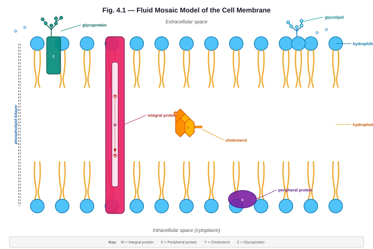
Generated diagram for Q4.
Fig. 4.1 shows the fluid mosaic model of a cell membrane.
Which labelled component is responsible for cell-cell recognition?
A. W B. X C. Y D. Z
5. Which of the following molecules is a monosaccharide?
A. Maltose B. Sucrose C. Lactose D. Fructose
6. During an enzyme-catalysed reaction, the rate of reaction decreases when the temperature exceeds the optimum. This is because:
A. the substrate molecules move too slowly to bind to the enzyme. B. the enzyme is denatured and the active site changes shape. C. the enzyme-substrate complex becomes too stable. D. the activation energy increases.
7. A solution of starch is placed in dialysis tubing and suspended in distilled water. After 30 minutes, which test would confirm that starch has not passed through the tubing?
A. Adding iodine solution to the water outside the tubing — a blue-black colour is observed. B. Adding iodine solution to the water outside the tubing — no blue-black colour is observed. C. Adding Benedict's solution to the water outside the tubing and heating — a brick-red precipitate forms. D. Adding biuret solution to the water outside the tubing — a violet colour is observed.
8. Which type of bond is primarily responsible for holding the two strands of DNA together?
A. Covalent bonds B. Ionic bonds C. Hydrogen bonds D. Peptide bonds
9. A protein is composed of 150 amino acids. How many water molecules were released during the condensation reactions that formed this single polypeptide chain?
A. 75 B. 149 C. 150 D. 151
10. Which of the following is a function of the Golgi apparatus?
A. Synthesising ribosomal RNA. B. Modifying, sorting, and packaging proteins for secretion. C. Breaking down damaged organelles. D. Producing ATP through oxidative phosphorylation.
Section B: Structured Questions [30 marks]
Answer all questions. Show your working where applicable.
11. Fig. 11.1 shows the structure of an animal cell as seen under an electron microscope.
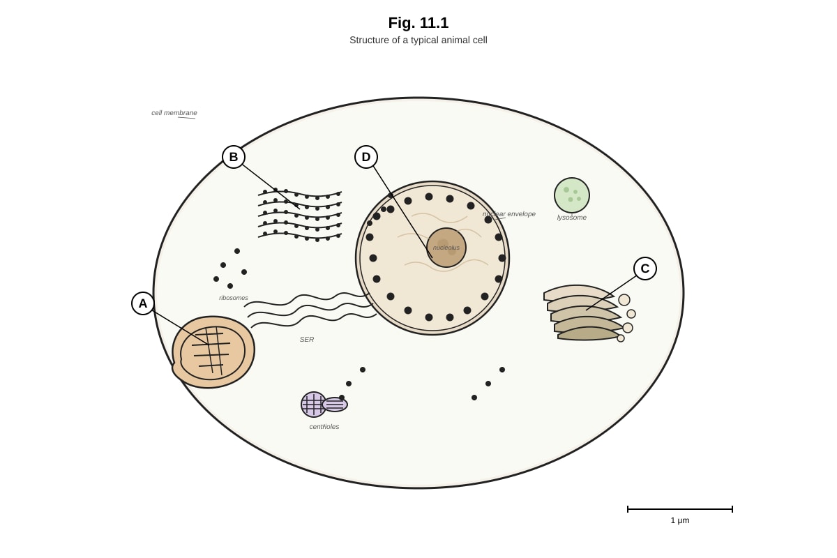
Generated diagram for Q11.
(a) Identify the organelles labelled A, B, C, and D. [4]
(b) State one structural difference between the rough endoplasmic reticulum and the smooth endoplasmic reticulum. [1]
(c) Explain why cells that secrete large amounts of digestive enzymes would have a well-developed Golgi apparatus. [2]
(d) Describe the function of the mitochondrion and explain how its structure is adapted to this function. [3]
[Total: 10 marks]
12. Fig. 12.1 shows the effect of temperature on the rate of an enzyme-catalysed reaction.
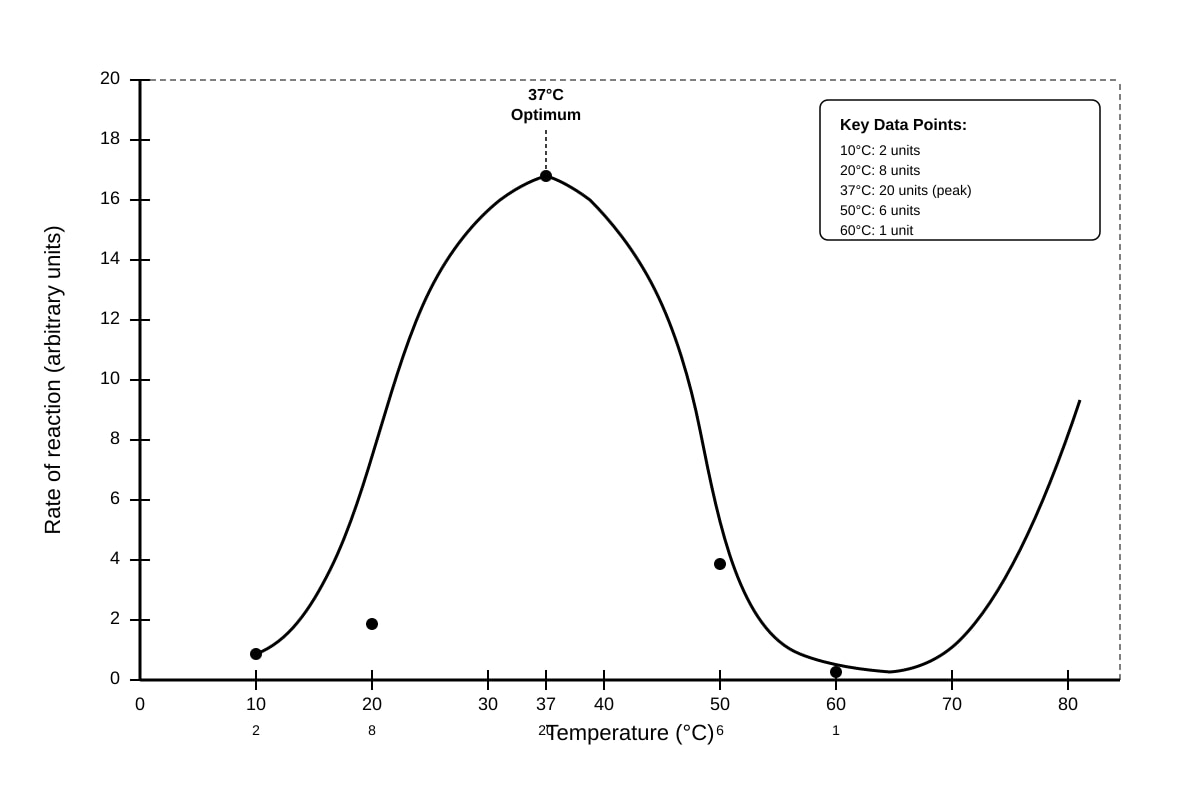
Generated graph for Q12.
(a) Describe the relationship between temperature and the rate of reaction as shown in Fig. 12.1. [3]
(b) Explain why the rate of reaction increases between 10°C and 37°C. [2]
(c) Explain why the rate of reaction decreases sharply above 37°C. [2]
(d) A student repeated the experiment using an enzyme from a thermophilic bacterium. Predict, with a reason, how the optimum temperature would differ from that shown in Fig. 12.1. [2]
[Total: 9 marks]
13. Table 13.1 shows the results of tests carried out on four unknown biological samples (P, Q, R, and S).
| Sample | Benedict's test (after heating) | Iodine test | Biuret test | Ethanol emulsion test |
|---|---|---|---|---|
| P | Blue | Brown-yellow | Violet | Cloudy white |
| Q | Brick-red | Brown-yellow | Blue | Clear |
| R | Blue | Blue-black | Blue | Clear |
| S | Blue | Brown-yellow | Blue | Cloudy white |
(a) Identify the biological molecule(s) present in each sample. [4]
(b) Explain why Benedict's test requires heating to produce a positive result. [2]
(c) Describe how you would carry out the ethanol emulsion test on an unknown sample. [3]
[Total: 9 marks]
14. Fig. 14.1 shows the process of osmosis in plant cells placed in solutions of different concentrations.
Image pending generation: diagram for Q14.
(a) With reference to Fig. 14.1, describe what has happened to the cells in terms of water movement. [3]
(b) Explain why animal cells would not become turgid in a hypotonic solution. [2]
(c) A potato cylinder was placed in distilled water for 2 hours. Explain, in terms of water potential, why the potato cylinder increased in mass. [3]
[Total: 8 marks]
Section C: Free Response [20 marks]
Answer two questions. Each question carries 10 marks. Write your answers in continuous prose, where appropriate.
15. (a) Describe the structure of DNA, including the arrangement of nucleotides, the types of bonds involved, and the significance of complementary base pairing. [6]
(b) Explain how the structure of DNA enables it to carry out its role in the storage and transmission of genetic information. [4]
[Total: 10 marks]
16. (a) Compare and contrast active transport and facilitated diffusion, giving one example of each in living organisms. [6]
(b) Explain how the cell membrane's structure enables it to be selectively permeable. [4]
[Total: 10 marks]
17. (a) Describe the structure and function of proteins, including the four levels of protein structure and the types of bonds that stabilise each level. [6]
(b) Explain how the structure of an enzyme relates to its function, and discuss how competitive and non-competitive inhibition affect enzyme activity. [4]
[Total: 10 marks]
End of Paper
Answers
TuitionGoWhere Practice Paper - Biology H1 A-Level
Answer Key — Cells & Biomolecules
Section A: Multiple Choice [10 marks]
1. B — Smooth endoplasmic reticulum [1]
The smooth endoplasmic reticulum (SER) is responsible for lipid synthesis and detoxification. The rough endoplasmic reticulum (RER) has ribosomes attached and is involved in protein synthesis. The Golgi apparatus modifies and packages proteins, and lysosomes digest cellular waste.
Common mistake: Students often confuse RER and SER functions. Remember: "Smooth = lipids, Rough = proteins."
2. C — a prokaryotic cell [1]
Prokaryotic cells (e.g., bacteria) lack membrane-bound organelles such as a nucleus, mitochondria, and endoplasmic reticulum. Plant, animal, and fungal cells are all eukaryotic and possess membrane-bound organelles.
3. C — It regulates membrane fluidity by restricting phospholipid movement at high temperatures and preventing tight packing at low temperatures. [1]
Cholesterol acts as a fluidity buffer. At high temperatures, it restricts the movement of phospholipids, reducing fluidity. At low temperatures, it prevents the fatty acid tails from packing too closely together, maintaining fluidity. It does not provide energy (A) or solely increase fluidity (B).
Common mistake: Selecting B — cholesterol does not simply increase fluidity; it regulates it in both directions.
4. C — Y [1]
Glycoproteins (carbohydrate chains attached to proteins on the extracellular surface of the membrane) are responsible for cell-cell recognition. In the diagram, Y should be the glycoprotein. Glycolipids also play a role in cell recognition, but glycoproteins are the primary markers.
Note: The specific letter answer depends on the diagram labels. In the described diagram, Y = glycoprotein. If the generated image uses different letters, the answer should point to the glycoprotein component.
5. D — Fructose [1]
Fructose is a monosaccharide (a single sugar unit). Maltose, sucrose, and lactose are all disaccharides (composed of two monosaccharide units).
Key concept: Mono- = one, Di- = two, Poly- = many sugar units.
6. B — the enzyme is denatured and the active site changes shape. [1]
Above the optimum temperature, the excessive kinetic energy disrupts the hydrogen bonds and other weak interactions that maintain the enzyme's tertiary structure. This causes the active site to lose its specific shape, so substrate molecules can no longer bind effectively. This is called denaturation and is usually irreversible.
Common mistake: Selecting D — the activation energy is a property of the reaction, not something that changes with temperature. The enzyme lowers the activation energy; when denatured, it can no longer do so.
7. B — Adding iodine solution to the water outside the tubing — no blue-black colour is observed. [1]
Starch is a large polysaccharide that cannot pass through the pores of dialysis tubing. If starch has not moved into the surrounding water, adding iodine solution will not produce the characteristic blue-black colour. A negative result (no colour change) confirms starch has remained inside the tubing.
Common mistake: Selecting A — a blue-black colour would indicate starch has passed through, which would mean the tubing was permeable to starch (unlikely).
8. C — Hydrogen bonds [1]
The two strands of DNA are held together by hydrogen bonds between complementary base pairs: adenine forms two hydrogen bonds with thymine, and guanine forms three hydrogen bonds with cytosine. Covalent bonds form the sugar-phosphate backbone within each strand. Peptide bonds are found in proteins, not DNA.
9. B — 149 [1]
Each condensation reaction between two amino acids releases one water molecule. For a polypeptide chain of n amino acids, the number of peptide bonds (and therefore water molecules released) is n − 1. For 150 amino acids: 150 − 1 = 149 water molecules.
Working: Number of water molecules = Number of amino acids − 1 = 150 − 1 = 149
Common mistake: Selecting C (150) — students forget that the number of bonds is always one less than the number of monomers in a linear chain.
10. B — Modifying, sorting, and packaging proteins for secretion. [1]
The Golgi apparatus receives proteins from the RER, modifies them (e.g., by adding carbohydrate groups), sorts them, and packages them into vesicles for transport to their final destinations (secretion, lysosomes, or the cell membrane). Ribosomal RNA is synthesised in the nucleolus (A), lysosomes break down damaged organelles (C), and mitochondria produce ATP (D).
Section B: Structured Questions [30 marks]
11.
(a) [4 marks — 1 mark each]
- A: Mitochondrion
- B: Rough endoplasmic reticulum (RER)
- C: Golgi apparatus
- D: Nucleus
Marking note: Accept "mitochondria" (plural). Spelling must be sufficiently correct for identification.
(b) [1 mark]
The rough endoplasmic reticulum has ribosomes attached to its cytoplasmic surface, whereas the smooth endoplasmic reticulum lacks ribosomes.
Alternative acceptable answer: The RER appears "rough" under EM due to bound ribosomes; the SER appears smooth.
(c) [2 marks]
Digestive enzymes are proteins. The Golgi apparatus modifies proteins (e.g., by glycosylation) received from the RER [1] and packages them into secretory vesicles for transport to the cell membrane for exocytosis [1].
Teaching note: Students should connect the function of the Golgi to the specific context — cells secreting large amounts of enzymes need extensive protein processing and packaging capacity.
(d) [3 marks]
The mitochondrion is the site of aerobic respiration, where ATP is produced [1]. Its structure is adapted in the following ways:
- The inner membrane is folded into cristae, providing a large surface area for the electron transport chain and ATP synthase enzymes [1].
- The matrix contains enzymes required for the Krebs (citric acid) cycle [1].
Additional credit-worthy points:
- The double membrane creates a compartment (intermembrane space) for the proton gradient.
- The small size allows rapid diffusion of substrates.
Marking note: 1 mark for function + 2 marks for structural adaptations linked to function.
12.
(a) [3 marks]
As temperature increases from 10°C to 37°C, the rate of reaction increases [1], reaching a maximum (optimum) at 37°C [1]. Above 37°C, the rate of reaction decreases sharply as temperature increases further [1].
Marking note: Students must describe all three phases: increase, peak, and decrease. Award 1 mark for each phase correctly described.
(b) [2 marks]
As temperature increases, both the enzyme and substrate molecules gain kinetic energy [1], resulting in more frequent and energetic collisions between enzyme and substrate, increasing the rate of formation of enzyme-substrate complexes [1].
Teaching note: This is a standard explanation linking kinetic energy to collision frequency and reaction rate. Students should mention both enzyme AND substrate molecules.
(c) [2 marks]
Above 37°C, the excessive thermal energy disrupts the hydrogen bonds and other weak interactions that maintain the enzyme's tertiary structure [1]. This causes the active site to change shape (denaturation), so substrate molecules can no longer bind effectively, and the rate of reaction decreases [1].
Common mistake: Students may say "the enzyme is killed" — enzymes are not living organisms and cannot be "killed." The correct term is "denatured."
(d) [2 marks]
The optimum temperature would be higher than 37°C [1], because enzymes from thermophilic bacteria are adapted to function at high temperatures and have more stable tertiary structures (e.g., more disulfide bonds or stronger hydrophobic interactions) that resist denaturation [1].
Acceptable alternative: The optimum would be around 60–80°C, as thermophilic bacteria live in hot environments such as hot springs.
13.
(a) [4 marks — 1 mark each]
- Sample P: Protein (biuret test positive = violet) AND lipid (ethanol emulsion test positive = cloudy white). Benedict's test negative (blue) and iodine test negative (brown-yellow) indicate no reducing sugar or starch.
- Sample Q: Reducing sugar (Benedict's test positive = brick-red). No protein, starch, or lipid detected.
- Sample R: Starch (iodine test positive = blue-black). No reducing sugar, protein, or lipid detected.
- Sample S: Lipid (ethanol emulsion test positive = cloudy white). No reducing sugar, protein, or starch detected.
Marking note: Award 1 mark per sample for correctly identifying all molecules present. If a student identifies only one molecule in a sample containing two, award ½ mark for that sample.
(b) [2 marks]
Benedict's test detects reducing sugars by reducing blue Cu2+ ions (in copper(II) sulfate) to brick-red Cu+ oxide (Cu2O) [1]. Heating provides the activation energy needed for this redox reaction to proceed at a detectable rate [1].
Teaching note: The reaction is a reduction of copper ions by the aldehyde or ketone group of the reducing sugar. Heat is required to overcome the activation energy barrier.
(c) [3 marks]
- Add ethanol (or alcohol) to the sample and shake thoroughly to dissolve any lipids present [1].
- Pour the ethanol mixture into a test tube containing water [1].
- If lipids are present, a cloudy white emulsion forms as the lipid comes out of solution in the water [1].
Alternative: Dissolve the sample in ethanol, then filter; add the filtrate to water — a cloudy white precipitate indicates the presence of lipid.
Marking note: The key steps are: (1) dissolve in ethanol, (2) add to water, (3) observe cloudy white emulsion.
14.
(a) [3 marks]
- Cell A (turgid): The cell was placed in a hypotonic solution (higher water potential than the cell). Water moved into the cell by osmosis [1].
- Cell B (incipient plasmolysis): The cell was placed in an isotonic solution (same water potential as the cell). There was no net movement of water [1].
- Cell C (plasmolysed): The cell was placed in a hypertonic solution (lower water potential than the cell). Water moved out of the cell by osmosis [1].
Teaching note: Students must link each cell state to the direction of water movement and the relative water potentials of the solution and cell contents.
(b) [2 marks]
Animal cells do not have a cell wall [1]. In a hypotonic solution, water enters the cell by osmosis, causing the cell to swell and eventually burst (lyse) rather than becoming turgid [1].
Key concept: Turgidity requires a rigid cell wall to resist the inward pressure of water. Without a cell wall, animal cells are vulnerable to osmotic lysis.
(c) [3 marks]
The water potential of distilled water is 0 kPa (highest possible) [1], which is higher (less negative) than the water potential inside the potato cells [1]. Therefore, water moved into the potato cells by osmosis, from a region of higher water potential to a region of lower water potential, causing the cells to gain mass [1].
Teaching note: Water potential (Ψ) is always negative or zero in biological systems. Distilled water has Ψ=0 kPa. Plant cell contents have Ψ<0 kPa due to dissolved solutes. Water moves from high Ψ to low Ψ.
Section C: Free Response [20 marks]
Answer any two questions. Marking descriptors below indicate the expected content and quality of response.
15.
(a) Describe the structure of DNA [6 marks]
Marking descriptors (indicative content):
- DNA is a double-stranded polynucleotide. Each nucleotide consists of a deoxyribose sugar, a phosphate group, and a nitrogenous base [1].
- The four nitrogenous bases are adenine (A), thymine (T), guanine (G), and cytosine (C) [1].
- Nucleotides are joined by phosphodiester bonds between the 3' carbon of one deoxyribose and the 5' carbon of the next, forming a sugar-phosphate backbone [1].
- The two strands run in opposite directions (antiparallel): one runs 5'→3' and the other 3'→5' [1].
- The two strands are held together by hydrogen bonds between complementary base pairs: A pairs with T (2 hydrogen bonds), G pairs with C (3 hydrogen bonds) [1].
- The molecule twists to form a double helix, with the bases stacked in the interior and the sugar-phosphate backbones on the outside [1].
Marking: 1 mark per valid point, up to 6 marks. Award marks for clearly labelled diagrams that show these features.
(b) Explain how the structure of DNA enables it to carry out its role in the storage and transmission of genetic information [4 marks]
Marking descriptors:
- The sequence of bases along the DNA strand encodes genetic information. The four bases can be arranged in any order, providing virtually unlimited variation in the information stored [1].
- Complementary base pairing ensures that during DNA replication, each strand can serve as a template for the synthesis of a new complementary strand, enabling accurate copying of genetic information [1].
- The double-stranded structure provides a backup copy of the genetic information — if one strand is damaged, the information can be repaired using the complementary strand as a template [1].
- The hydrogen bonds between base pairs are weak enough to allow the strands to be separated during replication and transcription, but strong enough to maintain the double helix structure under normal conditions [1].
Additional credit-worthy points:
- The sugar-phosphate backbone is strong and stable (covalent bonds), protecting the genetic information stored in the bases.
- The double helix is compact, allowing large amounts of genetic information to be stored in a small space within the nucleus.
Marking: 1 mark per valid point, up to 4 marks. Responses should demonstrate clear linkage between structure and function.
16.
(a) Compare and contrast active transport and facilitated diffusion [6 marks]
Marking descriptors:
Similarities (at least 1 required):
- Both involve transport across the cell membrane [1].
- Both may involve transmembrane proteins (carrier/channel proteins) [1].
Differences:
| Feature | Active Transport | Facilitated Diffusion |
|---|---|---|
| Energy requirement | Requires ATP | Does not require ATP |
| Direction of transport | Against concentration gradient (low → high) | Down concentration gradient (high → low) |
| Example | Sodium-potassium pump (Na+/K+-ATPase) pumping Na+ out of cells and K+ into cells | Glucose uptake via GLUT transporter proteins in red blood cells or intestinal epithelial cells |
- Active transport requires energy in the form of ATP to move substances against their concentration gradient [1].
- Facilitated diffusion is a passive process that moves substances down their concentration gradient and does not require energy [1].
- Active transport uses carrier proteins that undergo conformational changes driven by ATP hydrolysis [1].
- Facilitated diffusion uses channel proteins or carrier proteins to provide a pathway for specific molecules to cross the membrane [1].
Marking: Award marks for clear comparison, correct examples, and accurate description of energy requirements and direction of transport. Up to 6 marks.
(b) Explain how the cell membrane's structure enables it to be selectively permeable [4 marks]
Marking descriptors:
- The phospholipid bilayer forms the basic structure of the membrane. Small, non-polar molecules (e.g., O2, CO2) can pass through the hydrophobic core of the bilayer by simple diffusion [1].
- Large polar molecules and ions cannot pass through the hydrophobic core and require channel proteins or carrier proteins to cross the membrane [1].
- Channel proteins provide hydrophilic pores that allow specific ions or water molecules (via aquaporins) to pass through the membrane [1].
- Carrier proteins bind to specific molecules and undergo conformational changes to transport them across the membrane, ensuring that only certain substances are transported [1].
Additional credit-worthy points:
- Cholesterol regulates membrane fluidity, which affects the permeability of the membrane.
- Glycoproteins and glycolipids on the surface may play roles in cell recognition and signalling, indirectly influencing what enters or leaves the cell.
Marking: 1 mark per valid point, up to 4 marks. Responses should clearly link structural features to the selective permeability function.
17.
(a) Describe the structure and function of proteins [6 marks]
Marking descriptors:
Structure:
- Primary structure: The sequence of amino acids in a polypeptide chain, linked by peptide bonds [1].
- Secondary structure: Local folding of the polypeptide chain into alpha-helices or beta-pleated sheets, stabilised by hydrogen bonds between the backbone C=O and N–H groups [1].
- Tertiary structure: The overall three-dimensional shape of the protein, stabilised by interactions between R-groups including hydrogen bonds, ionic bonds, disulfide bonds (covalent), and hydrophobic interactions [1].
- Quaternary structure: The arrangement of two or more polypeptide subunits into a functional protein (e.g., haemoglobin has four subunits) [1].
Function (any two for 2 marks):
- Enzymes: catalyse biochemical reactions (e.g., amylase, catalase).
- Structural proteins: provide support (e.g., collagen in connective tissue, keratin in hair).
- Transport proteins: carry substances (e.g., haemoglobin transports oxygen).
- Hormones: chemical messengers (e.g., insulin regulates blood glucose).
- Antibodies: defence against pathogens (e.g., immunoglobulins).
- Receptor proteins: receive chemical signals on cell surfaces.
Marking: Up to 4 marks for structure (1 per level) + up to 2 marks for functions (1 per valid function with example).
(b) Explain how the structure of an enzyme relates to its function, and discuss competitive and non-competitive inhibition [4 marks]
Marking descriptors:
- Enzymes are proteins with a specific three-dimensional shape. The active site is a region on the enzyme with a specific shape and chemical properties that is complementary to the substrate [1].
- The substrate binds to the active site to form an enzyme-substrate complex. The specificity of the active site determines which substrate the enzyme can act on (lock-and-key or induced fit model) [1].
- Competitive inhibition: The inhibitor molecule has a similar shape to the substrate and competes for the active site. It blocks the substrate from binding. The effect can be overcome by increasing substrate concentration [1].
- Non-competitive inhibition: The inhibitor binds to a site other than the active site (allosteric site), causing a change in the shape of the enzyme and active site. The substrate can no longer bind effectively. Increasing substrate concentration does not overcome this type of inhibition [1].
Additional credit-worthy points:
- Competitive inhibitors often resemble the substrate structurally.
- Non-competitive inhibition reduces the maximum rate of reaction (Vmax) but does not affect the Km (affinity).
- Competitive inhibition increases the apparent Km but Vmax remains the same if sufficient substrate is added.
Marking: 1 mark per valid point, up to 4 marks. Award credit for clearly labelled diagrams showing inhibition mechanisms.
End of Answer Key
Free quiz and exam paper access
Enter your details to view this paper
Your access is remembered on this device.