From Real Exams Exam Paper
A Level H1 Biology Practice Paper 5
Free A Level H1 Biology Practice Paper 5, HY3 Exam version, with questions, answers, and A Level-style practice for Singapore students.
These static practice materials are generated from the site's syllabus and paper-generation workflow, with source and model context shown so students and parents can evaluate the material before use.
Questions
Free quiz and exam paper access
Enter your details to view this paper
Your access is remembered on this device.
Answers
TuitionGoWhere Exam Practice (AI) – Biology H1 A-Level
Practice Paper: Cells & Biomolecules (Version 5 of 5) – Answer Key
Section A (20 marks)
1. [1] One feature: Cells are the smallest unit of life (or: all cells come from pre-existing cells / living organisms are composed of cells).
Teaching note: Cell theory has three tenets; any one accepted.
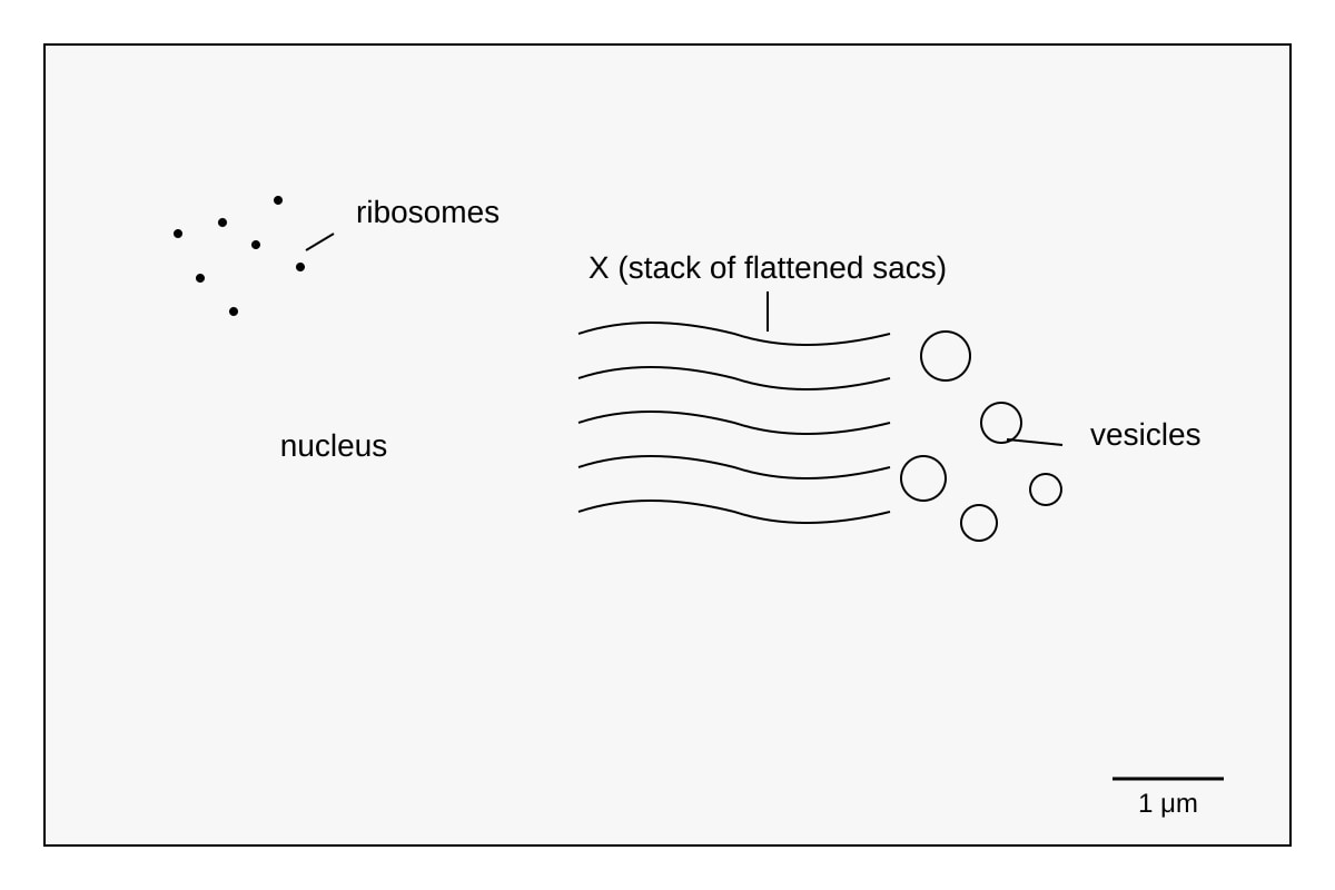
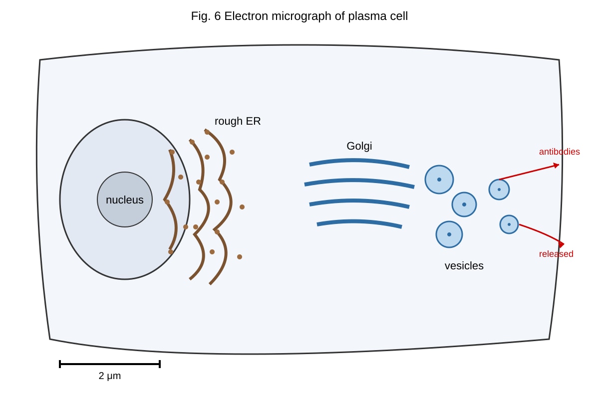
2. [2] Structure X = Golgi body (Golgi apparatus). Function: modifies, sorts, and packages proteins (e.g. enzymes, hormones) into vesicles for secretion.
Marking: 1 for name, 1 for function linked to secretory role.
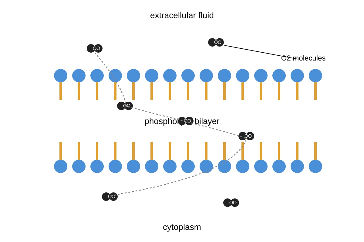
3. [2] Phospholipids form a bilayer; hydrophilic heads face outward to aqueous environments, hydrophobic tails face inward away from water.
Marking: 1 bilayer, 1 orientation of heads/tails.
4. [2] Oxygen moves by simple diffusion directly through the lipid bilayer down its concentration gradient; no protein needed as it is small and non-polar.
Marking: mechanism + through lipid core.
5. [1] Glycosidic bond (α-1,4 or α-1,6 in starch).
6. [1] Zygotic (totipotent) stem cell (or totipotent stem cell).
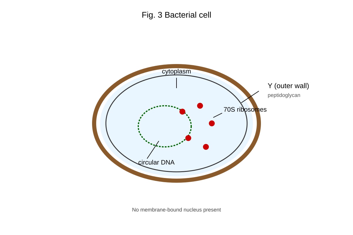
7. [2] Y = peptidoglycan cell wall; differs from plant wall as it is made of peptidoglycan not cellulose.
Marking: 1 name, 1 difference.
8. [2] Induced-fit: enzyme active site changes shape slightly to fit substrate, forming enzyme–substrate complex, lowering activation energy.
Marking: change shape + complex/energy.
9. [1] Cholesterol reduces membrane fluidity at high temp and prevents crystallisation at low temp (stabilises membrane).
10. [2] Example: maltose = glucose + glucose (or sucrose = glucose + fructose; lactose = glucose + galactose).
Marking: 1 example, 1 monomers.
Section B (25 marks)
11. [4 total]
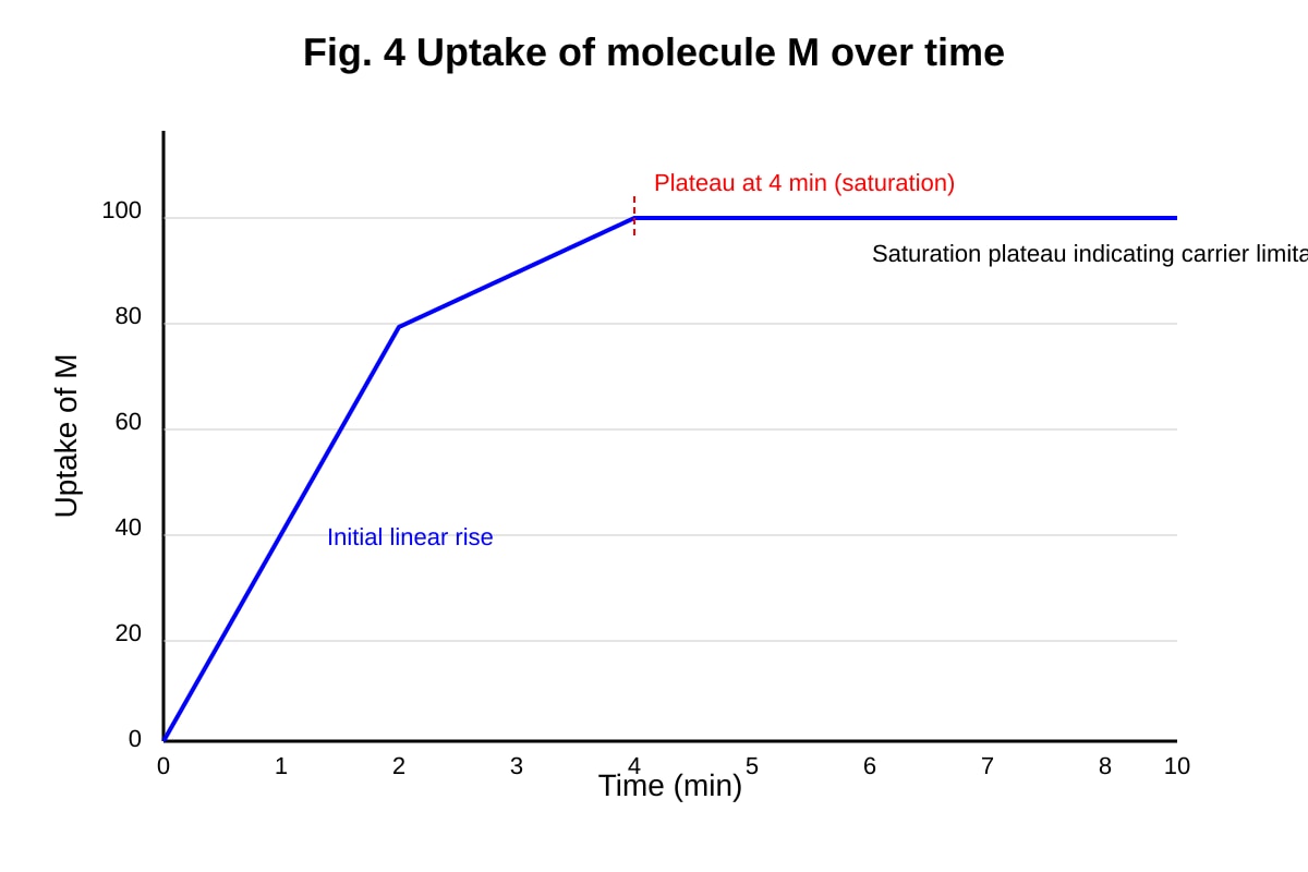
(a) [1] Facilitated diffusion (or carrier-mediated diffusion).
(b) [2] Graph plateaus → saturation of carriers; shows protein carriers limited, not simple diffusion.
(c) [1] Channel or carrier protein in membrane.
12. [4 total]
(a) [1] e.g. mitochondrion (or nucleus, Golgi, ER).
(b) [1] ATP production / aerobic respiration.
(c) [2] Bacterium has 70S ribosomes and circular DNA; can transcribe/translate proteins without membrane-bound organelles.
13. [4 total]
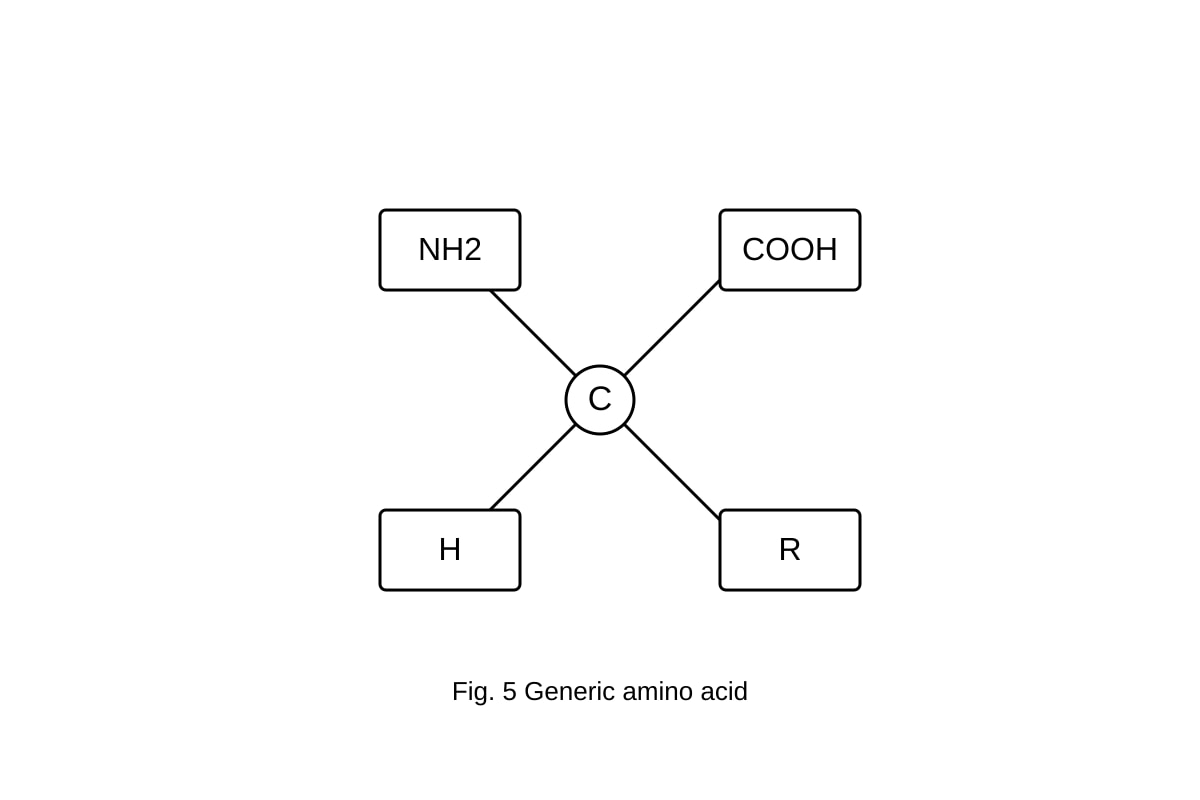
(a) [1] NH2 circled, COOH boxed.
(b) [2] Condensation: –OH from carboxyl of one + –H from amino of next removed as H2O; peptide bond forms.
(c) [1] Condensation (dehydration).
14. [6 total]
(a) [2] Optimum = 40 °C (peak rate).
(b) [2] Above 40 °C, enzyme denatures: H-bonds/ionic bonds break, active site lost.
(c) [2] % decrease = (38–8)/38 ×100 = 78.9%. Working: 30/38×100.
15. [3] Golgi receives proteins from rough ER, modifies (e.g. adds carbs), packages antibodies into vesicles for exocytosis.
Marking: receive 1, modify 1, package/secret 1.
16. [3] Cellulose = β-glucose polymers with β-1,4 glycosidic bonds; straight chains cross-linked by H-bonds → microfibrils; provides tensile strength.
Marking: monomer 1, bond/structure 1, strength link 1.
17. [3 total]
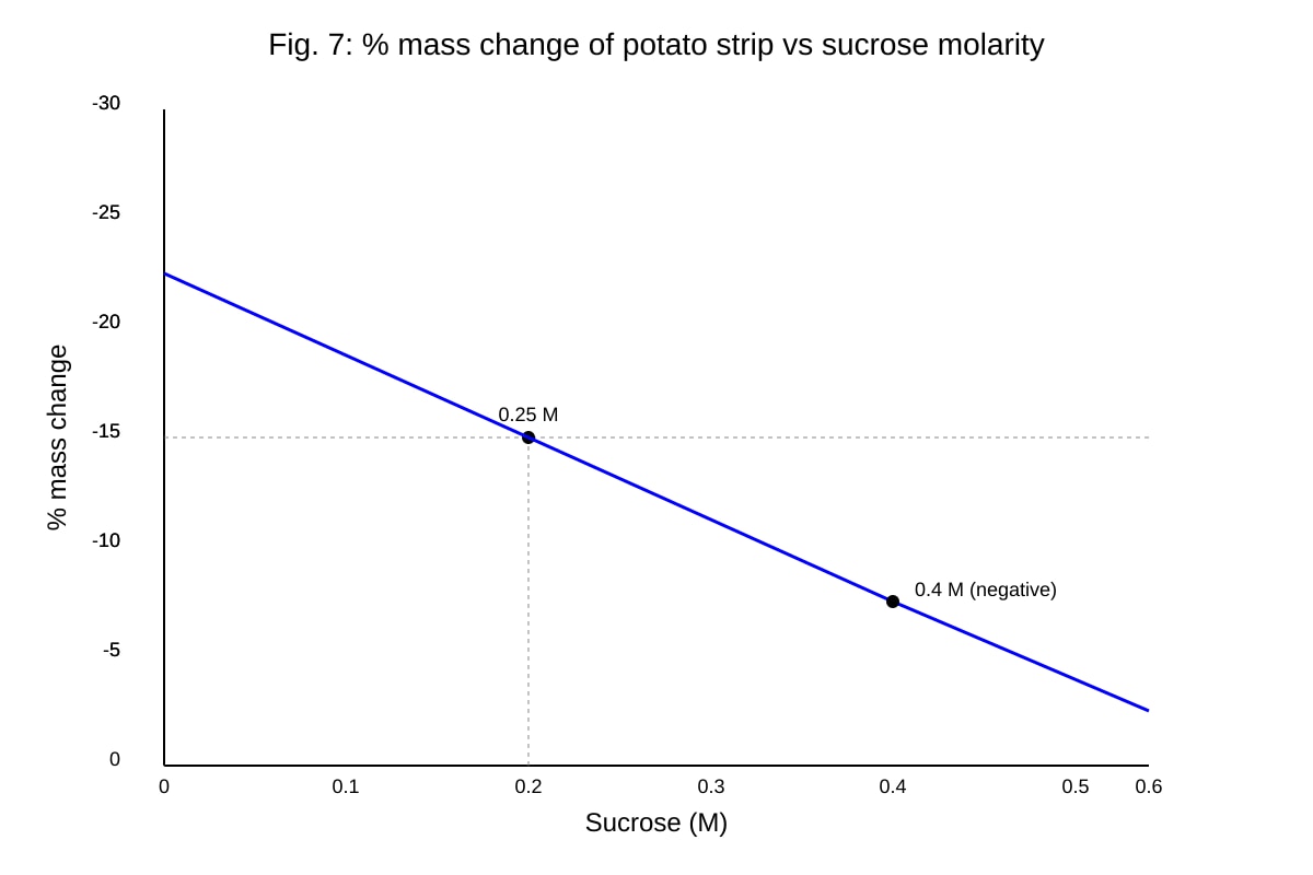
(a) [1] 0.25 M (isotonic, no net change).
(b) [2] At 0.4 M, solution hypertonic → water leaves by osmosis → mass decreases.
Section C (15 marks)
18. [7]
Structure: glycerol + 2 fatty acids + phosphate group (polar head, 2 non-polar tails). [2]
Fluid mosaic: bilayer with proteins embedded; hydrophobic tails give fluid core, heads face water; cholesterol restricts/permits movement. [3]
Properties: amphipathic nature lets self-assemble; fluidity allows fusion, transport. [2]
19. [8]
Primary: sequence of amino acids (2×α+2×β chains). [2]
Secondary: α-helix/β-sheet via H-bonds. [1]
Tertiary: folding, H/ionic/disulfide bonds. [2]
Quaternary: 4 subunits + haem group; Fe²⁺ binds O₂ reversibly. [3]
Marking descriptors: each level described + link to O₂ transport.