From Real Exams Exam Paper
A Level H1 Biology Practice Paper 3
Free A Level H1 Biology Practice Paper 3, LongCat Exam version, with questions, answers, and A Level-style practice for Singapore students.
These static practice materials are generated from the site's syllabus and paper-generation workflow, with source and model context shown so students and parents can evaluate the material before use.
Questions
TuitionGoWhere Practice Paper - Biology H1 A-Level
TuitionGoWhere Secondary School (AI)
Subject: Biology Level: A-Level H1 (8876) Paper: Practice Paper — Paper 2 Style Version: 3 of 5 Duration: 2 hours Total Marks: 75
Name: ___________________________ Class: ___________________________ Date: ___________________________
Instructions
- Write your name, class, and date in the spaces provided above.
- Answer ALL questions in the spaces provided.
- Write in dark blue or black pen.
- You may use a pencil for any diagrams or graphs.
- Do not use correction fluid.
- The number of marks for each question or part question is shown in brackets [ ].
- The total marks for this paper is 75.
Section A: Structured Questions [35 marks]
Answer ALL questions.
Question 1
(a) State two structural differences between a prokaryotic cell and a eukaryotic cell.
.......................................................................................................................................................................................
.......................................................................................................................................................................................
.......................................................................................................................................................................................
.......................................................................................................................................................................................[2]
(b) Explain why prokaryotic cells are generally smaller than eukaryotic cells.
.......................................................................................................................................................................................
.......................................................................................................................................................................................
.......................................................................................................................................................................................
.......................................................................................................................................................................................[2]
[Total: 4 marks]
Question 2
Generated diagram for Q2.
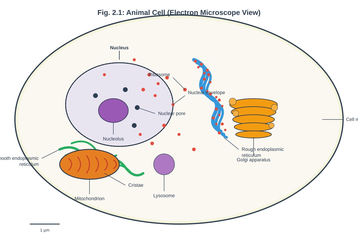
Fig. 2.1 shows an animal cell as seen under an electron microscope.
(a) Identify the organelles labelled A (nucleus), B (rough endoplasmic reticulum), and C (Golgi apparatus) in Fig. 2.1.
A: .......................................................................................................................
B: .......................................................................................................................
C: .......................................................................................................................[3]
(b) Describe the function of the organelle labelled B (rough endoplasmic reticulum).
.......................................................................................................................................................................................
.......................................................................................................................................................................................
.......................................................................................................................................................................................[2]
(c) State one structural feature of the mitochondrion that adapts it for its function.
.......................................................................................................................................................................................
.......................................................................................................................................................................................[1]
[Total: 6 marks]
Question 3
A student carried out an experiment to investigate the effect of temperature on the permeability of beetroot cell membranes. Beetroot discs of equal size were placed in distilled water at different temperatures for 10 minutes. The colour intensity of the solution, which indicates the amount of red pigment (betacyanin) released from the cells, was measured using a colorimeter.
The results are shown in Table 3.1.
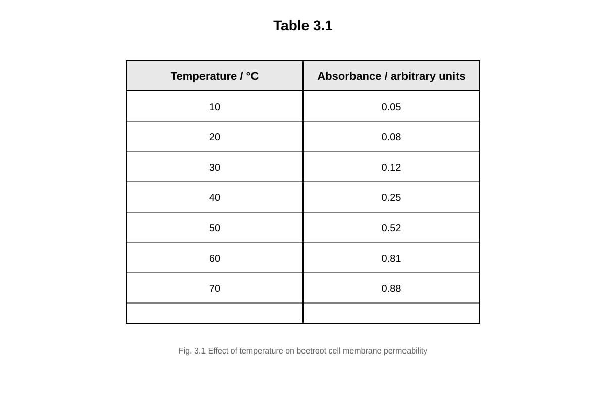
Generated table for Q3.
Table 3.1
(a) Describe the trend shown in Table 3.1.
.......................................................................................................................................................................................
.......................................................................................................................................................................................
.......................................................................................................................................................................................
.......................................................................................................................................................................................[2]
(b) Explain why the absorbance increases significantly between 40 °C and 60 °C.
.......................................................................................................................................................................................
.......................................................................................................................................................................................
.......................................................................................................................................................................................
.......................................................................................................................................................................................
.......................................................................................................................................................................................
.......................................................................................................................................................................................[3]
(c) Suggest why the absorbance values level off above 60 °C.
.......................................................................................................................................................................................
.......................................................................................................................................................................................
.......................................................................................................................................................................................[2]
[Total: 7 marks]
Question 4
(a) Describe the structure of the cell (plasma) membrane according to the fluid mosaic model.
.......................................................................................................................................................................................
.......................................................................................................................................................................................
.......................................................................................................................................................................................
.......................................................................................................................................................................................
.......................................................................................................................................................................................
.......................................................................................................................................................................................[3]
(b) Explain how the structure of the phospholipid bilayer allows the membrane to be selectively permeable.
.......................................................................................................................................................................................
.......................................................................................................................................................................................
.......................................................................................................................................................................................
.......................................................................................................................................................................................
.......................................................................................................................................................................................[3]
[Total: 6 marks]
Question 5
(a) Distinguish between facilitated diffusion and active transport across the cell membrane.
.......................................................................................................................................................................................
.......................................................................................................................................................................................
.......................................................................................................................................................................................
.......................................................................................................................................................................................
.......................................................................................................................................................................................[3]
(b) Explain why cells need to carry out active transport.
.......................................................................................................................................................................................
.......................................................................................................................................................................................
.......................................................................................................................................................................................
.......................................................................................................................................................................................[2]
[Total: 5 marks]
Question 6
A student observed two types of cells under an electron microscope. Cell X has a cell wall, chloroplasts, and a large central vacuole. Cell Y has centrioles and lysosomes but no cell wall.
(a) Identify cell type X and cell type Y.
X: .......................................................................................................................
Y: .......................................................................................................................[2]
(b) State two organelles that would be found in both cell X and cell Y.
.......................................................................................................................................................................................
.......................................................................................................................................................................................[2]
(c) Explain the role of centrioles in cell Y.
.......................................................................................................................................................................................
.......................................................................................................................................................................................
.......................................................................................................................................................................................[2]
[Total: 6 marks]
Section B: Biological Molecules [25 marks]
Answer ALL questions.
Question 7
(a) State the general formula of a monosaccharide.
.......................................................................................................................................................................................[1]
(b) Name the type of glycosidic bond found in starch.
.......................................................................................................................................................................................[1]
(c) Explain why starch is a suitable storage molecule in plants.
.......................................................................................................................................................................................
.......................................................................................................................................................................................
.......................................................................................................................................................................................
.......................................................................................................................................................................................[3]
[Total: 5 marks]
Question 8
Generated diagram for Q8.
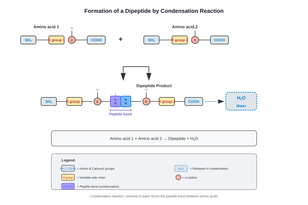
Fig. 8.1 shows the formation of a dipeptide.
(a) Name the type of reaction shown in Fig. 8.1.
.......................................................................................................................................................................................[1]
(b) Identify the bond labelled X in Fig. 8.1.
.......................................................................................................................................................................................[1]
(c) Describe the primary, secondary, and tertiary structure of a protein.
Primary structure: ...............................................................................................................................................................
.......................................................................................................................................................................................
Secondary structure: ...........................................................................................................................................................
.......................................................................................................................................................................................
Tertiary structure: ...............................................................................................................................................................
.......................................................................................................................................................................................
.......................................................................................................................................................................................[4]
[Total: 6 marks]
Question 9
(a) Describe the chemical composition of a triglyceride.
.......................................................................................................................................................................................
.......................................................................................................................................................................................
.......................................................................................................................................................................................[2]
(b) Explain how the properties of triglycerides make them suitable as energy storage molecules.
.......................................................................................................................................................................................
.......................................................................................................................................................................................
.......................................................................................................................................................................................
.......................................................................................................................................................................................
.......................................................................................................................................................................................[3]
(c) State one difference between a saturated fatty acid and an unsaturated fatty acid.
.......................................................................................................................................................................................
.......................................................................................................................................................................................[1]
[Total: 6 marks]
Question 10
(a) Name the three components of a nucleotide.
.......................................................................................................................................................................................
.......................................................................................................................................................................................
.......................................................................................................................................................................................[3]
(b) State two differences between DNA and RNA.
.......................................................................................................................................................................................
.......................................................................................................................................................................................
.......................................................................................................................................................................................
.......................................................................................................................................................................................[2]
[Total: 5 marks]
Question 11
Explain the importance of water as a biological molecule. In your answer, refer to at least three properties of water and explain how each property is important for living organisms.
.......................................................................................................................................................................................
.......................................................................................................................................................................................
.......................................................................................................................................................................................
.......................................................................................................................................................................................
.......................................................................................................................................................................................
.......................................................................................................................................................................................
.......................................................................................................................................................................................
.......................................................................................................................................................................................
.......................................................................................................................................................................................
.......................................................................................................................................................................................[5]
[Total: 5 marks]
Section C: Data-Based and Application Questions [15 marks]
Answer ALL questions.
Question 12
An enzyme-catalysed reaction was investigated at pH 7. The enzyme catalyses the breakdown of hydrogen peroxide (H2O2) into water and oxygen. The volume of oxygen gas produced was measured over time at different temperatures. The results are shown in Fig. 12.1.
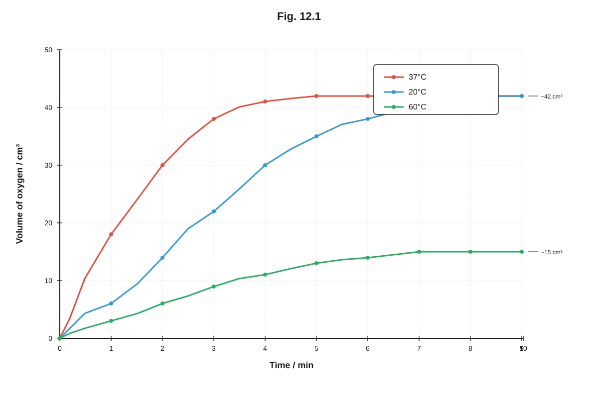
Generated graph for Q12.
Fig. 12.1
(a) Describe the effect of temperature on the rate of this enzyme-catalysed reaction.
.......................................................................................................................................................................................
.......................................................................................................................................................................................
.......................................................................................................................................................................................
.......................................................................................................................................................................................
.......................................................................................................................................................................................[3]
(b) Explain why the reaction at 60 °C produces a lower final volume of oxygen compared to the reaction at 37 °C.
.......................................................................................................................................................................................
.......................................................................................................................................................................................
.......................................................................................................................................................................................
.......................................................................................................................................................................................[2]
(c) Suggest why the reaction at 20 °C takes longer to reach the final volume compared to 37 °C, even though the final volume is the same.
.......................................................................................................................................................................................
.......................................................................................................................................................................................
.......................................................................................................................................................................................[2]
[Total: 7 marks]
Question 13
A student tested four unknown food samples (P, Q, R, and S) for the presence of biological molecules. The results are shown in Table 13.1.
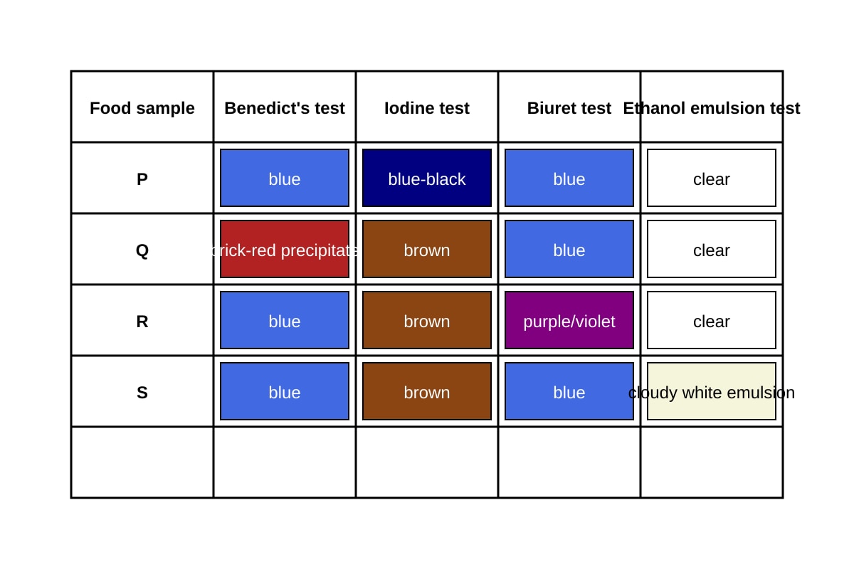
Generated table for Q13.
Table 13.1
(a) Identify which food sample contains:
(i) Starch
.......................................................................................................................[1]
(ii) Reducing sugar
.......................................................................................................................[1]
(iii) Protein
.......................................................................................................................[1]
(iv) Lipid
.......................................................................................................................[1]
(b) Explain the chemical basis of a positive Benedict's test.
.......................................................................................................................................................................................
.......................................................................................................................................................................................
.......................................................................................................................................................................................[2]
(c) Suggest one limitation of using the Biuret test to identify proteins.
.......................................................................................................................................................................................
.......................................................................................................................................................................................[1]
[Total: 7 marks]
End of Paper
Total Marks: 75
| Section | Marks |
|---|---|
| A: Structured Questions | 35 |
| B: Biological Molecules | 25 |
| C: Data-Based & Application | 15 |
| Total | 75 |
Answers
TuitionGoWhere Practice Paper — Biology H1 A-Level
Answer Key — Version 3 of 5
Section A: Structured Questions
Question 1
(a) State two structural differences between a prokaryotic cell and a eukaryotic cell. [2]
| Difference | Prokaryotic Cell | Eukaryotic Cell |
|---|---|---|
| 1 | No true nucleus (DNA is in the nucleoid region, not enclosed by a membrane) | Has a true nucleus enclosed by a double nuclear envelope |
| 2 | No membrane-bound organelles | Has membrane-bound organelles (e.g., mitochondria, ER, Golgi) |
Marking scheme: 1 mark per valid difference, maximum 2 marks. Accept any two from: absence of nuclear envelope, absence of membrane-bound organelles, presence of 70S ribosomes (vs 80S), circular DNA (vs linear chromosomes), smaller cell size, absence of mitochondria.
Teaching note: Prokaryotes (e.g., bacteria) lack internal compartmentalisation by membranes, while eukaryotes (e.g., animal and plant cells) have extensive membrane-bound organelles that allow different metabolic processes to occur in separate compartments.
(b) Explain why prokaryotic cells are generally smaller than eukaryotic cells. [2]
Prokaryotic cells have a smaller surface area-to-volume ratio constraint — as cells increase in volume, the surface area for exchange of materials increases more slowly. Without membrane-bound organelles to compartmentalise metabolic reactions, prokaryotes rely on diffusion across the cell surface for all transport. A smaller size ensures efficient diffusion of nutrients and waste products. Additionally, prokaryotes have simpler internal organisation and less genetic material, requiring less cytoplasmic volume.
Marking scheme: 1 mark for linking small size to efficient diffusion/exchange across the membrane. 1 mark for explaining the surface area-to-volume ratio constraint or the lack of compartmentalisation.
Common mistake: Students often state "prokaryotes are smaller because they are simpler" without explaining the functional reason (diffusion efficiency, SA:V ratio).
Question 2
(a) Identify the organelles labelled A, B, and C. [3]
- A: Nucleus
- B: Rough endoplasmic reticulum (RER)
- C: Golgi apparatus (or Golgi body)
Marking scheme: 1 mark each. Spelling must be correct for full credit.
(b) Describe the function of the organelle labelled B (rough endoplasmic reticulum). [2]
The rough endoplasmic reticulum is the site of protein synthesis (on its attached ribosomes). It also provides a transport pathway for proteins within the cell, folding and modifying proteins after synthesis. The ribosomes on its surface synthesise proteins that are either secreted from the cell or incorporated into membranes.
Marking scheme: 1 mark for protein synthesis (on ribosomes). 1 mark for transport/folding/modification of proteins. Accept "transports proteins to the Golgi apparatus" for the second mark.
Common mistake: Students may confuse RER with smooth ER. The key distinguishing feature is the presence of ribosomes on the RER surface.
(c) State one structural feature of the mitochondrion that adapts it for its function. [1]
The inner membrane is highly folded into cristae, which increases the surface area for the attachment of electron carriers and ATP synthase enzymes involved in aerobic respiration (oxidative phosphorylation).
Marking scheme: 1 mark for cristae / folded inner membrane. Accept also: "contains its own DNA and ribosomes for rapid protein production" or "double membrane creates a compartment for the proton gradient."
Question 3
(a) Describe the trend shown in Table 3.1. [2]
As temperature increases from 10 °C to 70 °C, the absorbance of the solution increases, indicating greater membrane permeability and more betacyanin pigment being released. The increase is gradual between 10 °C and 40 °C, but becomes much more rapid between 40 °C and 60 °C. Above 60 °C, the rate of increase slows and the absorbance approaches a maximum value.
Marking scheme: 1 mark for stating that absorbance increases with temperature. 1 mark for describing the pattern of increase (gradual at low temperatures, rapid at 40–60 °C, levelling off at high temperatures). Students must describe the trend, not just state "it increases."
(b) Explain why the absorbance increases significantly between 40 °C and 60 °C. [3]
At temperatures above approximately 40 °C, the phospholipid bilayer of the cell membrane gains increasing kinetic energy, causing the phospholipids to move more freely and creating gaps in the membrane. This increases membrane permeability. Additionally, the proteins embedded in the membrane (including transport proteins and channel proteins) begin to denature at these temperatures, losing their tertiary structure. The denatured proteins can no longer maintain the selective barrier function of the membrane, allowing the red betacyanin pigment to leak out of the vacuole and cell more rapidly.
Marking scheme: 1 mark for increased kinetic energy of phospholipids / increased fluidity of the bilayer. 1 mark for denaturation of membrane proteins. 1 mark for linking these changes to increased permeability / release of pigment.
Common mistake: Students may only mention phospholipid movement without mentioning protein denaturation, or vice versa. Both are needed for full marks at this level.
(c) Suggest why the absorbance values level off above 60 °C. [2]
Above 60 °C, the membrane is almost completely disrupted — most phospholipids are highly fluid and most membrane proteins are fully denatured. Therefore, nearly all the betacyanin pigment has already been released from the cells. Further increases in temperature cannot release more pigment because there is very little pigment remaining inside the cells. The absorbance approaches a maximum value.
Marking scheme: 1 mark for the membrane being fully disrupted / maximum permeability reached. 1 mark for all (or nearly all) pigment already being released.
Question 4
(a) Describe the structure of the cell (plasma) membrane according to the fluid mosaic model. [3]
The membrane consists of a phospholipid bilayer, where the hydrophilic phosphate heads face the aqueous environments on both sides of the membrane and the hydrophobic fatty acid tails face inwards, away from water. Protein molecules are embedded within (intrinsic/integral proteins) or attached to the surface (extrinsic/peripheral proteins) of the bilayer. Cholesterol molecules are found between the phospholipids, helping to regulate membrane fluidity. Glycoproteins and glycolipids may be present on the extracellular surface. The components are not fixed in position but can move laterally within the bilayer, giving the membrane its "fluid" nature.
Marking scheme: 1 mark for phospholipid bilayer with correct orientation of heads and tails. 1 mark for proteins embedded in or associated with the bilayer (intrinsic and/or extrinsic). 1 mark for cholesterol and/or the fluid nature of the membrane (lateral movement of components). Accept glycoproteins/glycolipids as an alternative to cholesterol for the third mark.
(b) Explain how the structure of the phospholipid bilayer allows the membrane to be selectively permeable. [3]
The hydrophobic interior of the phospholipid bilayer acts as a barrier to the passage of polar molecules, ions, and large hydrophilic molecules, which cannot easily dissolve in the nonpolar fatty acid tail region. Small, nonpolar molecules (e.g., oxygen, carbon dioxide) and small polar molecules (e.g., water) can pass through the bilayer by simple diffusion. Larger polar molecules and ions require transport proteins (channel proteins or carrier proteins) to cross the membrane. This selective passage of substances based on their size, polarity, and charge is what makes the membrane selectively (or partially) permeable.
Marking scheme: 1 mark for the hydrophobic interior acting as a barrier to polar/ionic/large molecules. 1 mark for small nonpolar molecules being able to pass through. 1 mark for the role of transport proteins in allowing specific molecules/ions to cross.
Common mistake: Students may state "the membrane is selectively permeable" without explaining the structural basis (hydrophobic core, transport proteins).
Question 5
(a) Distinguish between facilitated diffusion and active transport across the cell membrane. [3]
| Feature | Facilitated Diffusion | Active Transport |
|---|---|---|
| Energy requirement | Does not require ATP | Requires ATP |
| Direction of movement | Down the concentration gradient (high to low) | Against the concentration gradient (low to high) |
| Transport proteins used | Channel proteins or carrier proteins | Carrier proteins only |
Marking scheme: 1 mark for each valid distinction, maximum 3 marks. Key distinctions: energy requirement (ATP vs no ATP), direction relative to concentration gradient, and type of protein used.
**(b) Explain why cells need to carry out active transport. [2]]
Cells need active transport to accumulate substances at concentrations higher than their surroundings, which is essential for normal cell function. For example, nerve cells need to maintain a high concentration of potassium ions inside the cell and a high concentration of sodium ions outside the cell for nerve impulse transmission. Active transport also allows cells to take up essential nutrients (e.g., glucose, amino acids) from environments where their concentration is lower than inside the cell.
Marking scheme: 1 mark for moving substances against the concentration gradient. 1 mark for a valid example or explanation of why this is necessary (e.g., nutrient uptake, ion balance, nerve impulses).
Question 6
(a) Identify cell type X and cell type Y. [2]
- X: Plant cell
- Y: Animal cell
Marking scheme: 1 mark each. Cell X has a cell wall, chloroplasts, and a large central vacuole — all features of plant cells. Cell Y has centrioles and lysosomes but no cell wall — features of animal cells.
(b) State two organelles that would be found in both cell X and cell Y. [2]
Any two from: nucleus, mitochondria, endoplasmic reticulum (rough and/or smooth), Golgi apparatus, ribosomes, cell membrane.
Marking scheme: 1 mark each. Do not accept chloroplasts, cell wall, large central vacuole, or centrioles, as these are specific to one cell type only.
(c) Explain the role of centrioles in cell Y. [2]
Centrioles are involved in the formation of the spindle apparatus during cell division (mitosis and meiosis). They organise the microtubules of the spindle fibres, which attach to the centromeres of chromosomes and pull the sister chromatids apart to opposite poles of the cell during anaphase. This ensures that each daughter cell receives the correct number of chromosomes.
Marking scheme: 1 mark for spindle formation / organisation of microtubules. 1 mark for their role in chromosome separation during cell division.
Section B: Biological Molecules
Question 7
(a) State the general formula of a monosaccharide. [1]
(CH2O)n where n is typically 3–7 (most commonly n=5 or 6 for hexoses and pentoses).
Marking scheme: 1 mark for (CH2O)n. Accept C6H12O6 as a specific example (glucose).
(b) Name the type of glycosidic bond found in starch. [1]
1,4-glycosidic bond (and 1,6-glycosidic bonds at branch points in amylopectin).
Marking scheme: 1 mark for "1,4-glycosidic bond" or "alpha-1,4-glycosidic bond." Accept "glycosidic bond" alone only if the question does not require specificity, but at A-Level, the specific bond type is expected.
(c) Explain why starch is a suitable storage molecule in plants. [3]
Starch is insoluble in water, so it does not affect the water potential of the cell or cause osmotic problems. It is a large, compact molecule (amylose forms a helical structure and amylopectin is branched), so a large amount of energy can be stored in a small volume. Starch is also easily hydrolysed by enzymes (amylases) into glucose when the plant needs energy. The branched structure of amylopectin provides many ends for rapid enzymatic breakdown.
Marking scheme: 1 mark for insolubility (no osmotic effect). 1 mark for compact/large molecule (efficient storage). 1 mark for ease of hydrolysis / readily broken down when energy is needed.
Question 8
(a) Name the type of reaction shown in Fig. 8.1. [1]
Condensation reaction (or dehydration synthesis).
Marking scheme: 1 mark for "condensation reaction." Accept "dehydration synthesis."
(b) Identify the bond labelled X in Fig. 8.1. [1]
Peptide bond.
Marking scheme: 1 mark for "peptide bond." The peptide bond forms between the carboxyl group (–COOH) of one amino acid and the amino group (–NH₂) of another, releasing a water molecule.
(c) Describe the primary, secondary, and tertiary structure of a protein. [4]
Primary structure: The specific sequence of amino acids in the polypeptide chain, held together by peptide bonds. This sequence is determined by the genetic code.
Secondary structure: The local folding of the polypeptide chain into regular structures, primarily α-helices and β-pleated sheets, stabilised by hydrogen bonds between the peptide backbone (C=O and N–H groups).
Tertiary structure: The overall three-dimensional folding of the polypeptide chain, stabilised by interactions between the R groups of amino acids. These include hydrogen bonds, ionic bonds, disulfide bridges (covalent bonds between cysteine residues), and hydrophobic interactions.
Marking scheme: 1 mark for primary structure (sequence of amino acids / peptide bonds). 1 mark for secondary structure (α-helix / β-pleated sheet / hydrogen bonds in backbone). 2 marks for tertiary structure — 1 mark for 3D folding and 1 mark for naming at least two types of bonds/interactions between R groups (hydrogen bonds, ionic bonds, disulfide bridges, hydrophobic interactions).
Question 9
(a) Describe the chemical composition of a triglyceride. [2]
A triglyceride is composed of one glycerol molecule (a three-carbon alcohol) bonded to three fatty acid molecules. The bond between the glycerol and each fatty acid is an ester bond, formed by a condensation reaction with the elimination of three water molecules.
Marking scheme: 1 mark for one glycerol + three fatty acids. 1 mark for ester bonds / condensation reaction.
(b) Explain how the properties of triglycerides make them suitable as energy storage molecules. [3]
Triglycerides have a high energy content per gram because their fatty acid chains contain many C–H bonds, which release a large amount of energy when oxidised during respiration. They are insoluble in water, so they do not cause osmotic problems or affect the water potential of cells. They are also relatively light (low density) compared to the energy they provide, making them efficient for energy storage. Triglycerides are stored as fat droplets in adipose tissue.
Marking scheme: 1 mark for high energy content / many C–H bonds. 1 mark for insolubility in water (no osmotic effect). 1 mark for compact/lightweight / efficient storage.
(c) State one difference between a saturated fatty acid and an unsaturated fatty acid. [1]
Saturated fatty acids have no carbon–carbon double bonds (all C–C bonds are single bonds), while unsaturated fatty acid(s) have one or more carbon–carbon double bond(s) in their hydrocarbon chain.
Marking scheme: 1 mark for the presence/absence of C=C double bonds. Accept also: unsaturated fatty acids have kinks/bends in their chain; saturated fatty acids are straight-chained.
Question 10
(a) Name the three components of a nucleotide. [3]
- A pentose sugar (5-carbon sugar)
- A phosphate group
- A nitrogenous (organic) base
Marking scheme: 1 mark each.
(b) State two differences between DNA and RNA. [2]
| Feature | DNA | RNA |
|---|---|---|
| Sugar | Deoxyribose | Ribose |
| Bases | Adenine, Thymine, Guanine, Cytosine | Adenine, Uracil, Guanine, Cytosine |
| Structure | Double-stranded helix | Usually single-stranded |
| Stability | More stable | Less stable |
Marking scheme: 1 mark per valid difference, maximum 2 marks. Accept any two from: type of sugar, presence of thymine vs uracil, single vs double strand, length of molecule, stability.
Question 11
Explain the importance of water as a biological molecule. [5]
Water is essential for life due to its unique properties:
-
High specific heat capacity: Water can absorb or release a large amount of heat energy with only a small change in temperature. This helps organisms maintain a stable internal body temperature and provides a stable aquatic environment for aquatic organisms.
-
High latent heat of vaporisation: A large amount of energy is required to convert water from liquid to gas. This makes water an effective coolant — for example, sweating in mammals cools the body as water evaporates from the skin, absorbing heat energy.
-
Cohesion and surface tension: Water molecules are cohesive (attracted to each other) due to hydrogen bonding. This allows water to be pulled up through the xylem vessels in plants as a continuous column during transpiration. Surface tension also supports small organisms on water surfaces.
-
Universal solvent: Water is an excellent solvent for polar and ionic substances because of its polarity. This allows metabolic reactions to occur in aqueous solution within cells, and enables the transport of substances (e.g., glucose, ions, amino acids) in the blood and in the xylem/phloem.
-
High density of liquid water vs ice: Ice is less dense than liquid water and floats, insulating the water below and allowing aquatic life to survive under frozen surfaces in winter.
Marking scheme: 1 mark per property correctly described and linked to its biological importance, maximum 5 marks. Students must name the property AND explain its biological significance for each mark. Simply listing properties without biological context earns a maximum of 3 marks.
Section C: Data-Based and Application Questions
Question 12
(a) Describe the effect of temperature on the rate of this enzyme-catalysed reaction. [3]
At 37 °C, the reaction has the fastest initial rate (steepest gradient) and reaches the plateau (maximum volume of oxygen) in the shortest time (~5 minutes). At 20 °C, the initial rate is slower (more gradual gradient) but reaches the same final volume of oxygen (~42 cm³) as at 37 °C, taking longer (~8 minutes). At 60 °C, the initial rate is the slowest and the final volume of oxygen produced is much lower (~15 cm³), indicating that the enzyme has been denatured.
Marking scheme: 1 mark for describing the 37 °C curve (fastest rate, reaches plateau quickly). 1 mark for describing the 20 °C curve (slower rate, same final volume). 1 mark for describing the 60 °C curve (slowest rate, lower final volume / enzyme denatured).
(b) Explain why the reaction at 60 °C produces a lower final volume of oxygen compared to the reaction at 37 °C. [2]
At 60 °C, the enzyme (catalase) is denatured. The high temperature disrupts the hydrogen bonds and other interactions that maintain the enzyme's tertiary structure, causing the active site to change shape. The substrate (hydrogen peroxide) can no longer bind effectively to the denatured active site, so the enzyme cannot catalyse the reaction. Since the enzyme is permanently denatured, not all of the substrate is broken down, resulting in a lower final volume of oxygen.
Marking scheme: 1 mark for enzyme denaturation at high temperature. 1 mark for change in active site shape / substrate cannot bind / not all substrate broken down.
Common mistake: Students may say "the enzyme is killed" — enzymes are not living and cannot be "killed." The correct term is "denatured."
(c) Suggest why the reaction at 20 °C takes longer to reach the final volume compared to 37 °C, even though the final volume is the same. [2]
At 20 °C, the enzyme and substrate molecules have less kinetic energy than at 37 °C, so they move more slowly and collide less frequently. This means the rate of formation of enzyme–substrate complexes is lower, so the reaction proceeds more slowly. However, the enzyme is not denatured at 20 °C, so given enough time, all the substrate is eventually broken down, reaching the same final volume of oxygen as at 37 °C.
Marking scheme: 1 mark for lower kinetic energy / fewer collisions / slower rate at 20 °C. 1 mark for the enzyme not being denatured / all substrate eventually broken down.
Question 13
(a) Identify which food sample contains: [4]
(i) Starch: Sample P (positive iodine test — blue-black colour)
(ii) Reducing sugar: Sample Q (positive Benedict's test — brick-red precipitate)
(iii) Protein: Sample R (positive Biuret test — purple/violet colour)
(iv) Lipid: Sample S (positive ethanol emulsion test — cloudy white emulsion)
Marking scheme: 1 mark each.
(b) Explain the chemical basis of a positive Benedict's test. [2]
Benedict's solution contains copper(II) sulfate (Cu²⁺ ions) in an alkaline solution. Reducing sugars (such as glucose, maltose, and fructose) have a free aldehyde or ketone group that can act as a reducing agent. When heated with Benedict's solution, the reducing sugar reduces the blue Cu²⁺ ions to orange-red copper(I) oxide (Cu₂O), which precipitates out of solution. The colour change from blue → green → yellow → orange → brick-red indicates the presence and approximate concentration of reducing sugar.
Marking scheme: 1 mark for Cu²⁺ being reduced (by the reducing sugar). 1 mark for the formation of copper(I) oxide precipitate / colour change from blue to brick-red.
(c) Suggest one limitation of using the Biuret test to identify proteins. [1]
The Biuret test only indicates the presence of peptide bonds, so it cannot distinguish between different proteins. It also gives a positive result for any substance containing two or more peptide bonds (including some non-protein substances like urea in high concentrations). Additionally, it is a qualitative test and does not provide information about the concentration or type of protein present.
Marking scheme: 1 mark for any valid limitation. Accept: cannot identify specific proteins, only detects peptide bonds, does not give quantitative results, may give false positives with substances containing peptide bonds that are not proteins.
Mark Summary
| Question | Marks |
|---|---|
| 1 | 4 |
| 2 | 6 |
| 3 | 7 |
| 4 | 6 |
| 5 | 5 |
| 6 | 6 |
| Section A Total | 34 |
| 7 | 5 |
| 8 | 6 |
| 9 | 6 |
| 10 | 5 |
| 11 | 5 |
| Section B Total | 27 |
| 12 | 7 |
| 13 | 7 |
| Section C Total | 14 |
| Grand Total | 75 |
Note: Section A totals 34 marks, Section B totals 27 marks, Section C totals 14 marks. Grand total = 75 marks.
Free quiz and exam paper access
Enter your details to view this paper
Your access is remembered on this device.