From Real Exams Exam Paper
A Level H1 Biology Practice Paper 2
Free A Level H1 Biology Practice Paper 2, LongCat Exam version, with questions, answers, and A Level-style practice for Singapore students.
These static practice materials are generated from the site's syllabus and paper-generation workflow, with source and model context shown so students and parents can evaluate the material before use.
Questions
TuitionGoWhere Practice Paper - Biology H1 A-Level
TuitionGoWhere Secondary School (AI)
| Subject: | Biology |
| Level: | A-Level H1 |
| Paper: | Practice Paper — Paper 2 Style |
| Version: | 2 of 5 |
| Duration: | 2 hours |
| Total Marks: | 80 |
| Name: | ________________________ |
| Class: | ________________________ |
| Date: | ________________________ |
Instructions
- Write your answers in the spaces provided.
- Answer ALL questions.
- The number of marks for each question is shown in brackets [ ].
- You are advised to spend no more than 1 hour 40 minutes on Section A and B combined, and the remaining time on Section C.
- Where a question requires explanation or reasoning, answers must be clearly structured and use appropriate biological terminology.
Section A: Structured Questions [40 marks]
Answer ALL questions in this section.
Question 1 [4 marks]
(a) State two structural differences between a prokaryotic cell and a eukaryotic cell.
(i) _______________________________________________________________
(ii) _______________________________________________________________
[2 marks]
(b) Explain why the presence of membrane-bound organelles in eukaryotic cells is important for cellular function.
[2 marks]
Question 2 [5 marks]
Generated diagram for Q2.
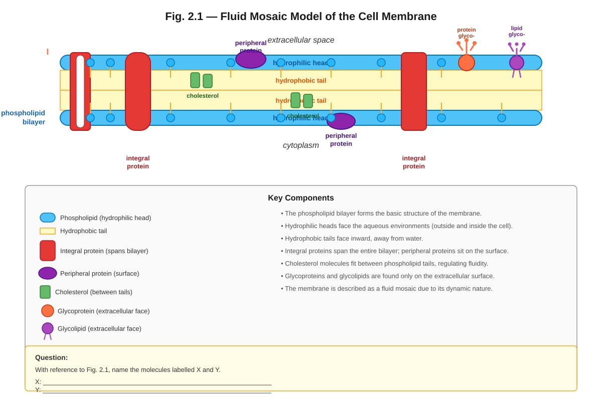
Fig. 2.1 shows the structure of a cell membrane.
(a) With reference to Fig. 2.1, name the molecules labelled X and Y.
X: _______________________________________________________________
Y: _______________________________________________________________
[2 marks]
(b) Explain how the structure of the phospholipid bilayer allows the membrane to act as a barrier to hydrophilic molecules.
[2 marks]
(c) State one function of cholesterol in the cell membrane.
[1 mark]
Question 3 [4 marks]
A student carried out an experiment to investigate the effect of temperature on the permeability of beetroot cell membranes. Beetroot cylinders of equal size were placed in distilled water at different temperatures for 10 minutes. The colour intensity of the solution, which indicates the amount of red pigment (betacyanin) released, was measured using a colorimeter.
The results are shown in the table below.
| Temperature (°C) | Absorbance of solution (arbitrary units) |
|---|---|
| 10 | 0.05 |
| 20 | 0.08 |
| 30 | 0.15 |
| 40 | 0.32 |
| 50 | 0.78 |
| 60 | 1.25 |
| 70 | 1.42 |
(a) Describe the trend shown in the results.
[2 marks]
(b) Explain the results obtained at 60 °C and 70 °C.
[2 marks]
Question 4 [5 marks]
(a) Define the term enzyme.
[2 marks]
(b) Explain the lock-and-key hypothesis of enzyme action.
[3 marks]
Question 5 [6 marks]
Generated graph for Q5.
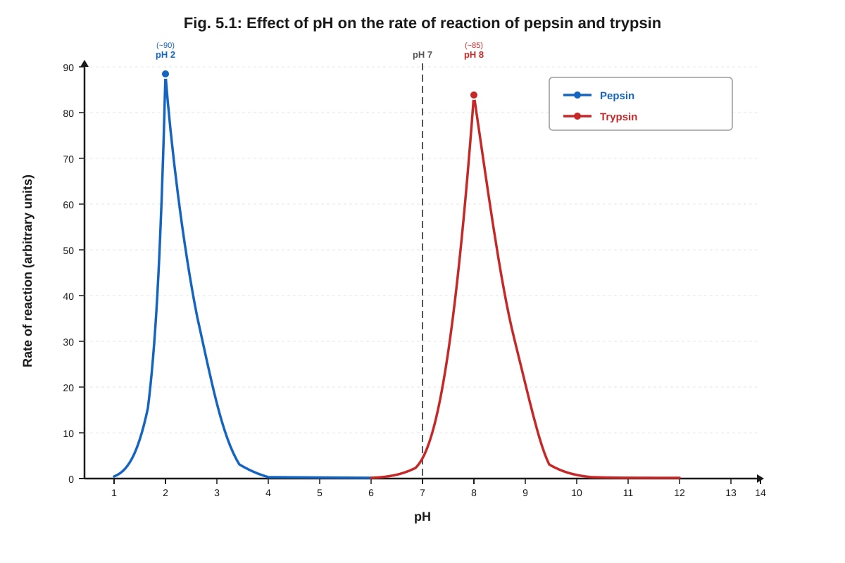
Fig. 5.1 shows the effect of pH on the rate of reaction of two enzymes, pepsin and trypsin.
(a) With reference to Fig. 5.1, state the optimum pH for each enzyme.
Pepsin: _______________________________________________________________
Trypsin: _______________________________________________________________
[2 marks]
(b) Explain why the rate of reaction decreases when the pH is above the optimum for trypsin.
[2 marks]
(c) Pepsin is found in the stomach. Explain how the environment of the stomach is suited to pepsin's function.
[2 marks]
Question 6 [4 marks]
Describe the structure of DNA. In your answer, refer to the following: sugar, phosphate, bases, and the double helix.
[4 marks]
Question 7 [5 marks]
(a) State two functions of proteins in living organisms.
(i) _______________________________________________________________
(ii) _______________________________________________________________
[2 marks]
(b) Describe how the structure of a protein determines its function. Include reference to primary, secondary, and tertiary structure.
[3 marks]
Question 8 [4 marks]
Distinguish between competitive and non-competitive enzyme inhibition.
[4 marks]
Question 9 [3 marks]
Explain why water is important as a biological molecule. Refer to two properties of water in your answer.
[3 marks]
Section B: Data-Based Questions [20 marks]
Answer ALL questions in this section.
Question 10 [10 marks]
Read the following passage and answer the questions that follow.
The process of osmosis is vital for the survival of all living cells. In plant cells, osmosis is responsible for the movement of water into and out of the cell, which affects the turgor pressure and overall rigidity of the plant tissue. When a plant cell is placed in a solution with a higher water potential than the cell's cytoplasm, water moves into the cell by osmosis. The cell swells and becomes turgid. However, the rigid cell wall prevents the cell from bursting. Conversely, when a plant cell is placed in a solution with a lower water potential than the cell's cytoplasm, water leaves the cell. The cell membrane pulls away from the cell wall — a process known as plasmolysis.
Animal cells, lacking a cell wall, respond differently to osmotic stress. In a hypotonic solution, animal cells swell and may burst (lysis). In a hypertonic solution, they shrink as water leaves the cell (crenation).
(a) Define water potential.
[2 marks]
(b) Explain why plant cells do not burst in a hypotonic solution, but animal cells do.
[3 marks]
(c) A student placed red blood cells in three different solutions: Solution A (distilled water), Solution B (0.9% NaCl), and Solution C (10% NaCl). Predict and explain what would happen to the red blood cells in each solution.
Solution A:
Solution B:
Solution C:
[5 marks]
Question 11 [10 marks]
Generated diagram for Q11.
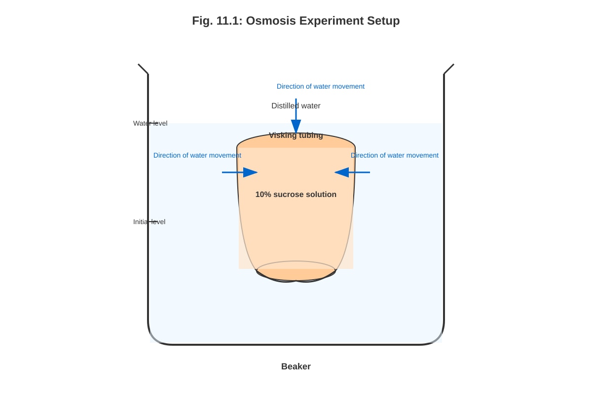
Fig. 11.1 shows an experiment to investigate osmosis.
(a) Explain why water moves into the Visking tubing from the beaker.
[2 marks]
(b) After 30 minutes, the mass of the Visking tubing increased. Explain this observation in terms of water potential.
[3 marks]
(c) The experiment was repeated with 20% sucrose solution inside the Visking tubing. Predict and explain how the results would differ from the original experiment.
[3 marks]
(d) State one limitation of using Visking tubing as a model for a cell membrane.
[2 marks]
Section C: Free Response [20 marks]
Answer ONE question in this section.
Question 12 [20 marks]
Enzymes are biological catalysts that play a central role in all metabolic processes.
(a) Describe the mechanism of enzyme action according to the induced-fit model. [5 marks]
(b) Explain how temperature and pH affect the rate of an enzyme-catalysed reaction. [8 marks]
(c) Discuss the importance of enzymes in two named industrial or medical applications. [7 marks]
OR
Question 13 [20 marks]
The structure of biological molecules is closely related to their function.
(a) Describe the structure and function of carbohydrates in living organisms. Include reference to monosaccharides, disaccharides, and polysaccharides. [7 marks]
(b) Compare and contrast the structure and function of lipids and proteins. [8 marks]
(c) Explain how the structure of DNA enables it to carry out its role as the genetic material. [5 marks]
END OF PAPER
Summary of Marks
| Section | Marks |
|---|---|
| Section A: Structured Questions (Q1–Q9) | 40 |
| Section B: Data-Based Questions (Q10–Q11) | 20 |
| Section C: Free Response (Q12 or Q13) | 20 |
| Total | 80 |
Answers
TuitionGoWhere Practice Paper - Biology H1 A-Level
Answer Key — Version 2 of 5
Subject: Biology | Level: A-Level H1 | Paper: Practice Paper — Paper 2 Style | Total Marks: 80
Section A: Structured Questions
Question 1 [4 marks]
(a) Two structural differences between prokaryotic and eukaryotic cells: [2 marks]
| Prokaryotic Cell | Eukaryotic Cell | |
|---|---|---|
| (i) | No membrane-bound nucleus; DNA is free in the cytoplasm (nucleoid region) | Has a membrane-bound nucleus containing DNA |
| (ii) | No membrane-bound organelles | Has membrane-bound organelles (e.g., mitochondria, ER, Golgi) |
Accept any two of the following (1 mark each, max 2):
- Prokaryotes have 70S ribosomes; eukaryotes have 80S ribosomes.
- Prokaryotic cells are smaller (1–5 μm); eukaryotic cells are larger (10–100 μm).
- Prokaryotes have a cell wall made of peptidoglycan; eukaryotic plant cells have a cell wall made of cellulose.
- Prokaryotes have circular DNA; eukaryotes have linear chromosomes.
Teaching note: Prokaryotes (e.g., bacteria) are simpler cells that lack internal membrane-bound compartments. Eukaryotes (e.g., animal and plant cells) have a nucleus enclosed by a nuclear envelope and various organelles that compartmentalise cellular functions.
(b) Importance of membrane-bound organelles: [2 marks]
- Membrane-bound organelles compartmentalise the cell, allowing different metabolic processes to occur simultaneously in separate environments without interference. [1 mark]
- Each organelle provides a specialised environment (e.g., specific pH, enzyme concentration) that is optimal for its particular function. For example, lysosomes maintain an acidic pH for hydrolytic enzymes, while the nucleus provides a protected environment for DNA. [1 mark]
- The membranes also provide a large surface area for reactions (e.g., cristae in mitochondria for ATP synthesis; thylakoid membranes in chloroplasts for photosynthesis). [Accept as part of either mark point]
Common mistake: Students often state that organelles "separate" processes without explaining why this is beneficial. The key idea is that incompatible reactions can occur at the same time, and each environment can be optimised independently.
Question 2 [5 marks]
(a) Molecules X and Y: [2 marks]
- X: Integral protein (or transmembrane protein) [1 mark]
- Y: Glycoprotein (or glycolipid — accept either if correctly identified from the diagram) [1 mark]
Note: The specific labels depend on the diagram. X should be the protein spanning the entire bilayer; Y should be the molecule on the extracellular surface with a carbohydrate chain attached.
Teaching note: Integral proteins are embedded within the phospholipid bilayer and may span the entire membrane (transmembrane proteins). Glycoproteins are proteins with carbohydrate chains attached, found on the extracellular surface, and are involved in cell recognition and signalling.
(b) How the phospholipid bilayer acts as a barrier to hydrophilic molecules: [2 marks]
- The phospholipid bilayer has a hydrophobic interior (the fatty acid tails) and hydrophilic exterior (the phosphate heads). [1 mark]
- Hydrophilic (polar or charged) molecules cannot pass through the hydrophobic core of the bilayer because they are repelled by the non-polar fatty acid tails. This makes the membrane selectively permeable. [1 mark]
Teaching note: The "like dissolves like" principle applies here. Hydrophilic molecules are polar or ionic and are insoluble in the non-polar hydrophobic interior. Only small non-polar molecules (e.g., O₂, CO₂) and very small polar molecules (e.g., water, slowly) can diffuse through the bilayer.
(c) One function of cholesterol: [1 mark]
- Cholesterol regulates membrane fluidity — it prevents the membrane from becoming too fluid at high temperatures and too rigid at low temperatures. [1 mark]
Accept any one of:
- Maintains membrane stability/fluidity.
- Reduces permeability to small water-soluble molecules.
- Prevents the phospholipid tails from packing too closely together.
Question 3 [4 marks]
(a) Description of trend: [2 marks]
- As temperature increases from 10 °C to 70 °C, the absorbance of the solution increases. [1 mark]
- The increase is gradual between 10 °C and 40 °C, but becomes much more rapid above 40 °C, with the greatest increase between 50 °C and 70 °C. [1 mark]
Marking note: Students must describe the overall trend AND note the change in rate (not just "it increases"). Award 1 mark for the general trend and 1 mark for describing the pattern of increase.
(b) Explanation of results at 60 °C and 70 °C: [2 marks]
- At high temperatures (60–70 °C), the phospholipid bilayer of the cell membrane becomes increasingly fluid as the kinetic energy of the phospholipids increases, disrupting the membrane structure. [1 mark]
- Additionally, the proteins in the membrane (including transport proteins) become denatured at these high temperatures, losing their tertiary structure. This increases membrane permeability, allowing the red pigment (betacyanin) to leak out of the cells more readily. [1 mark]
Teaching note: The cell membrane is made of a phospholipid bilayer with embedded proteins. Heat increases molecular motion, which disrupts the orderly arrangement of phospholipids and denatures proteins. Both effects increase membrane permeability, allowing pigment molecules that are normally retained inside the cell to escape into the surrounding solution.
Question 4 [5 marks]
(a) Definition of enzyme: [2 marks]
- An enzyme is a biological catalyst (a protein that speeds up chemical reactions in living organisms) without being consumed or permanently altered in the reaction. [2 marks]
Marking note: Award 1 mark for "biological catalyst" and 1 mark for stating that it is not used up / remains unchanged. Simply saying "speeds up reactions" without "biological" or "catalyst" earns only 1 mark.
(b) Lock-and-key hypothesis: [3 marks]
- The enzyme has a specific region called the active site, which has a complementary shape to the substrate molecule. [1 mark]
- The substrate fits into the active site of the enzyme like a key fits into a lock — the shapes are complementary. [1 mark]
- Once the substrate binds to the active site, an enzyme-substrate complex is formed, the reaction occurs, and the products are released. The enzyme remains unchanged and can be reused. [1 mark]
Teaching note: The lock-and-key model was proposed by Emil Fischer in 1894. It explains enzyme specificity — each enzyme catalyses only one (or a few) specific reaction(s) because only substrates with the correct shape can fit into the active site. This model has been largely superseded by the induced-fit model, but the lock-and-key hypothesis is still a valid concept at H1 level.
Question 5 [6 marks]
(a) Optimum pH: [2 marks]
- Pepsin: pH 2 [1 mark]
- Trypsin: pH 8 [1 mark]
(b) Why rate decreases above optimum pH for trypsin: [2 marks]
- Above the optimum pH, the excess OH⁻ ions (alkaline conditions) disrupt the ionic bonds and hydrogen bonds that maintain the enzyme's tertiary structure. [1 mark]
- This causes the active site to change shape (denaturation), so the substrate can no longer bind effectively to the enzyme. The enzyme-substrate complex cannot form, and the rate of reaction decreases. [1 mark]
Teaching note: Enzyme activity depends on the precise 3D shape of the active site. Changes in pH alter the charge on amino acid R-groups, breaking the ionic and hydrogen bonds that hold the protein in its specific shape. This is often irreversible at extreme pH values.
(c) Stomach environment suited to pepsin: [2 marks]
- The stomach produces hydrochloric acid (via parietal cells), which creates a highly acidic environment with a pH of approximately 1.5–2. [1 mark]
- This acidic pH matches the optimum pH of pepsin (pH 2), allowing the enzyme to function at maximum efficiency in breaking down proteins into peptides. [1 mark]
Additional point (not required for marks): Pepsin is secreted as an inactive precursor called pepsinogen, which is activated by the acidic environment. This prevents the enzyme from digesting the cells that produce it.
Question 6 [4 marks]
Description of DNA structure:
- DNA (deoxyribonucleic acid) is a double-stranded polynucleotide. Each strand consists of repeating nucleotide units. [1 mark]
- Each nucleotide is made up of three components: a deoxyribose sugar, a phosphate group, and a nitrogenous base (adenine, thymine, guanine, or cytosine). [1 mark]
- The two strands are held together by hydrogen bonds between complementary base pairs: adenine pairs with thymine (2 hydrogen bonds) and guanine pairs with cytosine (3 hydrogen bonds). [1 mark]
- The two strands run in opposite directions (antiparallel) and twist to form a double helix. The sugar-phosphate backbone is on the outside, and the bases project inwards. [1 mark]
Marking note: Award 1 mark for each clear, correct point. Students must mention all four specified terms (sugar, phosphate, bases, double helix) to access full marks. A well-labelled diagram can earn marks if it clearly shows the required features.
Teaching note: DNA's double-helical structure was discovered by Watson and Crick in 1953, building on X-ray crystallography data from Rosalind Franklin. The complementary base-pairing rule (A-T, G-C) is fundamental to DNA replication and transcription.
Question 7 [5 marks]
(a) Two functions of proteins: [2 marks]
Accept any two of the following (1 mark each):
- Enzymes — catalyse biochemical reactions (e.g., amylase, pepsin).
- Structural proteins — provide support (e.g., collagen in connective tissue, keratin in hair and nails).
- Transport proteins — transport molecules (e.g., haemoglobin transports oxygen).
- Hormones — chemical messengers (e.g., insulin regulates blood glucose).
- Antibodies — defence against pathogens (immunoglobulins).
- Motor proteins — movement (e.g., actin and myosin in muscle contraction).
(b) How protein structure determines function: [3 marks]
- Primary structure: The sequence of amino acids in the polypeptide chain, determined by the gene/DNA sequence. This sequence dictates how the protein will fold. [1 mark]
- Secondary structure: Local folding of the polypeptide chain into regular structures such as α-helices and β-pleated sheets, stabilised by hydrogen bonds between the backbone atoms. [1 mark]
- Tertiary structure: The overall 3D shape of the protein, formed by interactions between R-groups (ionic bonds, hydrogen bonds, disulfide bridges, hydrophobic interactions). The specific 3D shape, especially the shape of the active site (in enzymes) or binding site, determines the protein's function. If the tertiary structure is altered (denaturation), the protein loses its function. [1 mark]
Teaching note: The "sequence determines structure, structure determines function" principle is central to understanding proteins. Even a single amino acid substitution (e.g., glutamic acid → valine in sickle cell haemoglobin) can dramatically alter protein function.
Question 8 [4 marks]
Competitive vs. non-competitive inhibition:
| Feature | Competitive Inhibition | Non-competitive Inhibition |
|---|---|---|
| Where inhibitor binds | Binds to the active site of the enzyme | Binds to a site other than the active site (allosteric site) |
| Effect on enzyme | Competes with the substrate for the active site | Changes the shape of the enzyme (and thus the active site) |
| Effect of increasing substrate concentration | Inhibition can be overcome by increasing substrate concentration (substrate outcompetes inhibitor) | Inhibition cannot be overcome by increasing substrate concentration |
| Effect on Vmax | Vmax remains the same (at very high [S], the reaction can still reach the same maximum rate) | Vmax is reduced (the effective enzyme concentration is lowered) |
| Effect on Km | Km increases (apparent affinity of enzyme for substrate decreases) | Km remains the same |
Marking note: Award 1 mark for each valid distinguishing point, up to a maximum of 4 marks. Students may present the comparison in a table or in prose. Key points that must be addressed: site of binding, reversibility by substrate increase, and effect on enzyme shape.
Teaching note: Competitive inhibitors (e.g., malonate inhibiting succinate dehydrogenase) resemble the substrate structurally. Non-competitive inhibitors (e.g., heavy metal ions like lead or mercury) bind elsewhere and distort the enzyme's shape. Understanding these mechanisms is important for pharmacology — many drugs work as enzyme inhibitors.
Question 9 [3 marks]
Importance of water as a biological molecule (two properties):
Property 1: Water is a universal solvent [1–2 marks]
- Water is a polar molecule (due to the unequal sharing of electrons between O and H), which allows it to dissolve many ionic and polar substances. [1 mark]
- This makes water an excellent medium for metabolic reactions in the cytoplasm and body fluids. It also enables the transport of nutrients and waste products in blood and xylem. [1 mark]
Property 2: Water has a high specific heat capacity [1–2 marks]
- Water can absorb or release a large amount of heat energy with only a small change in temperature, due to the extensive hydrogen bonding between water molecules. [1 mark]
- This provides a stable internal environment for organisms (thermoregulation) and stabilises temperatures in aquatic habitats, allowing biochemical reactions to proceed at relatively constant rates. [1 mark]
Accept any two of the following properties (with explanation):
- High latent heat of vaporisation — effective cooling mechanism (sweating, transpiration).
- Cohesion and surface tension — enables capillary action in xylem; supports small organisms on water surfaces.
- High density of liquid water vs. ice — ice floats, insulating aquatic life below.
- Chemical reactivity — water participates in hydrolysis and condensation reactions.
Marking note: Award up to 2 marks per property: 1 mark for naming/stating the property and 1 mark for explaining its biological significance. Maximum 3 marks total.
Section B: Data-Based Questions
Question 10 [10 marks]
(a) Definition of water potential: [2 marks]
- Water potential (ψ) is a measure of the chemical free energy of water in a system, or the tendency of water to move from one area to another by osmosis. [1 mark]
- It is measured in kPa (kilopascals). Pure water at atmospheric pressure has a water potential of 0 kPa, which is the highest possible value. Adding solute lowers (makes more negative) the water potential. [1 mark]
Teaching note: Water always moves from a region of higher (less negative) water potential to a region of lower (more negative) water potential. This is analogous to how objects roll downhill — water "flows" down a water potential gradient.
(b) Why plant cells don't burst but animal cells do in hypotonic solution: [3 marks]
- In a hypotonic solution, water enters both plant and animal cells by osmosis (because the external solution has a higher water potential than the cell contents). [1 mark]
- In animal cells, there is no cell wall, so the cell continues to swell until the cell membrane can no longer withstand the internal pressure, and the cell bursts (lysis). [1 mark]
- In plant cells, the rigid cell wall (made of cellulose) provides structural support and resists further expansion. The cell becomes turgid (firm) but does not burst. The cell wall exerts an equal and opposite pressure (wall pressure) that balances the inward osmotic pressure. [1 mark]
(c) Predictions for red blood cells in three solutions: [5 marks]
Solution A (distilled water): [2 marks]
- Distilled water has a higher water potential (0 kPa) than the cytoplasm of red blood cells. [1 mark]
- Water enters the red blood cells by osmosis. The cells swell and eventually burst (haemolysis) because they lack a cell wall. [1 mark]
Solution B (0.9% NaCl): [1 mark]
- 0.9% NaCl is isotonic to red blood cells (same water potential as the cytoplasm). There is no net movement of water, and the cells maintain their normal shape. [1 mark]
Solution C (10% NaCl): [2 marks]
- 10% NaCl has a lower water potential (more negative) than the cytoplasm of red blood cells because of the high solute concentration. [1 mark]
- Water leaves the red blood cells by osmosis. The cells shrink and become crenated (wrinkled/spiky appearance). [1 mark]
Teaching note: This question tests understanding of water potential and osmosis in a medical/biological context. 0.9% saline is used in intravenous drips because it is isotonic to blood — it prevents red blood cells from lysing or crenating.
Question 11 [10 marks]
(a) Why water moves into the Visking tubing: [2 marks]
- The 10% sucrose solution inside the Visking tubing has a lower (more negative) water potential than the distilled water in the beaker. [1 mark]
- Water moves by osmosis from a region of higher water potential (distilled water in beaker) to a region of lower water potential (sucrose solution inside tubing) through the partially permeable Visking tubing. [1 mark]
(b) Explanation for increase in mass: [3 marks]
- The Visking tubing is partially permeable — it allows small water molecules to pass through but prevents larger sucrose molecules from passing through. [1 mark]
- Water molecules move into the Visking tubing by osmosis, from the distilled water (higher ψ) to the sucrose solution (lower ψ). [1 mark]
- As water enters the tubing, the volume and mass of the contents increase, causing the Visking tubing to gain mass. [1 mark]
(c) Prediction with 20% sucrose solution: [3 marks]
- The 20% sucrose solution has an even lower (more negative) water potential than the 10% sucrose solution. [1 mark]
- The water potential gradient between the distilled water (ψ = 0) and the 20% sucrose solution is steeper than with the 10% solution. [1 mark]
- Therefore, water will move into the Visking tubing faster and in greater quantity, resulting in a greater increase in mass compared to the original experiment. [1 mark]
(d) One limitation of Visking tubing as a model for a cell membrane: [2 marks]
Accept any one of the following (2 marks each, max 2):
- Visking tubing is not a living membrane — it does not have the dynamic, fluid properties of a real cell membrane (no fluid mosaic structure, no proteins, no active transport).
- Visking tubing only allows passage based on molecular size, whereas real cell membranes are selectively permeable and can regulate transport via carrier proteins, channel proteins, and active transport mechanisms.
- Visking tubing does not allow active transport — it only models passive movement (osmosis/diffusion).
- Real cell membranes can repair and adapt; Visking tubing cannot.
Marking note: Award 2 marks for a clearly stated limitation with a brief explanation. Award 1 mark for a valid limitation without explanation.
Section C: Free Response
Question 12 [20 marks]
(a) Induced-fit model of enzyme action: [5 marks]
- The induced-fit model proposes that the active site of the enzyme is not a rigid, pre-shaped structure (as in the lock-and-key model), but is flexible and can change shape. [1 mark]
- When the substrate approaches the enzyme, it induces a conformational change in the enzyme's active site, causing it to fit more closely around the substrate. [1 mark]
- This forms the enzyme-substrate complex. The close fit allows the enzyme to strain bonds in the substrate or provide a favourable microenvironment, thereby lowering the activation energy of the reaction. [1 mark]
- The reaction occurs, and the products are released. The enzyme returns to its original shape and is free to catalyse another reaction. [1 mark]
- This model better explains how enzymes can catalyse reactions involving substrates of slightly different shapes and accounts for the broad specificity of some enzymes. [1 mark]
Marking scheme: 1 mark per valid point, max 5. Must include: flexibility of active site, conformational change upon substrate binding, enzyme-substrate complex formation, lowering of activation energy, products released and enzyme unchanged.
(b) Effect of temperature and pH on enzyme-catalysed reactions: [8 marks]
Temperature: [4 marks]
- As temperature increases, the kinetic energy of both enzyme and substrate molecules increases, leading to more frequent and energetic collisions between enzyme and substrate. This increases the rate of formation of enzyme-substrate complexes. [1 mark]
- At the optimum temperature, the rate of reaction is at its maximum — the enzyme is working at peak efficiency. [1 mark]
- Above the optimum temperature, the excessive kinetic energy disrupts the hydrogen bonds, ionic bonds, and other weak interactions that maintain the enzyme's tertiary structure. [1 mark]
- The enzyme denatures — the active site changes shape permanently, and the substrate can no longer bind. The rate of reaction drops sharply. The enzyme is irreversibly inactivated. [1 mark]
pH: [4 marks]
- Enzymes have an optimum pH at which the rate of reaction is maximum. This is the pH at which the enzyme's active site has the correct shape and charge distribution for substrate binding. [1 mark]
- Changes in pH alter the concentration of H⁺ and OH⁻ ions, which affects the ionic charges on the R-groups of amino acids in the enzyme. [1 mark]
- This disrupts ionic bonds and hydrogen bonds that maintain the enzyme's tertiary structure, causing the active site to change shape. [1 mark]
- At extreme pH values, the enzyme is denatured and loses its function. Different enzymes have different optimum pH values depending on their environment (e.g., pepsin pH 2, trypsin pH 8). [1 mark]
Marking note: Award marks for clear, well-explained points. Diagrams showing rate vs. temperature/pH curves can earn marks if correctly labelled and annotated.
(c) Importance of enzymes in two named industrial or medical applications: [7 marks]
Application 1: Biological washing powders (Industrial) [3–4 marks]
- Biological washing powders contain proteases, lipases, and amylases that break down protein, fat, and carbohydrate stains on clothing at relatively low temperatures. [1 mark]
- These enzymes allow effective cleaning at lower temperatures (30–40 °C), saving energy and preventing damage to fabrics. [1 mark]
- The enzymes are often immobilised or encapsulated to prevent them from irritating the user's skin. [1 mark]
- This is more environmentally friendly than using harsh chemical detergents and high temperatures. [1 mark]
Application 2: Diagnosis of diseases / Blood glucose monitoring (Medical) [3–4 marks]
- Enzymes are used in biosensors and diagnostic kits. For example, glucose oxidase is used in blood glucose test strips to detect glucose levels in diabetic patients. [1 mark]
- Glucose oxidase catalyses the oxidation of glucose, producing a coloured product or electrical signal proportional to glucose concentration. [1 mark]
- Enzymes are also used in ELISA (Enzyme-Linked Immunosorbent Assay) to detect the presence of specific antibodies or antigens in blood samples, enabling diagnosis of diseases such as HIV. [1 mark]
- The high specificity of enzymes ensures accurate and reliable diagnostic results. [1 mark]
Accept other valid applications such as:
- Production of lactose-free milk using immobilised lactase.
- Use of penicillin acylase in antibiotic production.
- Use of PCR (Taq polymerase) in DNA amplification for forensic science.
- Use of restriction enzymes in genetic engineering.
Marking scheme for part (c):
- Name of application: 1 mark
- Enzyme(s) involved: 1 mark
- Explanation of how the enzyme is used: 1–2 marks
- Advantage/importance: 1 mark
- Per application: max 4 marks. Total: max 7 marks.
Question 13 [20 marks]
(a) Structure and function of carbohydrates: [7 marks]
Monosaccharides: [2 marks]
- Monosaccharides are the simplest carbohydrates (single sugar units), with the general formula (CH2O)n. Examples include glucose (C6H12O6), fructose, and galactose. [1 mark]
- They are the immediate energy source for cellular respiration. Glucose is the primary substrate for glycolysis and ATP production. They are also building blocks for larger carbohydrates. [1 mark]
Disaccharides: [2 marks]
- Disaccharides are formed by a condensation reaction between two monosaccharides, forming a glycosidic bond and releasing a water molecule. Examples: maltose (glucose + glucose), sucrose (glucose + fructose), lactose (glucose + galactose). [1 mark]
- They serve as transport sugars in organisms (e.g., sucrose is transported in the phloem of plants) and as energy sources that are hydrolysed into monosaccharides before use. [1 mark]
Polysaccharides: [3 marks]
- Polysaccharides are long chains of monosaccharide units joined by glycosidic bonds. Examples: starch (amylose and amylopectin in plants), glycogen (in animals), cellulose (in plant cell walls). [1 mark]
- Starch and glycogen serve as energy storage molecules. Glycogen is highly branched, allowing rapid hydrolysis to release glucose when energy is needed. Starch is the main storage carbohydrate in plants. [1 mark]
- Cellulose is a structural polysaccharide. It forms long, unbranched chains held together by hydrogen bonds into strong microfibrils, providing rigidity to plant cell walls. It is insoluble and has high tensile strength. [1 mark]
Marking note: Award marks for correct identification of each type, correct examples, and clear explanation of function. Well-labelled diagrams showing glycosidic bond formation can earn marks.
(b) Compare and contrast lipids and proteins: [8 marks]
Similarities: [2 marks]
- Both are organic macromolecules essential for life, containing the element carbon, hydrogen, and oxygen. [1 mark]
- Both are formed by condensation reactions (ester bonds in lipids/triglycerides; peptide bonds in proteins) and broken down by hydrolysis. [1 mark]
Differences: [6 marks]
| Feature | Lipids | Proteins |
|---|---|---|
| Basic units | Glycerol + fatty acids (for triglycerides); not strictly polymers | Amino acids (20 different types) |
| Bond type | Ester bonds | Peptide bonds |
| Elements | C, H, O (some contain P, e.g., phospholipids) | C, H, O, N (some contain S) |
| Structural levels | No distinct levels of organisation | Primary, secondary, tertiary, quaternary |
| Solubility | Insoluble in water (hydrophobic); soluble in organic solvents | Many are soluble in water (hydrophilic R-groups on surface) |
| Functions | Energy storage (high energy density — 38 kJ/g), insulation, protection of organs, cell membrane structure (phospholipids), hormone production (steroids) | Enzymes, structural (collagen), transport (haemoglobin), defence (antibodies), hormones (insulin), movement (actin/myosin) |
| Test | Ethanol emulsion test (turns cloudy white) | Biuret test (violet/purple colour) |
Marking note: Award 1 mark per valid comparison point, up to 6 marks for differences and 2 marks for similarities. Students may present the comparison in a table or prose. Key points: building blocks, bonds, elements, solubility, functions, and tests.
(c) How DNA structure enables it to function as genetic material: [5 marks]
- DNA is a double-stranded molecule, which provides a backup copy of genetic information. If one strand is damaged, the other strand can serve as a template for repair. [1 mark]
- The complementary base pairing (A-T, G-C) ensures accurate replication during cell division. Each strand can serve as a template for the synthesis of a new complementary strand (semi-conservative replication). [1 mark]
- The sugar-phosphate backbone is strong and stable (covalent bonds), protecting the genetic information stored in the bases from chemical damage. [1 mark]
- The sequence of bases along the DNA strand encodes genetic information. The vast number of possible sequences (4^n, where n = number of base pairs) allows DNA to store enormous amounts of information. [1 mark]
- The double helix is a compact structure that allows a large amount of genetic information to be stored in a small space within the nucleus. DNA can also be tightly coiled (with histone proteins in eukaryotes) for efficient packaging. [1 mark]
Additional point (not required): DNA is relatively stable compared to RNA (due to the absence of the 2'-OH group on deoxyribose), making it more suitable for long-term storage of genetic information.
Marking scheme: 1 mark per valid point, max 5. Must include reference to: double-stranded nature, complementary base pairing, stability of backbone, information storage capacity, and compact structure.
Mark Summary
| Question | Marks |
|---|---|
| Q1 | 4 |
| Q2 | 5 |
| Q3 | 4 |
| Q4 | 5 |
| Q5 | 6 |
| Q6 | 4 |
| Q7 | 5 |
| Q8 | 4 |
| Q9 | 3 |
| Section A Total | 40 |
| Q10 | 10 |
| Q11 | 10 |
| Section B Total | 20 |
| Q12 or Q13 | 20 |
| Section C Total | 20 |
| Grand Total | 80 |
Free quiz and exam paper access
Enter your details to view this paper
Your access is remembered on this device.