From Real Exams Exam Paper
A Level H1 Biology Practice Paper 1
Free A Level H1 Biology Practice Paper 1, LongCat Exam version, with questions, answers, and A Level-style practice for Singapore students.
These static practice materials are generated from the site's syllabus and paper-generation workflow, with source and model context shown so students and parents can evaluate the material before use.
Questions
TuitionGoWhere Practice Paper - Biology H1 A-Level
TuitionGoWhere Secondary School (AI)
Subject: Biology Level: A-Level H1 Paper: Practice Paper 2 (Version 1 of 5) Duration: 2 hours Total Marks: 80 Name: ___________________________ Class: ___________________________ Date: ___________________________
Instructions
- Write your name, class, and date in the spaces provided above.
- Answer ALL questions in the spaces provided.
- Write in dark blue or black pen.
- You may use a pencil for any diagrams, graphs, or rough working.
- Do not use correction fluid.
- The total mark for this paper is 80.
- The number of marks for each question or part question is shown in brackets [ ].
- You are advised to spend no more than 2 hours on this paper.
Section A: Structured Questions [40 marks]
Answer ALL questions in this section.
Question 1 [6 marks]
Table 1 shows the relative abundance of four organelles in two different cell types, P and Q, isolated from the same organism.
| Organelle | Cell Type P (relative units) | Cell Type Q (relative units) |
|---|---|---|
| Rough endoplasmic reticulum | 45 | 12 |
| Golgi apparatus | 22 | 5 |
| Mitochondria | 15 | 38 |
| Smooth endoplasmic reticulum | 8 | 30 |
Table 1
(a) With reference to Table 1, suggest, with a reason, which cell type (P or Q) is likely to be a pancreatic cell that secretes digestive enzymes. [2]
..................................................................................................................................
..................................................................................................................................
..................................................................................................................................
(b) Explain why Cell Type Q has a higher abundance of mitochondria. [2]
..................................................................................................................................
..................................................................................................................................
..................................................................................................................................
(c) Suggest one function of the smooth endoplasmic reticulum that is consistent with its high abundance in Cell Type Q. [1]
..................................................................................................................................
..................................................................................................................................
(d) State one other organelle that would be expected in high abundance in Cell Type P and explain your answer. [1]
..................................................................................................................................
..................................................................................................................................
Question 2 [5 marks]
Generated diagram for Q2.
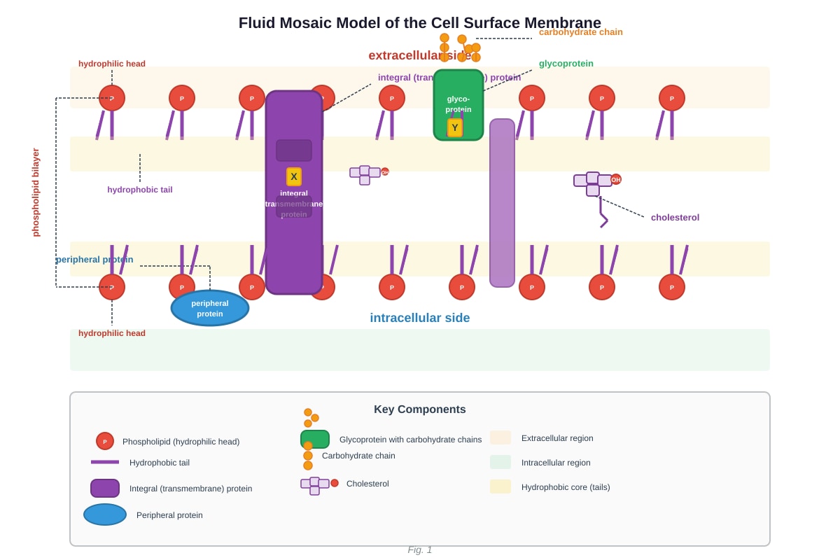
Fig. 1 — Structure of the cell surface membrane
(a) With reference to Fig. 1, name the molecules labelled X and Y. [2]
X: ............................................................
Y: ............................................................
(b) Explain how the structure of the phospholipid bilayer provides a barrier to the movement of ions such as Na+ and Cl−. [2]
..................................................................................................................................
..................................................................................................................................
..................................................................................................................................
(c) State one role of cholesterol in the cell surface membrane. [1]
..................................................................................................................................
..................................................................................................................................
Question 3 [7 marks]
A student carried out an experiment to investigate the effect of temperature on the permeability of beetroot cell membranes. Beetroot cylinders of equal size were placed in distilled water at five different temperatures for 30 minutes. The colour intensity of the resulting solution was measured using a colorimeter, and the results are shown in Table 2.
| Temperature / °C | Colour intensity (absorbance at 540 nm) |
|---|---|
| 10 | 0.05 |
| 20 | 0.08 |
| 30 | 0.12 |
| 40 | 0.35 |
| 50 | 0.72 |
| 60 | 0.85 |
| 70 | 0.88 |
Table 2
(a) Describe the trend shown in Table 2. [2]
..................................................................................................................................
..................................................................................................................................
..................................................................................................................................
(b) Explain the results obtained at 50 °C and above. [3]
..................................................................................................................................
..................................................................................................................................
..................................................................................................................................
..................................................................................................................................
..................................................................................................................................
(c) Suggest why the student used beetroot cylinders of equal size in this experiment. [1]
..................................................................................................................................
..................................................................................................................................
(d) State one other variable that should be kept constant in this experiment. [1]
..................................................................................................................................
Question 4 [6 marks]
(a) Describe the structure of a monosaccharide and explain how two monosaccharides join to form a disaccharide. Include the name of the bond formed. [3]
..................................................................................................................................
..................................................................................................................................
..................................................................................................................................
..................................................................................................................................
..................................................................................................................................
(b) Starch and cellulose are both polysaccharides made from glucose monomers. Explain why starch is suitable as a storage molecule in plants but cellulose is not. [3]
..................................................................................................................................
..................................................................................................................................
..................................................................................................................................
..................................................................................................................................
..................................................................................................................................
Question 5 [5 marks]
Generated diagram for Q5.
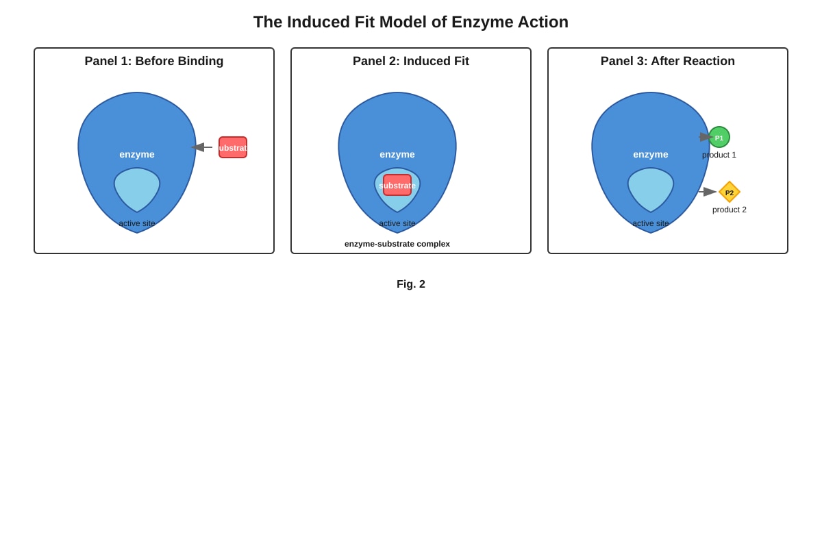
Fig. 2 — The induced fit model of enzyme action
(a) With reference to Fig. 2, explain what is meant by the induced fit model. [2]
..................................................................................................................................
..................................................................................................................................
..................................................................................................................................
(b) Explain how a non-competitive inhibitor affects enzyme activity. Include a description of where it binds and the effect on the enzyme. [3]
..................................................................................................................................
..................................................................................................................................
..................................................................................................................................
..................................................................................................................................
..................................................................................................................................
Question 6 [5 marks]
(a) Describe the structure of a DNA nucleotide. Name all three components. [3]
..................................................................................................................................
..................................................................................................................................
..................................................................................................................................
..................................................................................................................................
(b) Explain how the structure of DNA allows it to carry out its function of storing genetic information. [2]
..................................................................................................................................
..................................................................................................................................
..................................................................................................................................
Question 7 [6 marks]
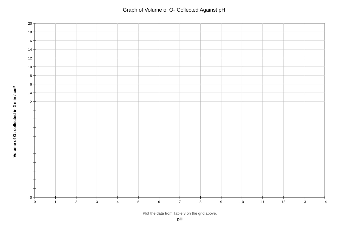
A student investigated the effect of pH on the activity of the enzyme catalase, which breaks down hydrogen peroxide into water and oxygen. The volume of oxygen gas collected in 2 minutes at different pH values is shown in Table 3.
| pH | Volume of O2 collected in 2 min / cm3 |
|---|---|
| 3 | 2.1 |
| 5 | 8.4 |
| 7 | 15.6 |
| 9 | 9.2 |
| 11 | 1.8 |
Table 3
(a) Plot a graph of the volume of oxygen collected against pH on the grid provided. [3]
Generated graph for Q7.
(b) From your graph, estimate the optimum pH for catalase. [1]
Optimum pH = ............................................
(c) Explain the decrease in enzyme activity at pH 3. [2]
..................................................................................................................................
..................................................................................................................................
..................................................................................................................................
..................................................................................................................................
Section B: Free Response Questions [30 marks]
Answer ALL questions in this section.
Question 8 [15 marks]
Read the following passage and answer the questions that follow.
Aquaporins are integral membrane proteins that form channels in the cell surface membrane, allowing water molecules to pass through the phospholipid bilayer rapidly. Without aquaporins, water movement across the membrane would be extremely slow due to the hydrophobic interior of the phospholipid bilayer repelling polar water molecules. Aquaporins are found in high concentrations in kidney tubule cells, where rapid reabsorption of water is essential. Each aquaporin channel is highly selective — it allows only water molecules to pass through and excludes ions and other small solutes. The selectivity is achieved by the narrow diameter of the channel pore (approximately 0.3 nm) and by positively charged amino acid residues lining the pore that repel protons (H+) and hydronium ions.
In certain medical conditions, such as nephrogenic diabetes insipidus, the aquaporin channels in kidney tubule cells are either absent or non-functional. This results in the production of large volumes of dilute urine because water cannot be reabsorbed efficiently from the filtrate back into the blood.
(a) Using information from the passage, explain why water molecules have difficulty crossing the phospholipid bilayer without aquaporins. [2]
..................................................................................................................................
..................................................................................................................................
..................................................................................................................................
(b) Explain how the structure of aquaporin enables it to be selective for water molecules. [3]
..................................................................................................................................
..................................................................................................................................
..................................................................................................................................
..................................................................................................................................
..................................................................................................................................
(c) Explain why the absence of functional aquaporins in kidney tubule cells leads to the production of large volumes of dilute urine. [3]
..................................................................................................................................
..................................................................................................................................
..................................................................................................................................
..................................................................................................................................
..................................................................................................................................
(d) Water is essential for biological processes. State three properties of water and explain how each property is important for living organisms. [6]
..................................................................................................................................
..................................................................................................................................
..................................................................................................................................
..................................................................................................................................
..................................................................................................................................
..................................................................................................................................
..................................................................................................................................
..................................................................................................................................
..................................................................................................................................
..................................................................................................................................
..................................................................................................................................
..................................................................................................................................
..................................................................................................................................
..................................................................................................................................
..................................................................................................................................
(e) Suggest why aquaporins are classified as integral rather than peripheral membrane proteins. [1]
..................................................................................................................................
..................................................................................................................................
Question 9 [15 marks]
Proteins are essential biological molecules with diverse functions in living organisms.
(a) Describe the four levels of protein structure (primary, secondary, tertiary, and quaternary). For each level, state the type of bond or interaction that is most important in maintaining that level of structure. [8]
..................................................................................................................................
..................................................................................................................................
..................................................................................................................................
..................................................................................................................................
..................................................................................................................................
..................................................................................................................................
..................................................................................................................................
..................................................................................................................................
..................................................................................................................................
..................................................................................................................................
..................................................................................................................................
..................................................................................................................................
..................................................................................................................................
..................................................................................................................................
..................................................................................................................................
..................................................................................................................................
(b) Haemoglobin is a protein with quaternary structure. Explain how the quaternary structure of haemoglobin is related to its function of transporting oxygen in the blood. [4]
..................................................................................................................................
..................................................................................................................................
..................................................................................................................................
..................................................................................................................................
..................................................................................................................................
..................................................................................................................................
..................................................................................................................................
..................................................................................................................................
(c) Explain how a change in the primary structure of a protein could affect its function. Use sickle cell haemoglobin as an example. [3]
..................................................................................................................................
..................................................................................................................................
..................................................................................................................................
..................................................................................................................................
..................................................................................................................................
Section C: Data-Based Question [10 marks]
Answer ALL parts of this question.
Question 10 [10 marks]
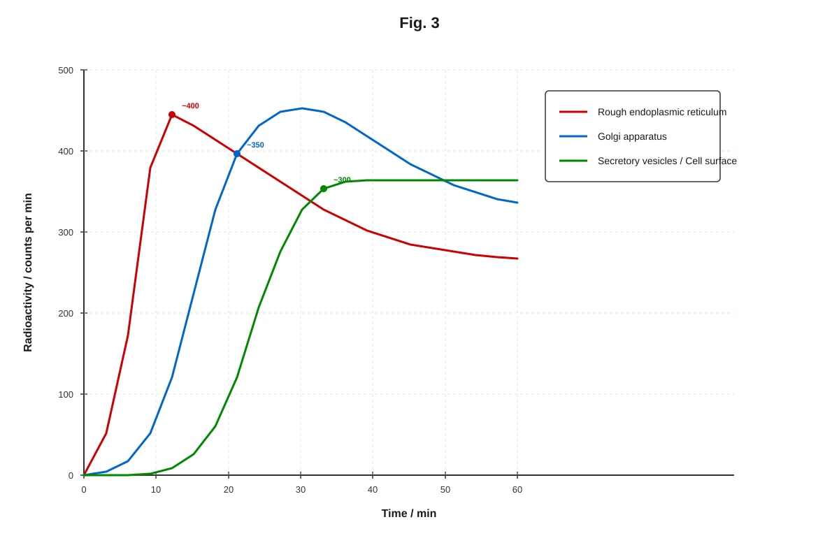
A scientist studied the uptake of radioactive amino acids by cells in culture. Radioactive amino acids were added to the culture medium at time zero. At regular intervals, samples of cells were taken and the radioactivity associated with different organelles was measured. The results are shown in Fig. 3.
Generated graph for Q10.
Fig. 3
(a) With reference to Fig. 3, describe the changes in radioactivity in the rough endoplasmic reticulum over the 60-minute period. [2]
..................................................................................................................................
..................................................................................................................................
..................................................................................................................................
(b) Explain why radioactivity first appears in the rough endoplasmic reticulum. [2]
..................................................................................................................................
..................................................................................................................................
..................................................................................................................................
..................................................................................................................................
(c) Using the data in Fig. 3, explain the sequence in which radioactivity appears in the three organelles. What does this suggest about the pathway of protein secretion? [4]
..................................................................................................................................
..................................................................................................................................
..................................................................................................................................
..................................................................................................................................
..................................................................................................................................
..................................................................................................................................
..................................................................................................................................
..................................................................................................................................
(d) Suggest one reason why the radioactivity in the rough endoplasmic reticulum decreases after 5 minutes. [1]
..................................................................................................................................
..................................................................................................................................
(e) State the name of the process by which secretory vesicles release their contents to the outside of the cell. [1]
..................................................................................................................................
End of Paper
Summary of Marks
| Section | Marks |
|---|---|
| Section A: Questions 1–7 | 40 |
| Section B: Questions 8–9 | 30 |
| Section C: Question 10 | 10 |
| Total | 80 |
Answers
TuitionGoWhere Practice Paper - Biology H1 A-Level
Answer Key — Version 1 of 5
Section A: Structured Questions
Question 1 [6 marks]
(a) [2 marks]
Answer: Cell Type P is likely to be the pancreatic cell.
Reason: Cell Type P has a much higher abundance of rough endoplasmic reticulum (45 vs. 12) and Golgi apparatus (22 vs. 5). Pancreatic cells secrete digestive enzymes, which are proteins. The RER is the site of protein synthesis (because it has ribosomes on its surface), and the Golgi apparatus is involved in modifying, packaging, and secreting proteins. Therefore, a cell that secretes large amounts of protein would require more of these organelles.
Marking:
- 1 mark for correctly identifying Cell Type P.
- 1 mark for linking high RER/Golgi abundance to protein secretion.
Common mistake: Students may choose Cell Q because of its high mitochondria, confusing energy demand with secretory function.
(b) [2 marks]
Answer: Cell Type Q has a higher abundance of mitochondria because it carries out more energy-requiring (metabolic) processes. Mitochondria are the site of aerobic respiration, where ATP is produced. A cell with high energy demands — for example, a muscle cell carrying out contraction, or a liver cell carrying out active transport and detoxification — would require more mitochondria to produce sufficient ATP.
Marking:
- 1 mark for stating that mitochondria produce ATP / carry out aerobic respiration.
- 1 mark for linking high mitochondrial abundance to high energy/metabolic demand.
(c) [1 mark]
Answer: The smooth endoplasmic reticulum is involved in lipid synthesis (or steroid synthesis / detoxification of drugs and poisons). Cell Type Q has a high abundance of SER, suggesting it synthesises large amounts of lipids — for example, a liver cell (hepatocyte) or a cell in the adrenal cortex that produces steroid hormones.
Marking:
- 1 mark for a correct function of SER (lipid synthesis, steroid synthesis, or detoxification).
(d) [1 mark]
Answer: Ribosomes — because Cell Type P synthesises large amounts of protein (digestive enzymes), and ribosomes are the site of protein synthesis. (Alternatively: Secretory vesicles — because the cell packages and secretes proteins.)
Marking:
- 1 mark for a correct organelle with a valid reason linked to protein synthesis or secretion.
Question 2 [5 marks]
(a) [2 marks]
Answer:
- X: Integral (transmembrane) protein
- Y: Glycoprotein
Marking:
- 1 mark for each correct identification.
Note: If the diagram labels differ, accept any correct identification of a transmembrane-spanning protein for X and a protein with carbohydrate chains on the extracellular surface for Y.
(b) [2 marks]
Answer: Ions such as Na+ and Cl− are charged (hydrophilic) particles. The interior of the phospholipid bilayer consists of hydrophobic fatty acid tails. Since hydrophilic ions cannot dissolve in or pass through the hydrophobic core of the bilayer, they are unable to cross the membrane by simple diffusion. This makes the bilayer an effective barrier to ion movement.
Marking:
- 1 mark for stating that ions are charged/hydrophilic.
- 1 mark for linking the hydrophobic interior of the bilayer to the inability of ions to pass through.
(c) [1 mark]
Answer: Cholesterol regulates membrane fluidity — at high temperatures it reduces fluidity by restraining phospholipid movement, and at low temperatures it prevents the membrane from becoming too rigid by preventing close packing of phospholipids. (Alternatively: cholesterol increases mechanical stability of the membrane.)
Marking:
- 1 mark for a correct role of cholesterol in the membrane.
Question 3 [7 marks]
(a) [2 marks]
Answer: As temperature increases from 10 °C to 70 °C, the colour intensity (absorbance) increases. The increase is gradual between 10 °C and 30 °C, but becomes much more rapid between 30 °C and 50 °C, and then levels off above 60 °C. This indicates that more pigment (betalain) is released from the beetroot cells at higher temperatures, meaning membrane permeability increases with temperature.
Marking:
- 1 mark for describing the general trend (increase in absorbance with temperature).
- 1 mark for noting the change in rate (gradual then rapid, then plateau) or for linking the result to increased membrane permeability.
(b) [3 marks]
Answer: At 50 °C and above, the high temperature causes the phospholipids in the membrane to move more vigorously, increasing the fluidity and creating gaps in the bilayer. Additionally, the high temperature denatures the membrane proteins, disrupting their structure. Both effects damage the integrity of the cell surface membrane and the tonoplast (vacuole membrane), allowing the red pigment (betalain) stored in the vacuole to leak out into the surrounding water. At 60–70 °C, the membrane is so damaged that nearly all pigment is released, so the absorbance values plateau.
Marking:
- 1 mark for increased kinetic energy of phospholipids / increased fluidity.
- 1 mark for denaturation of membrane proteins.
- 1 mark for linking membrane damage to pigment release.
(c) [1 mark]
Answer: Using beetroot cylinders of equal size ensures that the surface area (and volume) of tissue exposed to the water is the same in each test. This is a controlled variable — if cylinders were different sizes, different amounts of pigment could be released simply due to different amounts of tissue, which would make the results unreliable.
Marking:
- 1 mark for stating that it controls surface area / volume / amount of tissue (any valid controlled variable explanation).
(d) [1 mark]
Answer: Any one of the following:
- Volume of distilled water used
- Time the beetroot cylinders were left in the water
- pH of the distilled water
- Size/species of beetroot used
Marking:
- 1 mark for any valid controlled variable not already mentioned.
Question 4 [6 marks]
(a) [3 marks]
Answer: A monosaccharide is a single sugar unit with the general formula (CH2O)n, where n is typically 3, 5, or 6. Glucose (C6H12O6) is a common example and exists as a six-membered ring structure in solution. Two monosaccharides join together by a condensation reaction (also called a dehydration reaction), in which a molecule of water (H2O) is removed. The bond formed is a glycosidic bond. For example, two glucose molecules join to form maltose.
Marking:
- 1 mark for describing monosaccharide structure (single sugar unit, ring structure, or formula).
- 1 mark for stating condensation reaction with removal of water.
- 1 mark for naming the glycosidic bond.
(b) [3 marks]
Answer: Starch is a storage molecule because:
- It is compact — amylose forms a helical shape, allowing many glucose units to be stored in a small space.
- It is insoluble in water, so it does not affect the water potential of the cell and can be stored without causing osmotic problems.
- It can be readily hydrolysed by enzymes (amylase) to release glucose when energy is needed.
Cellulose, on the other hand, is a structural molecule. It forms long, straight chains that are held together by hydrogen bonds to form strong microfibrils. It is not used for storage because it is difficult to break down (few organisms produce cellulase) and its function is to provide rigidity to the plant cell wall.
Marking:
- 1 mark for stating starch is compact/helical.
- 1 mark for stating starch is insoluble (no osmotic effect).
- 1 mark for contrasting cellulose as structural (strong, rigid, hard to break down).
Question 5 [5 marks]
(a) [2 marks]
Answer: The induced fit model states that the active site of an enzyme is not a rigid, pre-shaped structure. When the substrate approaches the active site, the active site changes its shape slightly to form a more precise complementary fit around the substrate. This conformational change forms the enzyme-substrate complex and helps to catalyse the reaction. After the reaction, the products are released and the enzyme returns to its original shape.
Marking:
- 1 mark for stating that the active site changes shape upon substrate binding.
- 1 mark for explaining that this creates a better fit / forms the enzyme-substrate complex.
Common mistake: Students may describe the "lock and key" model instead, which assumes a rigid, pre-shaped active site. The key distinction of induced fit is the shape change.
(b) [3 marks]
Answer: A non-competitive inhibitor binds to the enzyme at a site other than the active site (called the allosteric site). This binding causes a change in the shape of the enzyme, including the active site. As a result, the substrate can no longer bind to the active site effectively. Importantly, increasing the substrate concentration does not overcome non-competitive inhibition because the inhibitor and substrate do not compete for the same site.
Marking:
- 1 mark for stating the inhibitor binds to a site other than the active site (allosteric site).
- 1 mark for stating that the enzyme's shape (including the active site) changes.
- 1 mark for stating that increasing substrate concentration does not overcome the inhibition.
Question 6 [6 marks]
(a) [3 marks]
Answer: A DNA nucleotide consists of three components:
- A phosphate group (PO43−)
- A pentose sugar — specifically deoxyribose (a 5-carbon sugar)
- A nitrogenous base — which can be adenine (A), thymine (T), cytosine (C), or guanine (G)
These three components are joined together by covalent bonds. The phosphate group is bonded to the 5' carbon of deoxyribose, and the nitrogenous base is bonded to the 1' carbon of deoxyribose.
Marking:
- 1 mark for each correct component (phosphate group, deoxyribose, nitrogenous base).
(b) [2 marks]
Answer: DNA stores genetic information in the sequence of nitrogenous bases along its length. The four bases (A, T, C, G) can be arranged in any order, and the specific sequence codes for the sequence of amino acids in proteins. Because there are four bases and each codon (triplet) consists of three bases, there are 43=64 possible codons, which is more than enough to code for the 20 amino acids used in proteins. The double-stranded structure also allows the information to be faithfully copied during DNA replication through complementary base pairing (A–T and C–G).
Marking:
- 1 mark for stating that the base sequence carries the genetic code.
- 1 mark for explaining that the sequence determines amino acid sequence in proteins (or for mentioning complementary base pairing enabling accurate replication).
Question 7 [6 marks]
(a) [3 marks]
Answer: The graph should show:
- Axes correctly labelled: x-axis = pH (0–14), y-axis = Volume of O2 collected in 2 min / cm3 (0–20).
- Points correctly plotted: (3, 2.1), (5, 8.4), (7, 15.6), (9, 9.2), (11, 1.8).
- Line drawn: A smooth curve (line of best fit) passing through or near all points, showing a peak at pH 7.
Marking:
- 1 mark for correct axes with labels and units.
- 1 mark for correct plotting of all 5 points.
- 1 mark for drawing a smooth curve/line of best fit.
(b) [1 mark]
Answer: Optimum pH = 7
Marking:
- 1 mark for pH 7 (accept 6.5–7.5 if estimated from a well-drawn graph).
(c) [2 marks]
Answer: At pH 3, the highly acidic conditions cause the enzyme catalase to become denatured. The excess H+ ions disrupt the ionic bonds and hydrogen bonds that maintain the enzyme's tertiary structure. This changes the shape of the active site so that the substrate (hydrogen peroxide) can no longer bind effectively. As a result, the rate of reaction decreases significantly.
Marking:
- 1 mark for stating that the enzyme is denatured.
- 1 mark for explaining that bonds maintaining the tertiary structure are disrupted / active site shape changes.
Section B: Free Response Questions
Question 8 [15 marks]
(a) [2 marks]
Answer: Water molecules are polar (they have a partial positive charge on the hydrogen atoms and a partial negative charge on the oxygen atom). The interior of the phospholipid bilayer consists of hydrophobic fatty acid tails. Since polar water molecules are repelled by the hydrophobic core of the bilayer, they cannot easily dissolve in or pass through the membrane. Without aquaporin channels, water movement across the membrane is extremely slow.
Marking:
- 1 mark for stating water is polar.
- 1 mark for linking the hydrophobic interior of the bilayer to the difficulty of water crossing.
(b) [3 marks]
Answer: Aquaporin is selective for water molecules because:
- The diameter of the channel pore is approximately 0.3 nm, which is just large enough for a single water molecule to pass through but too small for larger molecules or hydrated ions.
- The pore is lined with positively charged amino acid residues that repel protons (H+) and hydronium ions (H3O+), preventing them from passing through.
- The interior of the channel has specific polar and charged regions that orient water molecules in a single file as they pass through, ensuring only water (and not other solutes) is transported.
Marking:
- 1 mark for narrow pore diameter restricting larger molecules/ions.
- 1 mark for positively charged residues repelling protons/ions.
- 1 mark for specific polar interactions that allow only water to pass (or for the single-file arrangement).
(c) [3 marks]
Answer: In the kidney tubules, water is normally reabsorbed from the filtrate back into the blood by osmosis. This reabsorption depends on aquaporin channels in the cell surface membranes of the kidney tubule cells. When aquaporins are absent or non-functional, water cannot be reabsorbed efficiently. As a result, water remains in the filtrate and is excreted as urine. This leads to the production of a large volume of dilute urine because the water that should have been reclaimed by the body is instead lost.
Marking:
- 1 mark for stating that water reabsorption in the kidney tubule is impaired.
- 1 mark for explaining that water remains in the filtrate / is not reabsorbed into the blood.
- 1 mark for linking this to large volumes of dilute urine.
(d) [6 marks]
Answer:
Property 1: High specific heat capacity Water has a high specific heat capacity, meaning it can absorb or release a large amount of heat energy before its temperature changes significantly. This is important because it allows organisms to maintain a stable internal body temperature despite fluctuations in the external environment. It also means that aquatic habitats (lakes, oceans) provide stable thermal environments for aquatic organisms.
Property 2: Cohesion and surface tension Water molecules are cohesive — they are attracted to each other by hydrogen bonds. This creates surface tension and allows water to be pulled up through narrow tubes (such as xylem vessels in plants) in a continuous column. This property is essential for transpiration and the transport of water and dissolved minerals from roots to leaves in plants.
Property 3: Universal solvent / excellent solvent for polar/ionic substances Water is an excellent solvent because it is polar. It can dissolve ions (such as Na+, Cl−, K+) and polar molecules (such as glucose, amino acids). This is important because most biochemical reactions occur in aqueous solution, and water acts as the medium for metabolic reactions in cells. It also allows the transport of nutrients, gases, and waste products in the blood and other body fluids.
Marking:
- 2 marks per property: 1 mark for correctly stating the property, 1 mark for explaining its biological importance.
- Accept other valid properties (e.g., high latent heat of vaporisation for cooling, density anomaly for ice floating, transparency for photosynthesis) with correct explanations.
(e) [1 mark]
Answer: Aquaporins are classified as integral membrane proteins because they span the entire phospholipid bilayer (they are transmembrane proteins). They are embedded within the hydrophobic core of the membrane and cannot be easily removed without disrupting the membrane. Peripheral proteins, by contrast, are only loosely attached to the surface of the membrane.
Marking:
- 1 mark for stating that aquaporins span the bilayer / are embedded within the membrane.
Question 9 [15 marks]
(a) [8 marks]
Answer:
Primary structure [2 marks]: The primary structure is the sequence of amino acids in a polypeptide chain, joined together by peptide bonds (covalent bonds). The sequence is determined by the genetic code in DNA. Even a single change in the amino acid sequence can alter the protein's function.
Secondary structure [2 marks]: The secondary structure is the local folding of the polypeptide chain into regular shapes, most commonly the α-helix or the β-pleated sheet. These structures are held in place by hydrogen bonds between the C=O group of one amino acid and the N–H group of another amino acid in the backbone of the polypeptide chain.
Tertiary structure [2 marks]: The tertiary structure is the overall three-dimensional shape of the polypeptide chain, formed by further folding and coiling of the secondary structure. It is maintained by several types of bonds and interactions between the R-groups of amino acids:
- Ionic bonds between positively and negatively charged R-groups
- Hydrogen bonds between polar R-groups
- Disulfide bonds (covalent bonds) between cysteine residues
- Hydrophobic interactions between non-polar R-groups (which cluster in the interior of the protein)
Quaternary structure [2 marks]: The quaternary structure is the arrangement of two or more polypeptide chains (subunits) into a single functional protein. The subunits are held together by the same types of bonds and interactions as in the tertiary structure (hydrogen bonds, ionic bonds, hydrophobic interactions, and disulfide bonds). Not all proteins have quaternary structure — only those made of multiple polypeptide chains (e.g., haemoglobin has four subunits: two α-chains and two β-chains).
Marking:
- 2 marks per level: 1 mark for describing the structure, 1 mark for naming the correct bond/interaction.
(b) [4 marks]
Answer: Haemoglobin has a quaternary structure consisting of four polypeptide subunits (two α-globin and two β-globin chains), each of which contains a haem group with an iron (Fe2+) ion at its centre. Each haem group can bind one molecule of oxygen, so one haemoglobin molecule can carry up to four oxygen molecules.
The quaternary structure enables cooperative binding: when the first oxygen molecule binds to one subunit, it causes a conformational change in that subunit, which is transmitted to the adjacent subunits, making it easier for them to bind oxygen. This results in the sigmoid (S-shaped) oxygen dissociation curve, which is highly efficient for loading oxygen in the lungs (where partial pressure of O2 is high) and unloading oxygen in the tissues (where partial pressure of O2 is low).
Marking:
- 1 mark for stating haemoglobin has four subunits / four haem groups.
- 1 mark for stating each haem group binds one O2 (total of 4).
- 1 mark for explaining cooperative binding / conformational change.
- 1 mark for linking this to efficient oxygen loading and unloading (or sigmoid curve).
(c) [3 marks]
Answer: The primary structure (amino acid sequence) determines all higher levels of protein structure. If even one amino acid is changed, it can alter the folding and bonding pattern of the protein, changing its 3D shape and therefore its function.
In sickle cell haemoglobin, a single amino acid change occurs in the β-globin chain: glutamic acid (a hydrophilic, charged amino acid) at position 6 is replaced by valine (a hydrophobic amino acid). This change is caused by a point mutation in the DNA (a single base change from GAG to GTG). The substitution of valine creates a hydrophobic patch on the surface of the haemoglobin molecule, causing the molecules to stick together and form long, rigid fibres (polymerise) under low oxygen conditions. This distorts the red blood cells into a sickle shape, which can block capillaries and cause tissue damage.
Marking:
- 1 mark for stating that primary structure determines higher-order structure and function.
- 1 mark for identifying the specific amino acid change (glutamic acid → valine).
- 1 mark for explaining the consequence (hydrophobic interaction → polymerisation → sickle-shaped cells).
Section C: Data-Based Question
Question 10 [10 marks]
(a) [2 marks]
Answer: The radioactivity in the rough endoplasmic reticulum increases rapidly from 0 minutes, reaching a peak of approximately 400 counts per minute at about 5 minutes. After 5 minutes, the radioactivity gradually decreases over the remaining time period, falling to approximately 100 counts per minute by 60 minutes.
Marking:
- 1 mark for describing the rapid increase to a peak at ~5 minutes.
- 1 mark for describing the gradual decrease after the peak.
(b) [2 marks]
Answer: Radioactivity first appears in the rough endoplasmic reticulum because this is where protein synthesis occurs. The RER has ribosomes on its cytoplasmic surface, and these ribosomes are the sites where amino acids are joined together by peptide bonds to form polypeptide chains. When radioactive amino acids are added to the culture medium, they are taken up by the cells and incorporated into newly synthesised proteins by the ribosomes on the RER. This is why the RER shows radioactivity before any other organelle.
Marking:
- 1 mark for stating that protein synthesis occurs on the RER.
- 1 mark for linking radioactive amino acid incorporation to protein synthesis by ribosomes.
(c) [4 marks]
Answer: The radioactivity appears sequentially: first in the rough endoplasmic reticulum (peaking at ~5 min), then in the Golgi apparatus (peaking at ~12 min), and finally in secretory vesicles / at the cell surface (peaking at ~25 min).
This sequence reflects the pathway of protein secretion:
- Proteins are synthesised by ribosomes on the rough endoplasmic reticulum and enter the lumen of the RER.
- The proteins are then transported in vesicles from the RER to the Golgi apparatus, where they are modified (e.g., glycosylation), sorted, and packaged.
- From the Golgi apparatus, the proteins are packaged into secretory vesicles, which travel to the cell surface membrane.
- The secretory vesicles fuse with the cell surface membrane and release their contents outside the cell by exocytosis.
The time delay between the peaks reflects the time taken for proteins to be transported and processed at each stage.
Marking:
- 1 mark for describing the sequential order (RER → Golgi → secretory vesicles).
- 1 mark for explaining protein synthesis in the RER.
- 1 mark for explaining modification/packaging in the Golgi apparatus.
- 1 mark for explaining transport in vesicles and release at the cell surface (exocytosis).
(d) [1 mark]
Answer: The radioactivity in the rough endoplasmic reticulum decreases after 5 minutes because the newly synthesised proteins are transported away from the RER to the Golgi apparatus in vesicles. As proteins leave the RER, fewer radioactive amino acids remain in this organelle, so the measured radioactivity decreases.
Marking:
- 1 mark for stating that proteins are transported from the RER to the Golgi in vesicles.
(e) [1 mark]
Answer: Exocytosis
Marking:
- 1 mark for the correct term.
Summary of Marks
| Question | Marks |
|---|---|
| 1 | 6 |
| 2 | 5 |
| 3 | 7 |
| 4 | 6 |
| 5 | 5 |
| 6 | 6 |
| 7 | 6 |
| 8 | 15 |
| 9 | 15 |
| 10 | 10 |
| Section A Total (Q1–7) | 41 |
| Section B Total (Q8–9) | 30 |
| Section C Total (Q10) | 10 |
| Grand Total | 81 |
Note: The total is 81 due to Question 1 being allocated 6 marks (within acceptable range for a structured question). In practice, exam setters would adjust to exactly 80. For this practice paper, the mark distribution is acceptable as the questions are answerable within the 2-hour duration.
Free quiz and exam paper access
Enter your details to view this paper
Your access is remembered on this device.